 E-submission
 TOTA
TOTA TOTS
TOTS
Articles
- Page Path
- HOME > J Musculoskelet Trauma > Volume 26(2); 2013 > Article
-
Original Article
- Salvage Re-Fixation for the Failed Fixation of Pertrochanteric Fracture
- Kyung-Jae Lee, M.D., Byung-Woo Min, M.D., Ki-Cheor Bae, M.D., Dong-Hu Kim, M.D., Kyoung-Lag Lee, M.D.
-
Journal of the Korean Fracture Society 2013;26(2):105-111.
DOI: https://doi.org/10.12671/jkfs.2013.26.2.105
Published online: April 22, 2013
Department of Orthopedic Surgery, Keimyung University School of Medicine, Daegu, Korea.
- Address reprint requests to: Byung-Woo Min, M.D. Department of Orthopedic Surgery, Keimyung University School of Medicine, 56 Dalseong-ro, Jung-gu, Daegu 700-712, Korea. Tel: 82-53-250-7267, Fax: 82-53-250-7205, min@dsmc.or.kr
• Received: October 12, 2012 • Revised: November 24, 2012 • Accepted: November 24, 2012
Copyright © 2013 The Korean Fracture Society
- 1,446 Views
- 14 Download
- 1 Crossref
Abstract
-
Purpose
- The purpose of this study is to evaluate the clinical and radiological result in patients who got salvage re-fixation for the failed fixation of pertrochanteric fracture retrospectively.
-
Materials and Methods
- Between 1992 and 2009, 21 patients who could be followed-up for more than 1 year after salvage re-fixation for the failed fixation of pertrochanteric fracture were enrolled in this study. There were 16 men and 5 women. The mean age was 53 years (19-84 years) at the index surgery and the mean follow-up was 6.4 years. We evaluated the clinical and radiographic results and postoperative complications.
-
Results
- Walking ability and pain were improved in all cases and the mean leg length discrepancy was improved from 2.5 cm (0-10 cm) preoperatively to 1 cm (0-4 cm) at the latest follow-up. Nineteen patients (90.5%) out of 21 patients achieved bony union at the final evaluation and the mean union time was 4 months (3-7 months). There were 2 cases of non-union who had not received bone graft as a complication.
-
Conclusion
- The clinical and radiological results of the salvage re-fixation for the failed fixation of pertrchanteric fracture were satisfactory in our study. Secure component fixation for the mechanical stability and proper bone graft for the improvement of bone biology are mandatory to improve the result.
- 1. Apel DM, Patwardhan A, Pinzur MS, Dobozi WR. Axial loading studies of unstable intertrochanteric fractures of the femur. Clin Orthop Relat Res, 1989;(246):156-164.
- 2. Bannister GC, Gibson AG, Ackroyd CE, Newman JH. The fixation and prognosis of trochanteric fractures. A randomized prospective controlled trial. Clin Orthop Relat Res, 1990;(254):242-246.
- 3. Barquet A, Mayora G, Fregeiro J, López L, Rienzi D, Francescoli L. The treatment of subtrochanteric nonunions with the long gamma nail: twenty-six patients with a minimum 2-year follow-up. J Orthop Trauma, 2004;18:346-353.
- 4. Bridle SH, Patel AD, Bircher M, Calvert PT. Fixation of intertrochanteric fractures of the femur. A randomised prospective comparison of the gamma nail and the dynamic hip screw. J Bone Joint Surg Br, 1991;73:330-334.
- 5. Chang JS, Oh HK. Blade plate fixation for failed internal fixation of intertrochanteric hip fractures. J Korean Hip Soc, 2006;18:182-188.
- 6. Choi CU, Kim YH, Song JM, Kim HS. Clinical study for the complications of unstable intertrochanteric femoral fracture. J Korean Orthop Assoc, 1993;28:683-693.
- 7. de Vries JS, Kloen P, Borens O, Marti RK, Helfet DL. Treatment of subtrochanteric nonunions. Injury, 2006;37:203-211.
- 8. Ferguson GM, Cabanela ME, Ilstrup DM. Total hip arthroplasty after failed intertrochanteric osteotomy. J Bone Joint Surg Br, 1994;76:252-257.
- 9. Galanakis IA, Steriopoulos KA, Dretakis EK. Correct placement of the screw or nail in trochanteric fractures. Effect of the initial placement in the migration. Clin Orthop Relat Res, 1995;(313):206-213.
- 10. Gotfried Y. The lateral trochanteric wall: a key element in the reconstruction of unstable pertrochanteric hip fractures. Clin Orthop Relat Res, 2004;(425):82-86.
- 11. Haidukewych GJ, Berry DJ. Salvage of failed internal fixation of intertrochanteric hip fractures. Clin Orthop Relat Res, 2003;(412):184-188.
- 12. Haidukewych GJ, Berry DJ. Nonunion of fractures of the subtrochanteric region of the femur. Clin Orthop Relat Res, 2004;(419):185-188.
- 13. Haidukewych GJ, Berry DJ. Salvage of failed treatment of hip fractures. J Am Acad Orthop Surg, 2005;13:101-109.
- 14. Han SK, Kim YS, Lee KH, Kim YH, Park C, Choi NY. Treatment of unstable intertrochanteric fractures of the femur by wayne-county reduction in elderly patients. J Korean Hip Soc, 2001;13:338-345.
- 15. Hardy DC, Descamps PY, Krallis P, et al. Use of an intramedullary hip-screw compared with a compression hip-screw with a plate for intertrochanteric femoral fractures. A prospective, randomized study of one hundred patients. J Bone Joint Surg Am, 1998;80:618-630.
- 16. Hornby R, Evans JG, Vardon V. Operative or conservative treatment for trochanteric fractures of the femur. A randomised epidemiological trial in elderly patients. J Bone Joint Surg Br, 1989;71:619-623.
- 17. Hughston JC. Interochanteric fractures of the femur (hip). Orthop Clin North Am, 1974;5:585-600.
- 18. Jensen JS. Classification of trochanteric fractures. Acta Orthop Scand, 1980;51:803-810.
- 19. Jensen JS. Trochanteric fractures. An epidemiological, clinical and biomechanical study. Acta Orthop Scand Suppl, 1981;188:1-100.
- 20. Kim WY, Han CH, Park JI, Kim JY. Failure of intertrochanteric fracture fixation with a dynamic hip screw in relation to pre-operative fracture stability and osteoporosis. Int Orthop, 2001;25:360-362.
- 21. Koval KJ, Skovron ML, Aharonoff GB, Meadows SE, Zuckerman JD. Ambulatory ability after hip fracture. A prospective study in geriatric patients. Clin Orthop Relat Res, 1995;(310):150-159.
- 22. LaVelle DG. Canale ST. Fracture of hip. In: Campbell's operative orthopaedics, 2003;10th ed. Philadelphia, Mosby. 2873-2938.
- 23. Lorich DG, Geller DS, Nielson JH. Osteoporotic pertrochanteric hip fractures: management and current controversies. Instr Course Lect, 2004;53:441-454.
- 24. Madsen JE, Naess L, Aune AK, Alho A, Ekeland A, Strømsøe K. Dynamic hip screw with trochanteric stabilizing plate in the treatment of unstable proximal femoral fractures: a comparative study with the Gamma nail and compression hip screw. J Orthop Trauma, 1998;12:241-248.
- 25. Mainds CC, Newman RJ. Implant failures in patients with proximal fractures of the femur treated with a sliding screw device. Injury, 1989;20:98-100.
- 26. Mariani EM, Rand JA. Nonunion of intertrochanteric fractures of the femur following open reduction and internal fixation. Results of second attempts to gain union. Clin Orthop Relat Res, 1987;(218):81-89.
- 27. Mariani EM, Rand JA. Subcapital fractures after open reduction and internal fixation of intertrochanteric fractures of the hip. Report of three cases. Clin Orthop Relat Res, 1989;(245):165-168.
- 28. May JM, Chacha PB. Displacements of trochanteric fractures and their influence on reduction. J Bone Joint Surg Br, 1968;50:318-323.
- 29. Park J, Kim SG, Yoon HK, Yang KH. The treatment of nonisthmal femoral shaft nonunions with im nail exchange versus augmentation plating. J Orthop Trauma, 2010;24:89-94.
- 30. Pervez H, Parker MJ, Pryor GA, Lutchman L, Chirodian N. Classification of trochanteric fracture of the proximal femur: a study of the reliability of current systems. Injury, 2002;33:713-715.
- 31. Rha JD, Kim YH, Yoon SI, Park TS, Lee MH. Factors affecting sliding of the lag screw in intertrochanteric fractures. Int Orthop, 1993;17:320-324.
- 32. Said GZ, Farouk O, El-Sayed A, Said HG. Salvage of failed dynamic hip screw fixation of intertrochanteric fractures. Injury, 2006;37:194-202.
- 33. Sarathy MP, Madhavan P, Ravichandran KM. Nonunion of intertrochanteric fractures of the femur. Treatment by modified medial displacement and valgus osteotomy. J Bone Joint Surg Br, 1995;77:90-92.
- 34. Sexson SB, Lehner JT. Factors affecting hip fracture mortality. J Orthop Trauma, 1987;1:298-305.
- 35. Templeman D, Baumgaertner MR, Leighton RK, Lindsey RW, Moed BR. Reducing complications in the surgical treatment of intertrochanteric fractures. Instr Course Lect, 2005;54:409-415.
- 36. Thomas AP. Dynamic hip screws that fail. Injury, 1991;22:45-46.
- 37. White BL, Fisher WD, Laurin CA. Rate of mortality for elderly patients after fracture of the hip in the 1980's. J Bone Joint Surg Am, 1987;69:1335-1340.
- 38. Wiss DA. What's new in orthopaedic trauma. J Bone Joint Surg Am, 2001;83:1762-1772.
- 39. Wu CC. Locked nailing for shortened subtrochanteric nonunions: a one-stage treatment. Clin Orthop Relat Res, 2009;467:254-259.
- 40. Wu CC, Shih CH, Chen WJ, Tai CL. Treatment of cutout of a lag screw of a dynamic hip screw in an intertrochanteric fracture. Arch Orthop Trauma Surg, 1998;117:193-196.
REFERENCES
Fig. 1A diagram shows author's treatment algorism for the failed fixation of pertrochanteric fracture. DHS: Dynamic hip screw, IM: Intramudullary.

Fig. 2
A 64-years-old male with left hip pain.
(A) Preoperative radiograph shows the non-union of pertrochanteric fracture with broken angle blade plate.
(B) Immediate postoperative radiograph shows dynamic hip screw fixation due to the inferior head defect. During the re-fixation, bone graft was added due to the bone defect on the non-union site.
(C) Radiograph after one year shows solid fracture union.

Fig. 3
A 69-years-old male with left hip pain.
(A) Preoperative radiograph shows infected non-union of fracture site with large bone defect.
(B) After infection control, 90 degree angle blade plate was used due to the superior head defect. During the re-fixation, bone graft was done.
(C) One year after the operation, the fracture site was well united.
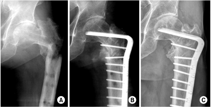
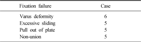
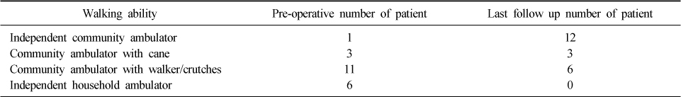
Figure & Data
REFERENCES
Citations
Citations to this article as recorded by 
- Salvage treatment of failed internal fixation of intertrochanteric fractures: What factors determine the failure of treatment?
Byung-Woo Min, Kyung-Jae Lee, Jong-Keon Oh, Chul-Hyun Cho, Jae-Woo Cho, Beom-Soo Kim
Injury.2020; 51(2): 367. CrossRef
 Cite
CiteSalvage Re-Fixation for the Failed Fixation of Pertrochanteric Fracture
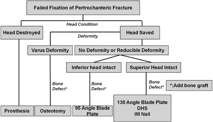
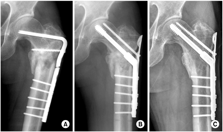

Fig. 1
A diagram shows author's treatment algorism for the failed fixation of pertrochanteric fracture. DHS: Dynamic hip screw, IM: Intramudullary.
Fig. 2
A 64-years-old male with left hip pain.
(A) Preoperative radiograph shows the non-union of pertrochanteric fracture with broken angle blade plate.
(B) Immediate postoperative radiograph shows dynamic hip screw fixation due to the inferior head defect. During the re-fixation, bone graft was added due to the bone defect on the non-union site.
(C) Radiograph after one year shows solid fracture union.
Fig. 3
A 69-years-old male with left hip pain.
(A) Preoperative radiograph shows infected non-union of fracture site with large bone defect.
(B) After infection control, 90 degree angle blade plate was used due to the superior head defect. During the re-fixation, bone graft was done.
(C) One year after the operation, the fracture site was well united.
Fig. 1
Fig. 2
Fig. 3
Salvage Re-Fixation for the Failed Fixation of Pertrochanteric Fracture
Aspects of Primary Fixation Failure
Change of Walking Ability
Table 1
Aspects of Primary Fixation Failure
Table 2
Change of Walking Ability

