 E-submission
 TOTA
TOTA TOTS
TOTS
Articles
- Page Path
- HOME > J Musculoskelet Trauma > Volume 24(2); 2011 > Article
-
Case Report
- Periprosthetic Fracture after Proximal Humeral Intramedullary Nail, Treated by Functional Bracing: A Case Report
- Jae-Hyuk Shin, M.D., Ho-Guen Chang, M.D., Young-Woo Kim, M.D., Nam-Kyou Rhee, M.D., Yong-Bok Park, M.D., Yong-Kuk Kim, M.D.
-
Journal of the Korean Fracture Society 2011;24(2):185-190.
DOI: https://doi.org/10.12671/jkfs.2011.24.2.185
Published online: April 13, 2011
Department of Orthopedic Surgery, Hangang Sacred Heart Hospital, College of Medicine, Hallym University, Seoul, Korea.
*Department of Orthopedic Surgery, Shin Hospital, Suwon, Korea.
- Address reprint requests to: Ho-Guen Chang, M.D. Department of Orthopedic Surgery, Hangang Sacred Heart Hospital, College of Medicine, Hallym University, 94-200, Yeongdeungpo-dong, Yeongdeungpo-gu, Seoul 150-037, Korea. Tel: 82-2-2639-5650, Fax: 82-2-2639-5654, hgchang@hallym.or.kr
• Received: August 29, 2010 • Revised: November 16, 2010 • Accepted: December 21, 2010
Copyright © 2011 The Korean Fracture Society
- 1,170 Views
- 3 Download
- 2 Crossref
Abstract
- Periprosthetic fracture following a proximal humeral intramedullary (IM) nailing is rarely reported neither for its occurrence nor for its treatment. Proximal humeral IM nail (Acumed, LLC, Hillsboro, OR, USA) has been increasingly reported of its successful treatment outcomes, yet there is paucity of data describing its complications. Here we report a 26 year-old female patient, who sustained a proximal humerus fracture which was initially successfully treated by proximal humeral IM nail, and was complicated by a periprosthetic fracture distal to the nail tip at postoperative 4 months. Serial application of U-shaped coaptation splint, hanging cast, and functional bracing resulted in satisfactory clinical outcome. Periprosthetic fracture after proximal humerus IM nail can occur by a low energy injury, which need to reminded in treating young and sports-active patients.
- 1. Boyd AD Jr, Thornhill TS, Barnes CL. Fractures adjacent to humeral prostheses. J Bone Joint Surg Am, 1992;74:1498-1504.Article
- 2. Chin PY, Sperling JW, Cofield RH, Schleck C. Complications of total shoulder arthroplasty: are they fewer or different? J Shoulder Elbow Surg, 2006;15:19-22.Article
- 3. Groh GI, Heckman MM, Wirth MA, Curtis RJ, Rockwood CA Jr. Treatment of fractures adjacent to humeral prostheses. J Shoulder Elbow Surg, 2008;17:85-89.Article
- 4. Kang HJ, Lee DY, Sung SY, Hahn SB. Intramedullary nailing for the fracture of proximal humerus. J Korean Fract Soc, 2004;17:271-276.Article
- 5. Kazakos K, Lyras DN, Galanis V, et al. Internal fixation of proximal humerus fractures using the Polarus intramedullary nail. Arch Orthop Trauma Surg, 2007;127:503-508.ArticlePDF
- 6. Kent ME, Sinopidis C, Brown DJ, Frostick SP. The locking compression plate in periprosthetic humeral fractures. A review of two cases. Injury, 2005;36:1241-1245.Article
- 7. Lin J, Hou SM, Hang YS. Locked nailing for displaced surgical neck fractures of the humerus. J Trauma, 1998;45:1051-1057.Article
- 8. Sarmiento A, Zagorski JB, Zych GA, Latta LL, Capps CA. Functional bracing for the treatment of fractures of the humeral diaphysis. J Bone Joint Surg Am, 2000;82:478-486.Article
- 9. Sarraf KM, Singh R, Corbett SA. Distal humeral plating of an intramedullary nail periprosthetic fracture using a miss-a-nail technique: a case report. Cases J, 2009;2:6704. ArticlePDF
- 10. Seo GY, Ahn JK, Kim YW, Kim JH, Rhee JH. Treatment of humerus shaft fractures using hanging arm cast with plastic cast. J Korean Soc Fract, 1991;4:8-14.Article
- 11. Tsiridis E, Haddad FS, Gie GA. The management of periprosthetic femoral fractures around hip replacements. Injury, 2003;34:95-105.Article
- 12. Tsiridis E, Spence G, Gamie Z, El Masry MA, Giannoudis PV. Grafting for periprosthetic femoral fractures: strut, impaction or femoral replacement. Injury, 2007;38:688-697.Article
- 13. Worland RL, Kim DY, Arredondo J. Periprosthetic humeral fractures: management and classification. J Shoulder Elbow Surg, 1999;8:590-594.Article
- 14. Wright TW, Cofield RH. Humeral fractures after shoulder arthroplasty. J Bone Joint Surg Am, 1995;77:1340-1346.Article
REFERENCES
Fig. 1
Initial radiographs of a 26-year-old woman shows proximal humerus three-part comminuted fracture, involving the metaphysis.
(A) Scapular lateral view.
(B) Humerus antero-posterior view, and (C) Shoulder Axial view.

Fig. 2
Postoperative radiographs show internal fixation of the proximal humerus fracture using the Polarus IM nail (Acumed). The nail head was sunk beneath the humeral head in order to avoid subacromial impingement.
(A) Antero-posterior view, and (B) Lateral view.

Fig. 3Periprosthetic fracture developed 4 months after the IM nailing. The fracture developed at 2 cm distal to the IM nail tip, spirally advancing distantly toward the metaphysis. Radiographs suggested union of the previously sustained proximal humerus fracture, and stable fixation of the IM nail.

Fig. 4The IM nail was removed and U-shaped coaptation splint was applied following careful reduction. The previous proximal fracture manifested callus formation and uneventful fracture healing.
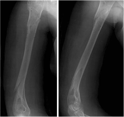
Fig. 5
(A) At 12 days after periprosthetic fracture hanging cast is applied.
(B) Initial hanging cast presents posterior angulation and varus angulation of the fracture site.
(C) The strap was lengthened and advanced toward volar aspect of the wrist to correct the posterior angulation and the varus angulation.


Figure & Data
REFERENCES
Citations
Citations to this article as recorded by 
- Locking compression plate fixation of periprosthetic distant humeral fracture after intramedullary nail for humeral shaft fracture: A case report
Mei-Ren Zhang, Kui Zhao, Jiang-Long Guo, Hai-Yun Chen
Trauma Case Reports.2022; 37: 100565. CrossRef - Distal Humeral Fixation of an Intramedullary Nail Periprosthetic Fracture
Hiren M. Divecha, Hans A. J. Marynissen, K. Erler, D. A. Fisher, J. Mayr, A. Sakamoto
Case Reports in Orthopedics.2013;[Epub] CrossRef
 Cite
CitePeriprosthetic Fracture after Proximal Humeral Intramedullary Nail, Treated by Functional Bracing: A Case Report
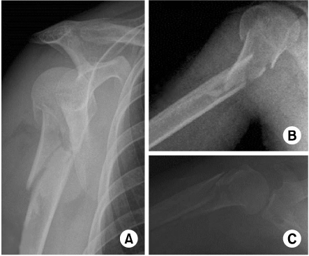
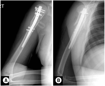
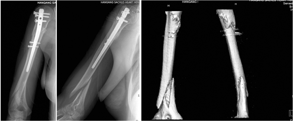

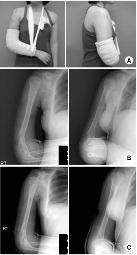
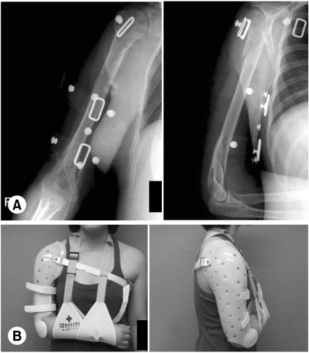
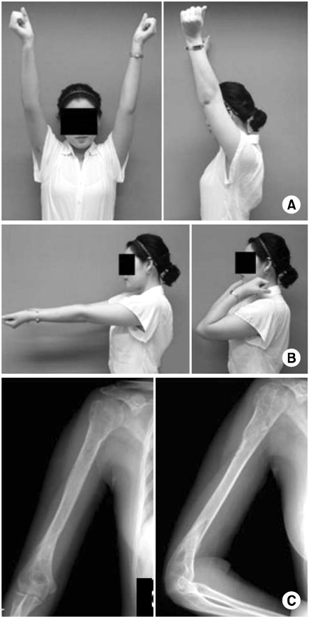
Fig. 1
Initial radiographs of a 26-year-old woman shows proximal humerus three-part comminuted fracture, involving the metaphysis.
(A) Scapular lateral view.
(B) Humerus antero-posterior view, and (C) Shoulder Axial view.
Fig. 2
Postoperative radiographs show internal fixation of the proximal humerus fracture using the Polarus IM nail (Acumed). The nail head was sunk beneath the humeral head in order to avoid subacromial impingement.
(A) Antero-posterior view, and (B) Lateral view.
Fig. 3
Periprosthetic fracture developed 4 months after the IM nailing. The fracture developed at 2 cm distal to the IM nail tip, spirally advancing distantly toward the metaphysis. Radiographs suggested union of the previously sustained proximal humerus fracture, and stable fixation of the IM nail.
Fig. 4
The IM nail was removed and U-shaped coaptation splint was applied following careful reduction. The previous proximal fracture manifested callus formation and uneventful fracture healing.
Fig. 5
(A) At 12 days after periprosthetic fracture hanging cast is applied.
(B) Initial hanging cast presents posterior angulation and varus angulation of the fracture site.
(C) The strap was lengthened and advanced toward volar aspect of the wrist to correct the posterior angulation and the varus angulation.
Fig. 6
At 3 weeks after periprosthetic fracture functional brace and arm sling were applied.
(A) The fracture angulation and alignment were tolerable.
(B) The elbow motion was gently allowed to prevent stiffness.
Fig. 7
By 40 days after periprosthetic fracture the patient presented ROM recovery both in the (A) shoulder joint, and in the (B) elbow joint.
(C) Radiographs show good alignment and callus formation suggesting successful union process.
Fig. 1
Fig. 2
Fig. 3
Fig. 4
Fig. 5
Fig. 6
Fig. 7
Periprosthetic Fracture after Proximal Humeral Intramedullary Nail, Treated by Functional Bracing: A Case Report

