 E-submission
 TOTA
TOTA TOTS
TOTS
Articles
- Page Path
- HOME > J Musculoskelet Trauma > Volume 24(2); 2011 > Article
-
Case Report
- Flexor Pollicis Longus Tendon Rupture as a Complication of a Closed Distal Radius Fracture: A Case Report
- Do-Young Kim, M.D., Eun-Min Seo, M.D., Woo-Dong Nam, M.D., Seung-Jae Park, M.D., Sang-Soo Lee, M.D.
-
Journal of the Korean Fracture Society 2011;24(2):191-194.
DOI: https://doi.org/10.12671/jkfs.2011.24.2.191
Published online: April 13, 2011
Institure for Skeletal Aging & Orthopedic Surgery, Hallym University College of Medicine, Chuncheon, Korea.
*Department of Orthopedic Surgery, Kangwon National University College of Medicine, Chuncheon, Korea.
- Address reprint requests to: Eun-min Seo, M.D. Department of Orthopedic Surgery, Chunchon Sacred Heart Hospital, Hallym University, 153, Gyo-dong, Chuncheon 200-704, Korea. Tel: 82-33-240-5781, Fax: 82-33-255-6244, seoem@hallym.or.kr
• Received: October 26, 2010 • Revised: December 11, 2010 • Accepted: January 25, 2011
Copyright © 2011 The Korean Fracture Society
- 1,141 Views
- 3 Download
- 1 Crossref
Abstract
- There are few reported cases of flexor pollicis longus tendon (FPL) rupture complicating a closed distal radius fracture. We report a case of FPL tendon rupture complicating a closed distal radius fracture. A 24-year-old male presented with a severe right wrist pain. He had a closed distal radius fracture that was treated by closed manual reduction. Three days later, he complained forearm pain and limitation of thumb motion. The physical examination revealed loss of active interphalangeal joint flexion of thumb. He was taken to the operating room. Intraoperatively, the FPL was found to be discontinuous at the level of the radius fracture site. The FPL was repaired by a modified Kessler technique, and the fracture was repaired with a volar plate. Clinicians must be cautious in possibility of tendon injury complicating a closed distal radius fracture and assessing patients with distal radius fracture following closed reduction.
- 1. Choi JY, Jung HJ, Kim HK, Lee JK, Chang IS. T-Plate fixation for fractures of distal radius. J Korean Fract Soc, 2004;17:350-358.Article
- 2. DiMatteo L, Wolf JM. Flexor carpi radialis tendon rupture as a complication of a closed distal radius fracture: a case report. J Hand Surg Am, 2007;32:818-820.Article
- 3. Hallett JP, Motta GR. Tendon ruptures in the hand with particular reference to attrition ruptures in the carpal tunnel. Hand, 1982;14:283-290.ArticlePDF
- 4. Kato N, Nemoto K, Arino H, Ichikawa T, Fujikawa K. Ruptures of flexor tendons at the wrist as a complication of fracture of the distal radius. Scand J Plast Reconstr Surg Hand Surg, 2002;36:245-248.Article
- 5. McMaster PE. Late ruptures of extensor and flexor pollicis longus tendons following Colles' fracture. J Bone Joint Surg Am, 1932;14:93-101.
- 6. McMaster PE. Tendon and muscle ruptures Clinical and experimental studies in the causes and location of subcutaneous ruptures. J Bone Joint Surg, 1933;15:705-722.
- 7. Roberts JO, Regan PJ, Roberts AH. Rupture of flexor pollicis longus as a complication of Colles' fracture: a case report. J Hand Surg Br, 1990;15:370-372.ArticlePDF
- 8. Suppaphol S, Woratanarat P, Channoom T. Flexor tendon rupture after distal radius fracture Report of 2 cases. J Med Assoc Thai, 2007;90:2695-2698.
- 9. Thomsen S, Falstie-Jensen S. Rupture of the flexor pollicis longus tendon associated with an ununited fracture of the scaphoid. J Hand Surg Am, 1988;13:220-222.Article
- 10. Yim SJ, Jeong YC, Yoon SR, Choi JG, Suh YS, Rah SK. Delayed rupture of the extensor pollicis longus due to fracture of the distal radius: a case report. J Korean Soc Fract, 1999;12:162-165.Article
REFERENCES
Fig. 1Anteroposterior and lateral radiographs of the right wrist show distal radius fracture with severe dorsal displacement and sharp anterior bony beak.

Fig. 2After manual reduction, anteroposterior and lateral radiographs of the right wrist show improvement of bony alignment but not perfect reduction.


Figure & Data
REFERENCES
Citations
Citations to this article as recorded by 
- Acute Rupture of Flexor Tendons as a Complication of Distal Radius Fracture
Youn Moo Heo, Sang Bum Kim, Kwang Kyoun Kim, Doo Hyun Kim, Won Keun Park
Journal of the Korean Orthopaedic Association.2015; 50(1): 60. CrossRef
 Cite
CiteFlexor Pollicis Longus Tendon Rupture as a Complication of a Closed Distal Radius Fracture: A Case Report
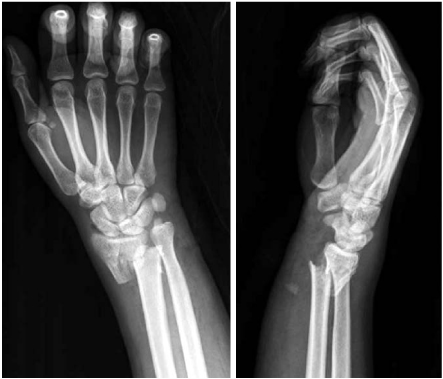
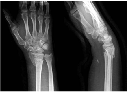
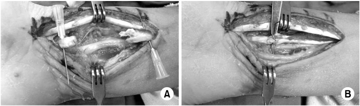
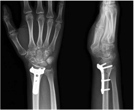
Fig. 1
Anteroposterior and lateral radiographs of the right wrist show distal radius fracture with severe dorsal displacement and sharp anterior bony beak.
Fig. 2
After manual reduction, anteroposterior and lateral radiographs of the right wrist show improvement of bony alignment but not perfect reduction.
Fig. 3
(A) Intraoperative photographs show rupture of the flexor pollicis longus tendon, (B) intraoperative photographs show that the flexor pollicis longus tendon is repaired by a modified Kessler technique.
Fig. 4
Eight weeks after surgery, anteroposterior and lateral radiographs of the right wrist show solid union.
Fig. 1
Fig. 2
Fig. 3
Fig. 4
Flexor Pollicis Longus Tendon Rupture as a Complication of a Closed Distal Radius Fracture: A Case Report

