 E-submission
 TOTA
TOTA TOTS
TOTS
Articles
- Page Path
- HOME > J Musculoskelet Trauma > Volume 24(2); 2011 > Article
-
Case Report
- Usefulness of Kyphoplasty in Sacral Insufficiency Fracture: A Case Report
- Soo Uk Chae, M.D., Yeung Jin Kim, M.D., Jung Hwan Yang, M.D., Ji Wan Lee, M.D.
-
Journal of the Korean Fracture Society 2011;24(2):174-177.
DOI: https://doi.org/10.12671/jkfs.2011.24.2.174
Published online: April 13, 2011
Department of Orthopeadic Surgery, School of Medicine, Wonkwang University, Iksan, Korea.
- Address reprint requests to: Jung Hwan Yang, M.D. Department of Orthopeadic Surgery, School of Medicine, Wonkwang University, 344-1, Shinyong-dong, Iksan 570-711, Korea. Tel: 82-63-472-5100, Fax: 82-63-472-5688, oschae68@hanmail.net
• Received: November 26, 2010 • Revised: December 22, 2010 • Accepted: March 3, 2011
Copyright © 2011 The Korean Fracture Society
- 1,035 Views
- 1 Download
- 1 Crossref
Abstract
- Kyphoplasty has recently attended as a potential treatment for sacral insufficiency fracture. We report a 85-years-old female patient with osteoporotic S1 insufficiency fracture with absence of trauma history treated with kyphoplasty which has no symptom improve with conservative treatment. Kyphoplasty is an effective and useful procedure in the treatment of the sacral insufficiency fracture, additionally reviewed of the literatures.
- 1. Atalay B, Caner H, Yilmaz C, Altinors N. Sacral kyphoplasty for relieving pain caused by sacral hemangioma. Spinal Cord, 2006;44:196-199.ArticlePDF
- 2. Bayley E, Srinivas S, Boszczyk BM. Clinical outcomes of sacroplasty in sacral insufficiency fractures: a review of the literature. Eur Spine J, 2009;18:1266-1271.ArticlePDF
- 3. Frey ME, Depalma MJ, Cifu DX, Bhagia SM, Carne W, Daitch JS. Percutaneous sacroplasty for osteoporotic sacral insufficiency fractures: a prospective, multicenter, observational pilot study. Spine J, 2008;8:367-373.Article
- 4. Garant M. Sacroplasty: a new treatment for sacral insufficiency fracture. J Vasc Interv Radiol, 2002;13:1265-1267.Article
- 5. Lee SE, Nam IH, Lee SS, Lee DH, Woo DH. Insufficiency fracture of the sacrum: a case report. J Korean Soc Spine Surg, 2001;8:172-175.Article
- 6. Lourie H. Spontaneous osteoporotic fracture of the sacrum. An unrecognized syndrome of the elderly. JAMA, 1982;248:715-717.Article
- 7. Lyders EM, Whitlow CT, Baker MD, Morris PP. Imaging and treatment of sacral insufficiency fractures. AJNR Am J Neuroradiol, 2010;31:201-210.Article
- 8. Smith DK, Dix JE. Percutaneous sacroplasty: long-axis injection technique. AJR Am J Roentgenol, 2006;186:1252-1255.Article
- 9. Waites MD, Mears SC, Richards AM, Mathis JM, Belkoff SM. A biomechanical comparison of lateral and posterior approaches to sacroplasty. Spine (Phila Pa 1976), 2008;33:E735-E738.Article
- 10. Whitlow CT, Yazdani SK, Reedy ML, Kaminsky SE, Berry JL, Morris PP. Investigating sacroplasty: technical considerations and finite element analysis of polymethylmethacrylate infusion into cadaveric sacrum. AJNR Am J Neuroradiol, 2007;28:1036-1041.Article
REFERENCES
Fig. 1Preoperative simple AP (A) and lateral (B) L-spine radiographs show the intervertebral disc space narrowing and spondylolisthesis between L4/L5.

Fig. 2Coronal image (A) and saggital image (B) of pelvic CT scan shows bilateral sacral insufficiency fracture with cortical discontinuity (white arrow). T2 (C) and T1-weighted saggital image (D) of lumbosacral MRI show perilesional edema (white arrow).


Figure & Data
REFERENCES
Citations
Citations to this article as recorded by 
- Pelvic Insufficiency Fracture in Severe Osteoporosis Patient
Woong Chae Na, Sang Hong Lee, Sung Jung, Hyun Woong Jang, Suenghwan Jo
Hip & Pelvis.2017; 29(2): 120. CrossRef
 Cite
CiteUsefulness of Kyphoplasty in Sacral Insufficiency Fracture: A Case Report
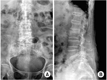
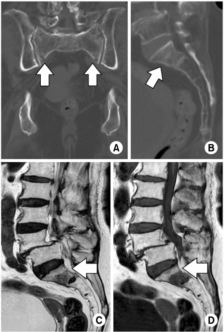
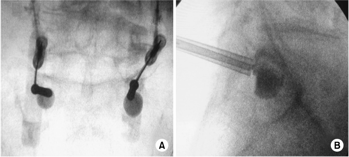
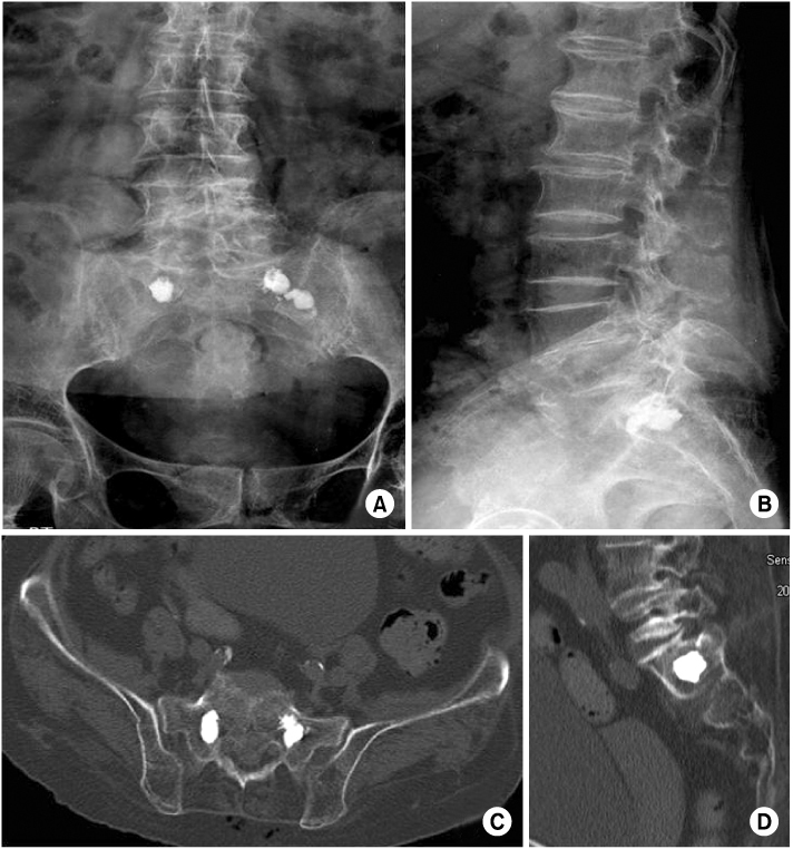
Fig. 1
Preoperative simple AP (A) and lateral (B) L-spine radiographs show the intervertebral disc space narrowing and spondylolisthesis between L4/L5.
Fig. 2
Coronal image (A) and saggital image (B) of pelvic CT scan shows bilateral sacral insufficiency fracture with cortical discontinuity (white arrow). T2 (C) and T1-weighted saggital image (D) of lumbosacral MRI show perilesional edema (white arrow).
Fig. 3
Under fluoscopy guidance with balloon assistance, intraoperative C-arm AP image (A) and lateral image (B) show balloon inflation at the sacral insufficiency fracture site.
Fig. 4
After fluoscopy-guided balloon-assisted sacroplasty, AP (A) and lateral (B) sacral radiographs, axial (C) and saggital (D) image of pelvic CT scan show the appearance of cement in the fracture area and no leakage of cement.
Fig. 1
Fig. 2
Fig. 3
Fig. 4
Usefulness of Kyphoplasty in Sacral Insufficiency Fracture: A Case Report

