 E-submission
 TOTA
TOTA TOTS
TOTS
Articles
- Page Path
- HOME > J Musculoskelet Trauma > Volume 29(1); 2016 > Article
-
Case Report
- Medial and Lateral Dual Plate Fixation for Osteoporotic Proximal Humerus Comminuted Fracture: 2 Case Reports
- Sam-Guk Park, M.D.
-
Journal of the Korean Fracture Society 2016;29(1):61-67.
DOI: https://doi.org/10.12671/jkfs.2016.29.1.61
Published online: January 19, 2016
Department of Orthopaedic Surgery, Yeungnam University Medical Center, Daegu, Korea.
- Address reprint requests to: Sam-Guk Park, M.D. Department of Orthopaedic Surgery, Yeungnam University Medical Center, 170 Hyeonchung-ro, Nam-gu, Daegu 42415, Korea. Tel: 82-53-620-3640, Fax: 82-53-628-4020, 70radiorth@naver.com
• Received: October 27, 2015 • Revised: November 23, 2015 • Accepted: November 23, 2015
Copyright © 2016 The Korean Fracture Society. All rights reserved.
This is an Open Access article distributed under the terms of the Creative Commons Attribution Non-Commercial License (http://creativecommons.org/licenses/by-nc/4.0) which permits unrestricted non-commercial use, distribution, and reproduction in any medium, provided the original work is properly cited.
- 1,093 Views
- 15 Download
- 3 Crossref
Abstract
- Some proximal humeral fractures in elderly patients are accompanied by medial metaphyseal comminution and quality of the bone is so poor that head preserving osteosynthesis seems to be amenable. In cases of medial metaphyseal comminution, lateral locking compression plate (LCP) fixation also has a tendency to become a matter of screw cut out or loss of fixation. The author reports on successful treatment of two osteoporotic proximal humeral fractures combined with medial meta-physeal comminution, with application of additional direct medial supporting plate fixation. Medial plate fixations were added when the fractures were still unstable after the conventional lateral LCP fixation and anterior circumflex humeral arteries had been ruptured before. The fixations were stable enough to start exercise immediately after surgery. The inclinations of the humeral neck were not changed until the last follow-up and clinical results were satisfactory without humeral head osteonecrosis which was a concern.
- 1. Oh JH, Kim YH. The current concepts in the treatment of proximal humerus fracture. J Korean Fract Soc, 2012;25:94-104.Article
- 2. Hawkins RJ, Bell RH, Gurr K. The three-part fracture of the proximal part of the humerus. Operative treatment. J Bone Joint Surg Am, 1986;68:1410-1414.Article
- 3. Jung HJ, Jeon IH, Chun JM. Arthroplasty for fractures of the proximal part of the humerus. J Korean Orthop Assoc, 2012;47:243-249.Article
- 4. Ricchetti ET, Warrender WJ, Abboud JA. Use of locking plates in the treatment of proximal humerus fractures. J Shoulder Elbow Surg, 2010;19:2 Suppl. 66-75.Article
- 5. Gardner MJ, Boraiah S, Helfet DL, Lorich DG. Indirect medial reduction and strut support of proximal humerus fractures using an endosteal implant. J Orthop Trauma, 2008;22:195-200.Article
- 6. He Y, He J, Wang F, et al. Application of additional medial plate in treatment of proximal humeral fractures with unstable medial column: a finite element study and clinical practice. Medicine (Baltimore), 2015;94:e1775.
- 7. Gerber C, Schneeberger AG, Vinh TS. The arterial vascularization of the humeral head. An anatomical study. J Bone Joint Surg Am, 1990;72:1486-1494.Article
- 8. Coudane H, Fays J, De La Selle H, Nicoud C, Pilot L. Arteriography after complex fractures of the upper extremity of the humerus bone: a prospective study-preliminary results. J Shoulder Elbow Surg, 2000;9:548.
- 9. Hertel R, Hempfing A, Stiehler M, Leunig M. Predictors of humeral head ischemia after intracapsular fracture of the proximal humerus. J Shoulder Elbow Surg, 2004;13:427-433.Article
- 10. Hettrich CM, Boraiah S, Dyke JP, Neviaser A, Helfet DL, Lorich DG. Quantitative assessment of the vascularity of the proximal part of the humerus. J Bone Joint Surg Am, 2010;92:943-948.Article
REFERENCES
Fig. 1

(A) Preoperative anteroposterior radiograph suggested 3 part humerus surgical neck fracture according to Neer classification. (B) Immediate postoperative anteroposterior radiograph showing satisfactory fracture alignment with medial & lateral plate fixation. (C) Radiograph taken 50 weeks postoperatively showing no noticeable neck-shaft angle change in comparison with immediate postoperative plain radiograph and healing.
Fig. 2

(A, B) Three dimensional computed tomography image showing comminuted fracture at great tubercle, lesser tubercle and medial metaphysis. (C) Two dimensional computed tomography image suggested the length of the metaphyseal head extension was 12 mm and integrity of medial hinge was disrupted.
Fig. 3
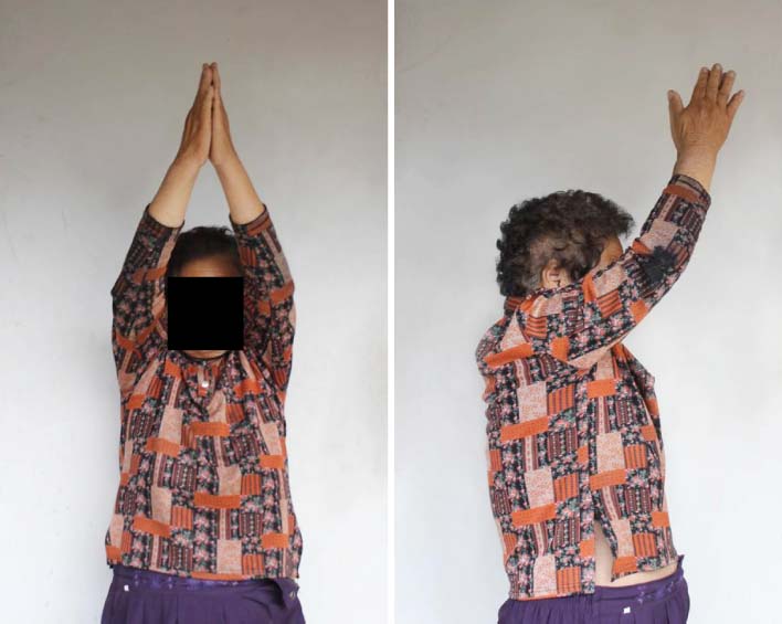
Photographs of the patient taken 3 years postoperatively showing abduction of right shoulder joint to 180 degrees and forward flexion to 140 degrees, the same as her left shoulder.
Fig. 4

(A) Preoperative anteroposterior plain radiograph showing the proximal humeral comminuted fracture (Neer type 3) with shoulder joint dislocation. (B) Immediate postoperative plain radiograph showing satisfactory fracture alignment with medial & lateral plate fixation and reduced shoulder joint. (C) Radiograph taken 54 weeks postoperatively showing no noticeable neckshaft angle change in comparison with immediate postoperative plain radiograph and healing.
Fig. 5

Photographs taken during the surgery. (A) Humerus head was dislocated anteriorly and metaphysis was severely comminuted with bone defect. (B) Unstable varus malalignment state after lateral locking compression plate fixation. (C) Stable acceptable alignment after additional medial buttressing plate fixation.
Figure & Data
REFERENCES
Citations
Citations to this article as recorded by 
- Dual-Plate Fixation for Proximal Humerus Fractures With Unstable Medial Column in Patients With Osteoporosis
Hyun-Gyu Seok, Sam-Guk Park
Journal of Orthopaedic Trauma.2023; 37(10): e387. CrossRef - The plate fixation strategy of complex proximal humeral fractures
Qi Sun, Xiaoming Wu, Lei Wang, Ming Cai
International Orthopaedics.2020; 44(9): 1785. CrossRef - Biomechanical evaluation of a novel dualplate fixation method for proximal humeral fractures without medial support
Yu He, Yaoshen Zhang, Yan Wang, Dongsheng Zhou, Fu Wang
Journal of Orthopaedic Surgery and Research.2017;[Epub] CrossRef
 Cite
CiteMedial and Lateral Dual Plate Fixation for Osteoporotic Proximal Humerus Comminuted Fracture: 2 Case Reports
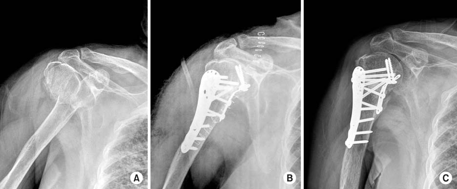
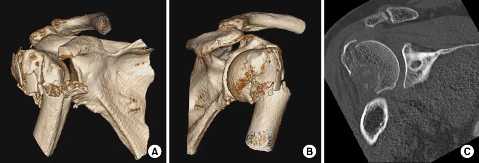

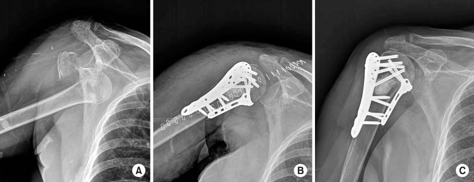
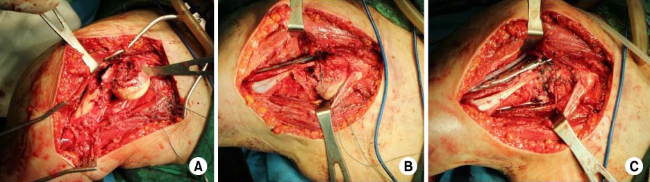
Fig. 1
(A) Preoperative anteroposterior radiograph suggested 3 part humerus surgical neck fracture according to Neer classification. (B) Immediate postoperative anteroposterior radiograph showing satisfactory fracture alignment with medial & lateral plate fixation. (C) Radiograph taken 50 weeks postoperatively showing no noticeable neck-shaft angle change in comparison with immediate postoperative plain radiograph and healing.
Fig. 2
(A, B) Three dimensional computed tomography image showing comminuted fracture at great tubercle, lesser tubercle and medial metaphysis. (C) Two dimensional computed tomography image suggested the length of the metaphyseal head extension was 12 mm and integrity of medial hinge was disrupted.
Fig. 3
Photographs of the patient taken 3 years postoperatively showing abduction of right shoulder joint to 180 degrees and forward flexion to 140 degrees, the same as her left shoulder.
Fig. 4
(A) Preoperative anteroposterior plain radiograph showing the proximal humeral comminuted fracture (Neer type 3) with shoulder joint dislocation. (B) Immediate postoperative plain radiograph showing satisfactory fracture alignment with medial & lateral plate fixation and reduced shoulder joint. (C) Radiograph taken 54 weeks postoperatively showing no noticeable neckshaft angle change in comparison with immediate postoperative plain radiograph and healing.
Fig. 5
Photographs taken during the surgery. (A) Humerus head was dislocated anteriorly and metaphysis was severely comminuted with bone defect. (B) Unstable varus malalignment state after lateral locking compression plate fixation. (C) Stable acceptable alignment after additional medial buttressing plate fixation.
Fig. 1
Fig. 2
Fig. 3
Fig. 4
Fig. 5
Medial and Lateral Dual Plate Fixation for Osteoporotic Proximal Humerus Comminuted Fracture: 2 Case Reports

