 E-submission
 TOTA
TOTA TOTS
TOTS
Articles
- Page Path
- HOME > J Musculoskelet Trauma > Volume 26(1); 2013 > Article
-
Case Report
- Interposition of Extensor Pollicis Longus Tendon in Smith's Fracture in a Child: A Case Report
- Seung-Ju Jeon, M.D., Haeng-Kee Noh, M.D., Do-Yeon Kim, M.D., Sung-Hoon Jung, M.D., Jun-Beum Shin, M.D., Ho-Seung Jeon, M.D.
-
Journal of the Korean Fracture Society 2013;26(1):65-68.
DOI: https://doi.org/10.12671/jkfs.2013.26.1.65
Published online: January 17, 2013
Department of Orthopaedic Surgery, Sung-Ae Hospital, Seoul, Korea.
*Department of Orthopaedic Surgery, Gwangmyeong Sung-Ae Hospital, Gwangmyeong, Korea.
- Address reprint requests to: Ho-Seung Jeon, M.D. Department of Othopaedic Surgery, Sung-Ae Hospital, 22, Yeouidaebang-ro 53-gil, Yeongdeungpo-gu, Seoul 150-960, Korea. Tel: 82-2-532-9554, Fax: 82-2-840-7755, hsjeonos@naver.com
• Received: June 21, 2012 • Revised: August 29, 2012 • Accepted: November 2, 2012
Copyright © 2013 The Korean Fracture Society
- 679 Views
- 0 Download
Abstract
- Entrapment of the extensor pollicis longus tendon is reported rarely on Smith's fractures in children. In our case, a 15 year old boy with Smith's fracture received treatment of closed reduction at another hospital. When he visited our hospital, a wide gap at the fracture site was detected on radiograph and the thumb movement was limited. We have doubt the entrapment of the soft tissue, especially the tendon. We decided on open reduction. In the operation field, entrapment of the extensor pollicis longus tendon at the gap of the fracture site was found through dorsal approach. In addition, fracture treatment with K-wire fixation after reduction of extensonr pollicis longus tendon reduction was done. Therefore, we report this case with a review of the literatures.
- 1. Cavanilles Walker JM, Masferrer Pino A, Alberti Fito G. Entrapment of the extensor pollicis longus tendon after a radial fracture in a child. J Hand Surg Eur Vol, 2012;37:182-183.ArticlePubMedPDF
- 2. Chung DW, Cho CH. Late rupture of extensor pollicis longus tendon after Colles' fracture. J Korean Soc Surg Hand, 1999;4:212-218.
- 3. Cooney WP 3rd, Dobyns JH, Linscheid RL. Complications of Colles' fractures. J Bone Joint Surg Am, 1980;62:613-619.ArticlePubMed
- 4. Han KJ, Khang SY, Moon JH. Entrapment of extensor pollicis longus tendon in the malunion of Smith's fracture. J Korean Soc Surg Hand, 2001;6:144-147.
- 5. Hunt DD. Dislocation of the extensor pollicis longus tendon in Smith's fracture of the radius. A case report. J Bone Joint Surg Am, 1969;51:991-994.PubMed
- 6. Itoh Y, Horiuchi Y, Takahashi M, Uchinishi K, Yabe Y. Extensor tendon involvement in Smith's and Galeazzi's fractures. J Hand Surg Am, 1987;12:535-540.ArticlePubMed
- 7. Oh IS, Lee KH, Ko SM, Kang HS, Kim CS. Rupture of the extensor pollicis longus tendon after Colle's fracture; the results of tendon transfer: case reports. J Korean Soc Surg Hand, 1999;4:51-54.
- 8. Rayan GM, Hayes M. Entrapment of the flexor digitorum profundus in the ulna with fracture of both bones of the forearm. Report of a case. J Bone Joint Surg Am, 1986;68:1102-1103.ArticlePubMed
- 9. Thomas WG, Kershaw CJ. Entrapment of extensor tendons in a Smith's fracture: brief report. J Bone Joint Surg Br, 1988;70:491. ArticlePubMedPDF
- 10. Uchida Y, Sugioka Y. Extensor tendon rupture associated with Smith's fracture. A case report. Acta Orthop Scand, 1990;61:374-375.ArticlePubMed
REFERENCES
Fig. 1
(A) Initial radiograph of the right wrist shows severe displacement of distal fracture fragment toward the volar side in Smith's fracture.
(B) Radiographs after reduction.
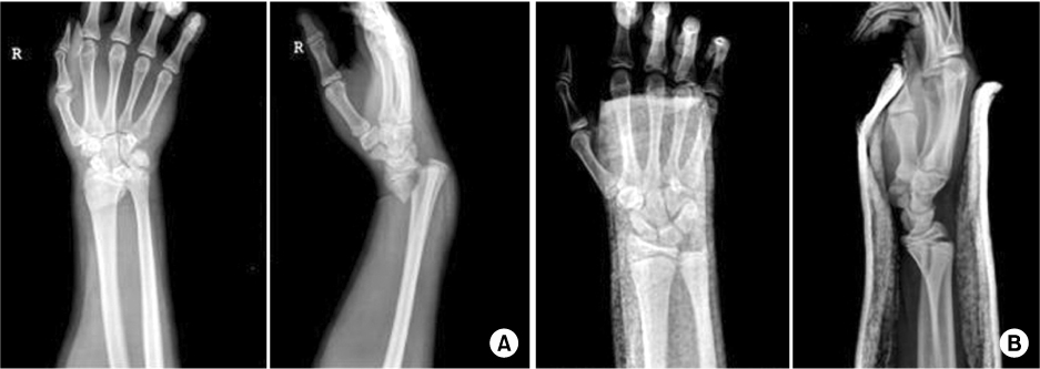
Fig. 2Preoperative computed tomography scan of the right wrist shows persistent gap at the fracture site after reduction.

Fig. 3The distal portion of extensor pollicis longus (EPL) tendon is found, but proximal portion is not seen (red arrow). EPL goes through a gap of fracture site and towards the dorsal side of the fracture site where it used to go through the proximal facture site.
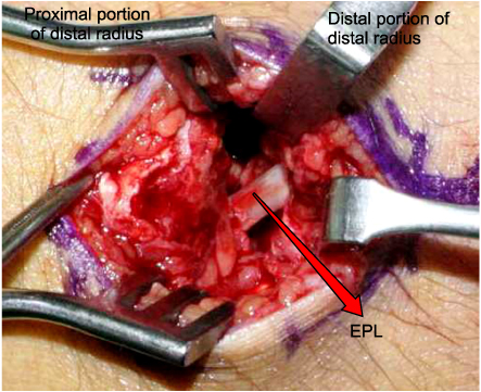

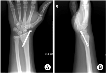
Figure & Data
REFERENCES
Citations
Citations to this article as recorded by 
 Cite
CiteInterposition of Extensor Pollicis Longus Tendon in Smith's Fracture in a Child: A Case Report

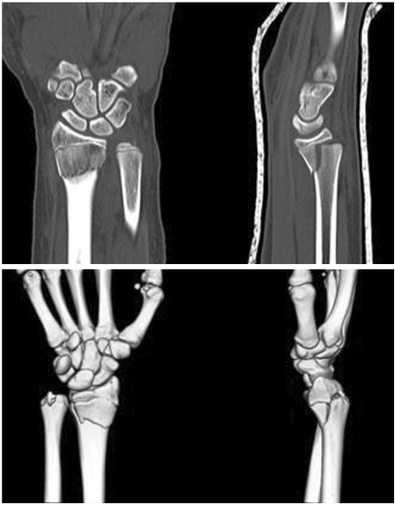

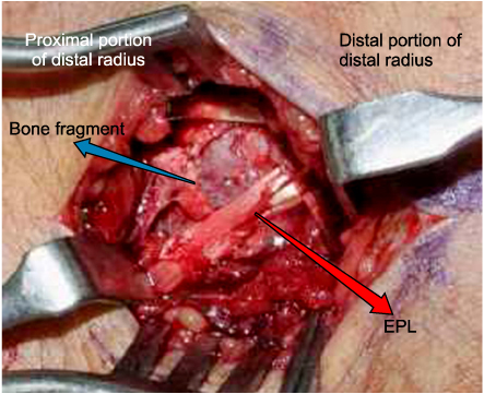

Fig. 1
(A) Initial radiograph of the right wrist shows severe displacement of distal fracture fragment toward the volar side in Smith's fracture.
(B) Radiographs after reduction.
Fig. 2
Preoperative computed tomography scan of the right wrist shows persistent gap at the fracture site after reduction.
Fig. 3
The distal portion of extensor pollicis longus (EPL) tendon is found, but proximal portion is not seen (red arrow). EPL goes through a gap of fracture site and towards the dorsal side of the fracture site where it used to go through the proximal facture site.
Fig. 4
The intraoperative photographs after reduction of the extensor pollicis longus (EPL) tendon (blue arrow). Fractured cortical bone fragment was reduced (red arrow). Entrapted EPL tendon was reduced from volar side to dorsal side.
Fig. 5
(A, B) Open reduction and internal fixation with Kirschner wires was done.
Fig. 1
Fig. 2
Fig. 3
Fig. 4
Fig. 5
Interposition of Extensor Pollicis Longus Tendon in Smith's Fracture in a Child: A Case Report

