 E-submission
 TOTA
TOTA TOTS
TOTS
Articles
- Page Path
- HOME > J Musculoskelet Trauma > Volume 23(4); 2010 > Article
-
Case Report
- False Negative Bone Scan in 56-year Old Man with L2 Compression Fracture Performed 78 Hours after Trauma: A Case Report
- Jeong-Gook Seo, M.D., Hae-Kyun Joo, M.D., Sung-Tae Kim, M.D.
-
Journal of the Korean Fracture Society 2010;23(4):386-390.
DOI: https://doi.org/10.12671/jkfs.2010.23.4.386
Published online: October 31, 2010
Department of Orthopedic Surgery, Seoul Paik Hospital, Inje University College of Medicine, Seoul, Korea.
- Address reprint requests to: Jeong-Gook Seo, M.D. Department of Orthopedic Surgery, Seoul Paik Hospital, Inje University College of Medicine, 85, Jeo-dong 2-ga, Jung-gu, Seoul 100-032, Korea. Tel: 82-2-2270-0025, Fax: 82-2-2270-0023, hd1404@hanafos.com
• Received: April 10, 2010 • Revised: June 29, 2010 • Accepted: August 17, 2010
Copyright © 2010 The Korean Fracture Society
- 843 Views
- 2 Download
Abstract
- It is very rare that the bone scan after 72 hours from the trauma doesn't exhibit the increased radio-nuclide uptake in the patient with fracture. The purpose of this study is to report the case that indicate the false negative finding in the bone scan performed after 78 hours from the trauma in the 56-year-old man with L2 compression fracture, including a review of the relevant literatures.
- 1. Jung JH, Kim JK, Jin W, et al. The value of radionuclide imaging as a screening test for the diagnosis of an acute thoracic spinal fractures. J Korean Soc Emerg Med, 2005;16:1-5.
- 2. Kaye M, Silverton S, Rosenthall L. Technetium-99m-pyrophosphate: studies in vivo and in vitro. J Nucl Med, 1975;16:40-45.
- 3. Kim BH, Im JI, Kim DJ, Park JY. Diganostic values of bone scan followed by CT scan in undetected pelvic bone fracture. J Korean Orthop Assoc, 1997;32:523-529.ArticlePDF
- 4. Kim HR, Thrall JH, Keyes JW Jr. Skeletal scintigraphy following incidental trauma. Radiology, 1979;130:447-451.Article
- 5. Lee E, Worsley DF. Role of radionuclide imaging in the orthopedic patient. Orthop Clin North Am, 2006;37:485-501.Article
- 6. Matheson GO, Clement DB, McKenzie DC, Taunton JE, Lloyd-Smith DR, MacIntyre JG. Stress fractures in athletes. A study of 320 cases. Am J Sports Med, 1987;15:46-58.ArticlePDF
- 7. Matin P. The appearance of bone scans following fractures, including immediate and long-term studies. J Nucl Med, 1979;20:1227-1231.
- 8. Milgrom C, Chisin R, Giladi M, et al. Negative bone scans in impending tibial stress fractures. A report of three cases. Am J Sports Med, 1984;12:488-491.ArticlePDF
- 9. Scott S, Alazraki N, Manaster B. Failure of bone scanning to detect fractures in a woman on chronic steroid therapy. Skeletal Radiol, 1984;12:204-207.ArticlePDF
- 10. Sterling JC, Webb RF Jr, Meyers MC, Calvo RD. False negative bone scan in a female runner. Med Sci Sports Exerc, 1993;25:179-185.Article
- 11. Subramanian G, McAfee JG. A new complex of 99mTc for skeletal imaging. Radiology, 1971;99:192-196.Article
- 12. Wen DY, Propeck T, Singh A. Femoral neck stress injury with negative bone scan. J Am Board Fam Pract, 2003;16:170-174.Article
- 13. Yao L, Lee JK. Occult intraosseous fracture: detection with MR imaging. Radiology, 1988;167:749-751.Article
REFERENCES
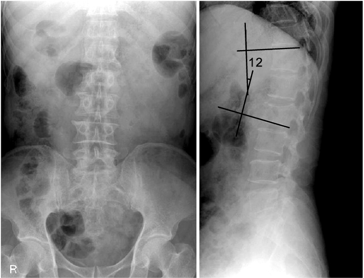


Figure & Data
REFERENCES
Citations
Citations to this article as recorded by 
 Cite
CiteFalse Negative Bone Scan in 56-year Old Man with L2 Compression Fracture Performed 78 Hours after Trauma: A Case Report

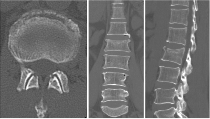
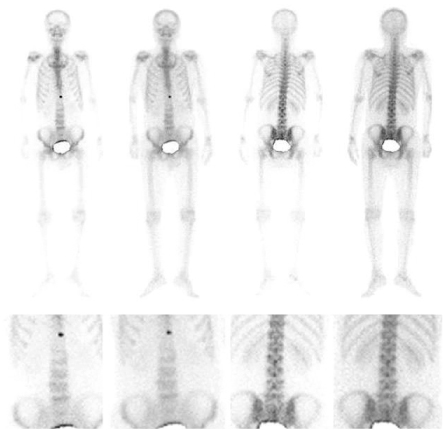
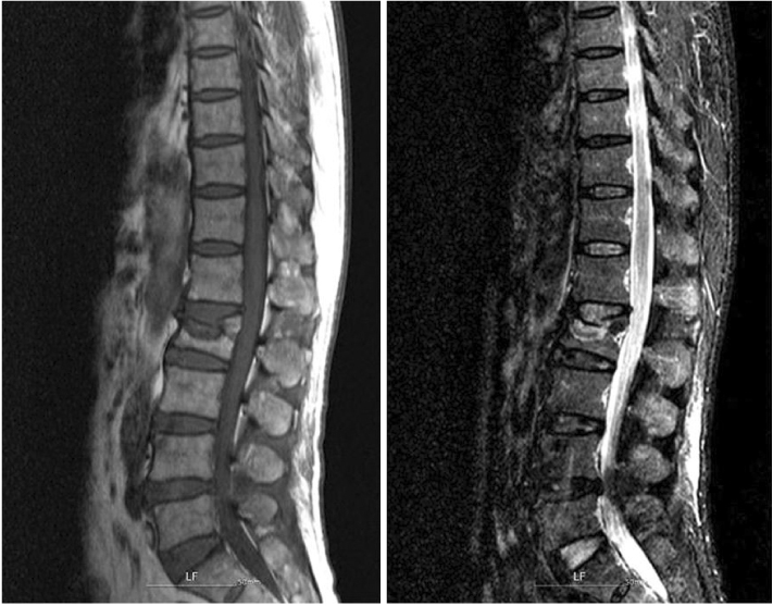
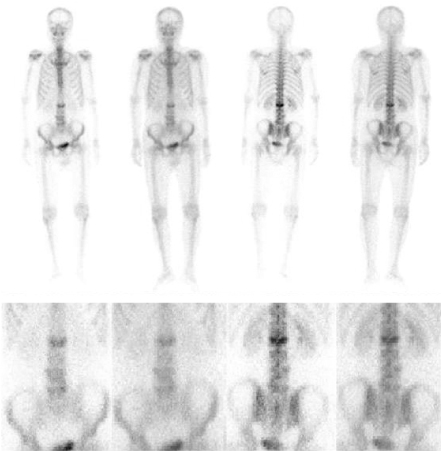
Fig. 1
Radiographs show anterior compression and kyphosis of L2 vertebral body in AP and lateral view.
Fig. 2
CT scan shows compression of L2 vertebral body.
Fig. 3
Bone scan performed at 78 hours of post-trauma shows no abnormal uptake in L2 vertebra.
Fig. 4
MRI performed at 2 months after trauma shows signals indicating recently developed compression fracture of L2 vertebra.
Fig. 5
Bone scan performed at 2 months after trauma shows increased uptake in L2 vertebra.
Fig. 1
Fig. 2
Fig. 3
Fig. 4
Fig. 5
False Negative Bone Scan in 56-year Old Man with L2 Compression Fracture Performed 78 Hours after Trauma: A Case Report

