 E-submission
 TOTA
TOTA TOTS
TOTS
Articles
- Page Path
- HOME > J Musculoskelet Trauma > Volume 23(4); 2010 > Article
-
Original Article
- Treatment of the Unstable Intertrochanteric Fracture with Proximal Femoral Nail Antirotation: Comparison with Compression Hip Screw with Trochanteric Stabilizing Plate
- Tae-Ho Kim, M.D., Jong-Oh Kim, M.D., Seung-Yup Lee, M.D., Geon-Ung Yun, M.D.
-
Journal of the Korean Fracture Society 2010;23(4):353-359.
DOI: https://doi.org/10.12671/jkfs.2010.23.4.353
Published online: October 31, 2010
Department of Orthopedic Surgery, Mokdong Hospital, Ewha Woman's University School of Medicine, Seoul, Korea.
*Department of Orthopedic Surgery, Sahmyook Medical Center, Seoul, Korea.
- Address reprint requests to: Jong-Oh Kim, M.D. Department of Orthopedic Surgery, Mok-dong Hospital, Ewha Woman's University School of Medicine, 911-1, Mok-dong, Yangcheon-gu, Seoul 158-710, Korea. Tel: 82-2-2650-5276, Fax: 82-2-2642-0349, ewhamdos@korea.com
• Received: April 21, 2010 • Accepted: August 11, 2010
Copyright © 2010 The Korean Fracture Society
- 1,423 Views
- 5 Download
- 6 Crossref
Abstract
-
Purpose
- To evaluate the effectiveness of Proximal Femoral Nail Anti-rotation (PFNA) for the treatment of unstable intertrochanteric fracture comparing with Compression Hip Screw (CHS) with Trochanteric Stabilizing Plate (TSP).
-
Materials and Methods
- With clinical study, 43 patients who were treated surgically for unstable intertrochanteric fractures were retrospectively evaluated. One group was treated with CHS and TSP (Group 1, 22 cases) and the other was treated with PFNA (Group 2, 21 cases). By postoperative radiograph and last follow up radiograph we measured Tip-apex distance, Cleveland index, Lag screw slippage, Neck-shaft angle change and Union time. And By retrospective medical record review, the clinical results were evaluated with the operation time, intraoperative estimated blood loss, amount of drainage, amount of transfusion, walking ability change and complication.
-
Results
- There was a lower operation time, intraoperative estimated blood loss, amount of drainage, amount of transfusion, lag screw slippage and neck shaft angle change in the Group 2 than in the Group 1 (p<0.05).
-
Conclusion
- PFNA showed better results than CHS with TSP in operation time, estimated blood loss, amount of drainage and transfusion, lag screw slippage and neck-shaft angle change.
- 1. Aune AK, Ekeland A, Odegaard B, Grøgaard B, Alho A. Gamma nail vs compression screw for trochanteric femoral fractures. 15 reoperations in a prospective, randomized study of 378 patients. Acta Orthop Scand, 1994;65:127-130.Article
- 2. Babst R, Renner N, Biedermann M, et al. Clinical results using the trochanter stabilizing plate (TSP): the modular extension of the dynamic hip screw (DHS) for internal fixation of selected unstable intertrochanteric fractures. J Orthop Trauma, 1998;12:392-399.Article
- 3. Bong MR, Patel V, Iesaka K, Egol KA, Kummer FJ, Koval KJ. Comparison of a sliding hip screw with a trochanteric lateral support plate to an intramedullary hip screw for fixation of unstable intertrochanteric hip fractures: a cadaver study. J Trauma, 2004;56:791-794.Article
- 4. Buciuto R, Uhlin B, Hammerby S, Hammer R. RAB-plate vs Richards CHS plate for unstable trochanteric hip fractures. A randomized study of 233 patients with 1-year follow-up. Acta Orthop Scand, 1998;69:25-28.Article
- 5. Cleveland M, Bosworth DM, Thompson FR, Wilson HJ Jr, Ishizuka T. A ten-year analysis of intertrochanteric fractures of the femur. J Bone Joint Surg Am, 1959;41:1399-1408.Article
- 6. Doppelt SH. The sliding compression screw--today's best answer for stabilization of intertrochanteric hip fractures. Orthop Clin North Am, 1980;11:507-523.Article
- 7. Fogagnolo F, Kfuri M Jr, Paccola CA. Intramedullary fixation of pertrochanteric hip fractures with the short AO-ASIF proximal femoral nail. Arch Orthop Trauma Surg, 2004;124:31-37.ArticlePDF
- 8. Forte ML, Virnig BA, Kane RL, et al. Geographic variation in device use for intertrochanteric hip fractures. J Bone Joint Surg Am, 2008;90:691-699.Article
- 9. Harrington P, Nihal A, Singhania AK, Howell FR. Intramedullary hip screw versus sliding hip screw for unstable intertrochanteric femoral fractures in the elderly. Injury, 2002;33:23-28.Article
- 10. Karunakar M, McLaurin TM, Morgan SJ, Egol KA. Improving outcomes after pertrochanteric hip fractures. Instr Course Lect, 2009;58:91-104.
- 11. Koval KJ, Cantu RV. Rookwood CA, Green DP. Intertrochanteric fractures. In: Fractures in adults, 2006;6th ed. Philadelphia, JB Lippincott. 1793-1825.Article
- 12. Koval KJ, Skovron ML, Aharonoff GB, Meadows SE, Zuckerman JD. Ambulatory ability after hip fracture. A prospective study in geriatric patients. Clin Orthop Relat Res, 1995;310:150-159.
- 13. Koval KJ, Zuckerman JD. Functional recovery after fracture of the hip. J Bone Joint Surg Am, 1994;76:751-758.Article
- 14. Kyle RF, Cabanela ME, Russell TA, et al. Fractures of the proximal part of the femur. Instr Course Lect, 1995;44:227-253.Article
- 15. Lee JY, Lee SY. Treatment of the proximal femoral extracapsular fracture with proximal femoral nail antirotation (PFNA): comparison with proximal femoral nail (PFN). J Korean Hip Soc, 2007;19:183-189.Article
- 16. Loch DA, Kyle RF, Bechtold JE, Kane M, Anderson K, Sherman RE. Forces required to initiate sliding in second-generation intramedullary nails. J Bone Joint Surg Am, 1998;80:1626-1631.Article
- 17. Madsen JE, Naess L, Aune AK, Alho A, Ekeland A, Strømsøe K. Dynamic hip screw with trochanteric stabilizing plate in the treatment of unstable proximal femoral fractures: a comparative study with the Gamma nail and compression hip screw. J Orthop Trauma, 1998;12:241-248.Article
- 18. Müller ME, Nazarian S. Classificaiton et documentation aoedes fractures femur. Rev Chir Orthop, 1981;67:297.
- 19. Nakata K, Ohzono K, Hiroshima K, Toge K. Serial change of sliding in intertrochanteric femoral fractures treated with sliding screw system. Arch Orthop Trauma Surg, 1994;113:276-280.ArticlePDF
- 20. Park JH, Park JW, Wang JH, Lee JW, Lee JI, Kim JG. Treatment of intertrochanteric fracture: comparison of proximal femoral nail and proximal femoral nail A. J Korean Fract Soc, 2008;21:103-109.Article
- 21. Park MS, Lim YJ, Kim YS, Kim KH, Cho HM. Treatment of the proximal femoral fractures with proximal femoral nail antirotation (PFNA). J Korean Fract Soc, 2009;22:91-97.Article
- 22. Rha JD, Kim YH, Yoon SI, Park TS, Lee MH. Factors affecting sliding of the lag screw in intertrochanteric fractures. Int Orthop, 1993;17:320-324.ArticlePDF
- 23. Simmermacher RK, Ljungqvist J, Bail H, et al. The new proximal femoral nail antirotation (PFNA) in daily practice: results of a multicentre clinical study. Injury, 2008;39:932-939.Article
- 24. Steinberg GG, Desai SS, Kornwitz NA, Sullivan TJ. The intertrochanteric hip fracture. A retrospective analysis. Orthopedics, 1988;11:265-273.Article
- 25. Templeman D, Baumgaertner MR, Leighton RK, Lindsey RW, Moed BR. Reducing complications in the surgical treatment of intertrochanteric fractures. Instr Course Lect, 2005;54:409-415.
- 26. Utrilla AL, Reig JS, Muñoz FM, Tufanisco CB. Trochanteric gamma nail and compression hip screw for trochanteric fractures: a randomized, prospective, comparative study in 210 elderly patients with a new design of the gamma nail. J Orthop Trauma, 2005;19:229-233.
REFERENCES
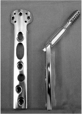


Fig. 4
(A) 73 year old female patient presented with intertrochanteric fracture as AO/ASIF classification A2.2.
(B) Postoperative radiograph shows that good reduction and well positioned PFNA blade.
(C) Postoperative 6 mons radiograph shows minimal sliding of PFNA blade and well united fracture site.
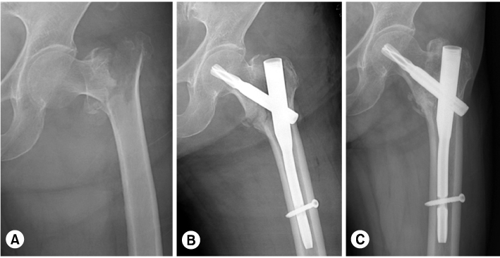
Fig. 5
(A) 84 year old female patient presented with intertrochanteric fracture as AO/ASIF classification A2.2.
(B) Postoperative radiograph shows that acceptable reduction but superior positioned lag screw.
(C) Postoperative 8 weeks radiograph shows cutting out of the lag screw.
(D) Reoperation was done with cemented bipolar hemiarthroplasty.
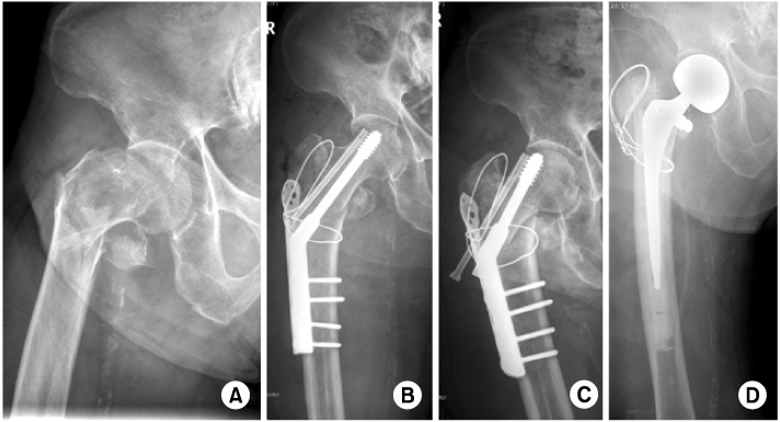
Fig. 6
(A) 73 year old female patient presented with intertrochanteric fracture as AO/ASIF classification A2.2.
(B) Postoperative radiograph shows that poor varus reduction and superior positioned PFNA blade.
(C) Postoperative 4 weeks radiograph shows cutting out of the PFNA blade.
(D) Reoperation was done with cemented bipolar hemiarthroplasty.

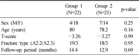
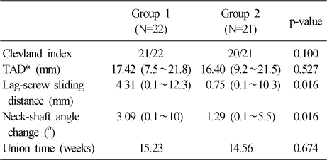
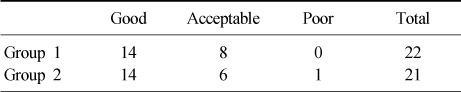
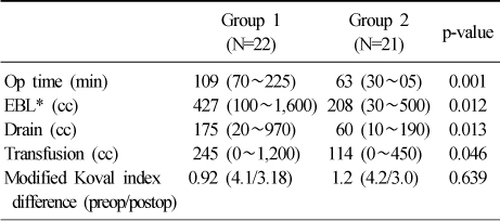
Figure & Data
REFERENCES
Citations
Citations to this article as recorded by 
- Results of Asian Type Gamma 3 Nail in Treatment of Trochanteric Fractures
Bing Zhe Huang, Yong Wook Park, Jin Su Park, Kyu Cheol Noh, Soung Yon Kim, Kook Jin Chung, Hong Kyun Kim, Hyong Nyun Kim, Yong Hyun Yoon, Ji Hyo Hwang
Journal of the Korean Fracture Society.2014; 27(3): 213. CrossRef - Comparative Study of Intertrochanteric Fracture Treated with the Proximal Femoral Nail Anti-Rotation and the Third Generation of Gamma Nail
Jae-Cheon Sim, Tae-Ho Kim, Ki-Do Hong, Sung-Sik Ha, Jong-Seong Lee
Journal of the Korean Fracture Society.2013; 26(1): 37. CrossRef - Comparative Study of Proximal Femoral Nail Antirotation and Zimmer Natural Nail for the Treatment of Stable Intertrochanteric Fractures
Jee-Hoon Kim, Oog-Jin Shon
Journal of the Korean Fracture Society.2013; 26(4): 305. CrossRef - A Comparison between Compression Hip Screw and Intramedullary Nail for the Treatment of AO/OTA A2.2 Intertrochanteric Femoral Fracture
Phil Hyun Chung, Suk Kang, Jong Pil Kim, Young Sung Kim, Ho Min Lee, Jong Hyun Kim
Journal of the Korean Fracture Society.2013; 26(1): 44. CrossRef - The Treatment of Intertrochanteric Femoral Fracture with Proximal Femoral Nail Antirotation
Jong Won Kim, Hyun Soo Park, Young Soo Jang, Jae Hyuk Choi, Sung Ju Bae, Chan Il Bae
Journal of the Korean Fracture Society.2012; 25(4): 257. CrossRef - Operative Treatment with Gamma 3 Nail in Femur Intertrochanteric Fracture
Ki-Do Hong, Jae-Chun Sim, Sung-Sik Ha, Tae-Ho Kim, Yoon-Ho Choi, Jong-Hyun Kim
Journal of the Korean Fracture Society.2011; 24(1): 7. CrossRef
 Cite
CiteTreatment of the Unstable Intertrochanteric Fracture with Proximal Femoral Nail Antirotation: Comparison with Compression Hip Screw with Trochanteric Stabilizing Plate

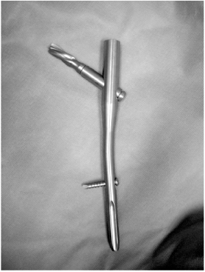
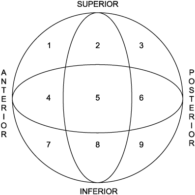


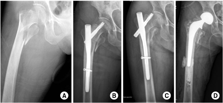
Fig. 1
Photographs of real implant of TSP (Trochanteric Stabilizing Plate).
Fig. 2
Photographs of real implant of PFNA (Proximal Femoral Nail Antirotation).
Fig. 3
Cleveland index.
Fig. 4
(A) 73 year old female patient presented with intertrochanteric fracture as AO/ASIF classification A2.2.
(B) Postoperative radiograph shows that good reduction and well positioned PFNA blade.
(C) Postoperative 6 mons radiograph shows minimal sliding of PFNA blade and well united fracture site.
Fig. 5
(A) 84 year old female patient presented with intertrochanteric fracture as AO/ASIF classification A2.2.
(B) Postoperative radiograph shows that acceptable reduction but superior positioned lag screw.
(C) Postoperative 8 weeks radiograph shows cutting out of the lag screw.
(D) Reoperation was done with cemented bipolar hemiarthroplasty.
Fig. 6
(A) 73 year old female patient presented with intertrochanteric fracture as AO/ASIF classification A2.2.
(B) Postoperative radiograph shows that poor varus reduction and superior positioned PFNA blade.
(C) Postoperative 4 weeks radiograph shows cutting out of the PFNA blade.
(D) Reoperation was done with cemented bipolar hemiarthroplasty.
Fig. 1
Fig. 2
Fig. 3
Fig. 4
Fig. 5
Fig. 6
Treatment of the Unstable Intertrochanteric Fracture with Proximal Femoral Nail Antirotation: Comparison with Compression Hip Screw with Trochanteric Stabilizing Plate
Demographic characteristics between the group 1 and 2
Comparison of radiographic results
*TAD: Tip apex distance.
Immediately postoperative reduction status
Comparison of clinical results
*EBL: Estimated blood loss.
Table 1
Demographic characteristics between the group 1 and 2
Table 2
Comparison of radiographic results
*TAD: Tip apex distance.
Table 3
Immediately postoperative reduction status
Table 4
Comparison of clinical results
*EBL: Estimated blood loss.

