 E-submission
 TOTA
TOTA TOTS
TOTS
Articles
- Page Path
- HOME > J Musculoskelet Trauma > Volume 29(1); 2016 > Article
-
Review Article
- Biomechanics of the Wrist
- Young Ho Shin, M.D., Young Ho Lee, M.D., Ph.D.
-
Journal of the Korean Fracture Society 2016;29(1):93-100.
DOI: https://doi.org/10.12671/jkfs.2016.29.1.93
Published online: January 19, 2016
Department of Orthopedic Surgery, Seoul National University College of Medicine, Seoul, Korea.
- Address reprint requests to: Young Ho Lee, M.D., Ph.D. Department of Orthopedic Surgery, Seoul National University Hospital, 101 Daehakro, Jongno-gu, Seoul 03080, Korea. Tel: 82-2-2072-0819, Fax: 82-2-740-2718, orthoyhl@snu.ac.kr
Copyright © 2016 The Korean Fracture Society. All rights reserved.
This is an Open Access article distributed under the terms of the Creative Commons Attribution Non-Commercial License (http://creativecommons.org/licenses/by-nc/4.0) which permits unrestricted non-commercial use, distribution, and reproduction in any medium, provided the original work is properly cited.
- 5,113 Views
- 179 Download
- 3 Crossref


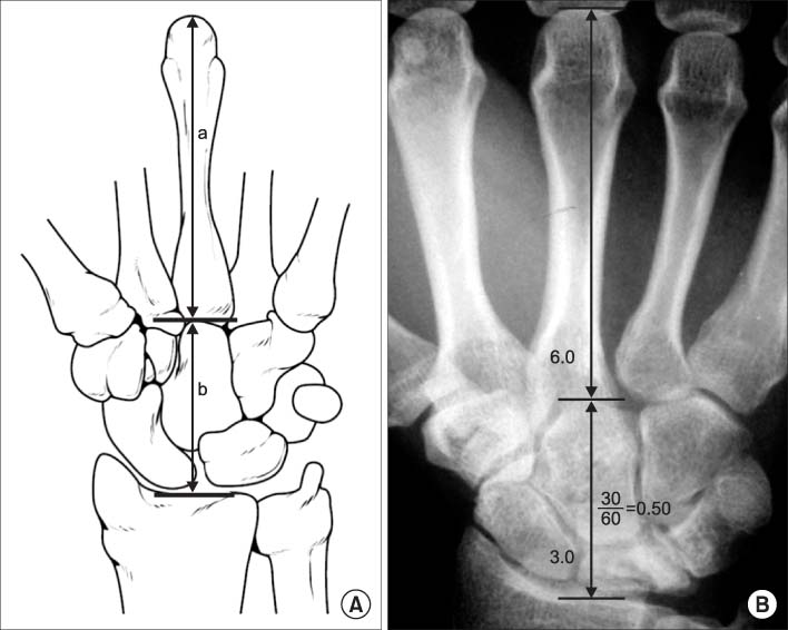


Figure & Data
REFERENCES
Citations
Citations to this article as recorded by 
- Fractal geometry in wrist biomechanics: A preliminary study with implications for arthroplasty and surgery
Lauren Gorelick, Amir Oron, Gil Gannot, Raphael Israeli
Hand Surgery and Rehabilitation.2025; 44(4): 102203. CrossRef - Association between carpal height ratio and ulnar variance in normal wrist radiography
Anas AR Altamimi, Monther A. Gharaibeh, Muntaser Abu Shokor, Moh’d S. Dawod, Mohammad N. Alswerki, Omar M. Al-Odat, Raghda H. Elkhaldi
BMC Musculoskeletal Disorders.2024;[Epub] CrossRef - Reliability and concurrent validity of a new iPhone® goniometric application for measuring active wrist range of motion: a cross‐sectional study in asymptomatic subjects
Mohammad Reza Pourahmadi, Ismail Ebrahimi Takamjani, Javad Sarrafzadeh, Mehrdad Bahramian, Mohammad Ali Mohseni‐Bandpei, Fatemeh Rajabzadeh, Morteza Taghipour
Journal of Anatomy.2017; 230(3): 484. CrossRef
 Cite
CiteBiomechanics of the Wrist
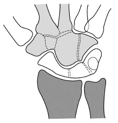
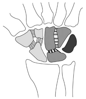
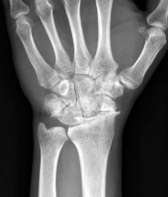

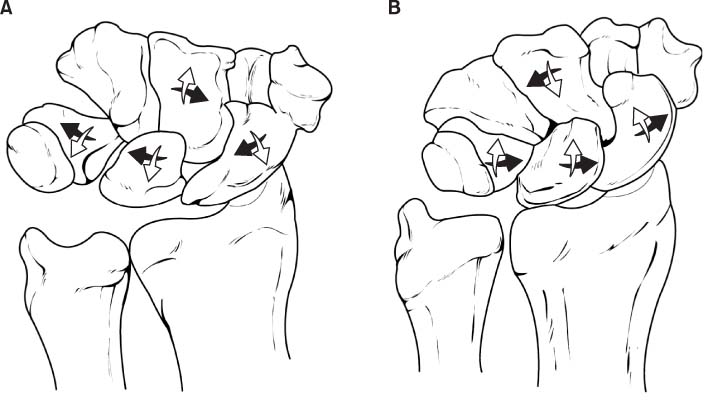
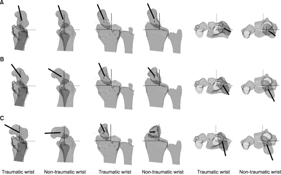
Fig. 1
Row concept of carpal bones. The joints with adjacent bones which do not have gross motion, considered as a single motor unit. The proximal row is composed of scaphoid, lunate, and triquetrum. The distal row is composed of trapezium, trapezoid, capitate, and hamate.
Fig. 2
Column concept of carpal bones. The lateral column is composed of scaphoid, trapezium, and trapezoid. The middle column is composed of lunate and capitate, and the medial column is composed of triquetrum and hamate.
Fig. 3
Scaphoid nonunion advanced collapse. Left wrist simple radiograph of a 53-year-old man. The advanced degenerative changes are evident around the radiocarpal joint with scaphoid nonunion.
Fig. 4
Carpal height ratio. (A) Carpal height ratio is calculated by dividing carpal height (b) with length of the 3rd metacarpal bone (a). (B) A simple radiograph of measurement of carpal height ratio.
Fig. 5
Movement of carpal rows during radial deviation and ulnar deviation. (A) During radial deviation of the wrist joint, the proximal carpal row is flexed volarly and deviated radially, and the distal carpal row is flexed dorsally. (B) During ulnar deviation of the wrist joint, the proximal carpal row is flexed dorsally and deviated ulnarly, and the distal carpal row is flexed volarly.
Fig. 6
Computed tomography based three dimensional kinematic comparison of the scaphoid during dart throwing motion between distal radius fracture malunion and contralateral normal side. (A) Radial deviation and dorsal tilt position. (B) Mid-range of dart throwing motion. (C) Ulnar deviation and volar tilt position. The orientations of the helical axes (bold line of each figure) of the scaphoid are different between two sides.
Fig. 1
Fig. 2
Fig. 3
Fig. 4
Fig. 5
Fig. 6
Biomechanics of the Wrist

