 E-submission
 TOTA
TOTA TOTS
TOTS
Articles
- Page Path
- HOME > J Musculoskelet Trauma > Volume 29(2); 2016 > Article
-
Review Article
- Diagnosis and Management of Ligament Injuries of the Wrist
- Ki-Tae Na, M.D., Joo-Yup Lee, M.D., Ph.D.
-
Journal of the Korean Fracture Society 2016;29(2):160-170.
DOI: https://doi.org/10.12671/jkfs.2016.29.2.160
Published online: April 19, 2016
Department of Orthopedic Surgery, The Catholic University of Korea, St. Vincent's Hospital, Suwon, Korea.
- Address reprint requests to: Joo-Yup Lee, M.D., Ph.D. Department of Orthopedic Surgery, The Catholic University of Korea, St. Vincent's Hospital, 93 Jungbu-daero, Paldal-gu, Suwon 16247, Korea. Tel: 82-31-249-8301, Fax: 82-31-254-7186, jylos1@gmail.com
Copyright © 2016 The Korean Fracture Society. All rights reserved.
This is an Open Access article distributed under the terms of the Creative Commons Attribution Non-Commercial License (http://creativecommons.org/licenses/by-nc/4.0) which permits unrestricted non-commercial use, distribution, and reproduction in any medium, provided the original work is properly cited.
- 1,739 Views
- 22 Download
Abstract
- The wrist joint is formed by the distal end of the radius and ulna proximally, and eight carpal bones distally. It has many ligaments to maintain stability of the complex bony structures. The incidence of ligament injuries of the wrist has increased due to sports activities. However, diagnosis and management of these injuries are sometimes difficult because of the anatomic complexity and variable injury patterns. Among them, scapholunate ligament injury and triangular fibrocartilage tears are the two most common injuries resulting in chronic disabling wrist pain. Thorough understanding of the wrist anatomy and physical and radiologic examination is mandatory for proper diagnosis and management of these conditions. This article will briefly discuss the wrist joint anatomy and biomechanics, and review the diagnosis and management of the scapholunate ligament injury and triangular fibrocartilage injury.
- 1. Mayfield JK, Johnson RP, Kilcoyne RF. The ligaments of the human wrist and their functional significance. Anat Rec, 1976;186:417-428.Article
- 2. Mayfield JK. Wrist ligamentous anatomy and pathogenesis of carpal instability. Orthop Clin North Am, 1984;15:209-216.Article
- 3. Berger RA. The anatomy of the ligaments of the wrist and distal radioulnar joints. Clin Orthop Relat Res, 2001;(383):32-40.Article
- 4. Melone CP Jr, Nathan R. Traumatic disruption of the triangular fibrocartilage complex. Pathoanatomy. Clin Orthop Relat Res, 1992;(275):65-73.
- 5. Atzei A. New trends in arthroscopic management of type 1-B TFCC injuries with DRUJ instability. J Hand Surg Eur Vol, 2009;34:582-591.ArticlePDF
- 6. Atzei A, Luchetti R. Foveal TFCC tear classification and treatment. Hand Clin, 2011;27:263-272.Article
- 7. Walsh JJ, Berger RA, Cooney WP. Current status of scapholunate interosseous ligament injuries. J Am Acad Orthop Surg, 2002;10:32-42.Article
- 8. Kitay A, Wolfe SW. Scapholunate instability: current concepts in diagnosis and management. J Hand Surg Am, 2012;37:2175-2196.Article
- 9. Hagert E, Hagert CG. Understanding stability of the distal radioulnar joint through an understanding of its anatomy. Hand Clin, 2010;26:459-466.Article
- 10. Rajan PV, Day CS. Scapholunate ligament insufficiency. J Hand Surg Am, 2015;40:583-585.Article
- 11. Watson HK, Ballet FL. The SLAC wrist: scapholunate advanced collapse pattern of degenerative arthritis. J Hand Surg Am, 1984;9:358-365.Article
- 12. O'Meeghan CJ, Stuart W, Mamo V, Stanley JK, Trail IA. The natural history of an untreated isolated scapholunate interosseus ligament injury. J Hand Surg Br, 2003;28:307-310.ArticlePDF
- 13. Watson HK, Ashmead D 4th, Makhlouf MV. Examination of the scaphoid. J Hand Surg Am, 1988;13:657-660.Article
- 14. Magee T. Comparison of 3-T MRI and arthroscopy of intrinsic wrist ligament and TFCC tears. AJR Am J Roentgenol, 2009;192:80-85.Article
- 15. Geissler WB. Arthroscopic management of scapholunate instability. J Wrist Surg, 2013;2:129-135.Article
- 16. Andersson JK, Garcia-Elias M. Dorsal scapholunate ligament injury: a classification of clinical forms. J Hand Surg Eur Vol, 2013;38:165-169.ArticlePDF
- 17. May MM, Lawton JN, Blazar PE. Ulnar styloid fractures associated with distal radius fractures: incidence and implications for distal radioulnar joint instability. J Hand Surg Am, 2002;27:965-971.Article
- 18. Nöbauer-Huhmann IM, Pretterklieber M, Erhart J, et al. Anatomy and variants of the triangular fibrocartilage complex and its MR appearance at 3 and 7T. Semin Musculoskelet Radiol, 2012;16:93-103.
- 19. Anderson ML, Skinner JA, Felmlee JP, Berger RA, Amrami KK. Diagnostic comparison of 1.5 Tesla and 3.0 Tesla preoperative MRI of the wrist in patients with ulnar-sided wrist pain. J Hand Surg Am, 2008;33:1153-1159.Article
- 20. Palmer AK. Triangular fibrocartilage complex lesions: a classification. J Hand Surg Am, 1989;14:594-606.Article
- 21. Nakamura T, Nakao Y, Ikegami H, Sato K, Takayama S. Open repair of the ulnar disruption of the triangular fibrocartilage complex with double three-dimensional mattress suturing technique. Tech Hand Up Extrem Surg, 2004;8:116-123.Article
- 22. Atzei A, Rizzo A, Luchetti R, Fairplay T. Arthroscopic foveal repair of triangular fibrocartilage complex peripheral lesion with distal radioulnar joint instability. Tech Hand Up Extrem Surg, 2008;12:226-235.Article
- 23. Chou KH, Sarris IK, Sotereanos DG. Suture anchor repair of ulnar-sided triangular fibrocartilage complex tears. J Hand Surg Br, 2003;28:546-550.ArticlePDF
REFERENCES
Fig. 1
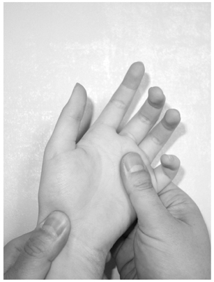
Watson's scaphoid shift test. The examiners grasp the wrist with their thumb over the scaphoid tubercle and they will feel a clunk when the patient's wrist is moved from ulnar to radial deviation.
Fig. 2

Radiologic findings of the scapholunate dissociation. A wide scapholunate gap, cortical ring sign in the posteroanterior view, and dorsal intercalated segmental instability pattern in the lateral view can be seen.
Fig. 3
Brunelli's scapholunate ligament reconstruction using flexor carpi radialis (FCR) tendon. (A) Visible gap between scaphoid and lunate. (B) Harvesting the half-slip of FCR tendon. (C) Tendon passed through the scaphoid bone tunnel. (D) Tendon sutured to the lunate and dorsal intercapal ligament.
Fig. 4

Magnetic resonance imaging findings of the triangular fibrocartilage complex (TFCC) tear. (A, B) Subluxation of the ulnar head and flexor carpi ulnaris tendon. (C, D) Foveal tear of the TFCC.

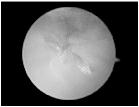
Figure & Data
REFERENCES
Citations
Citations to this article as recorded by 
 Cite
CiteDiagnosis and Management of Ligament Injuries of the Wrist

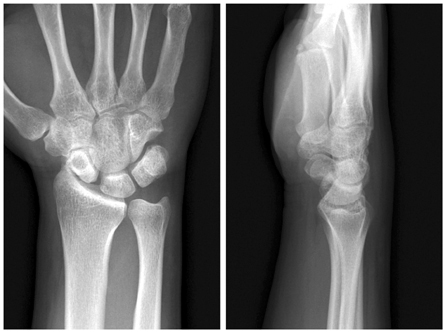

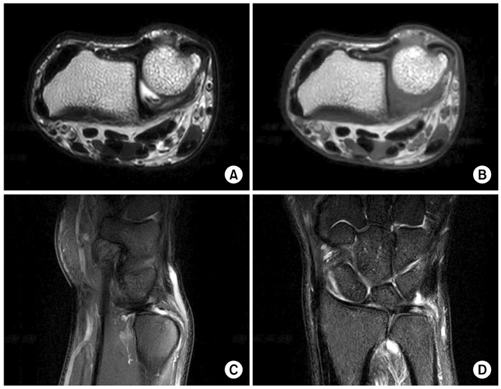
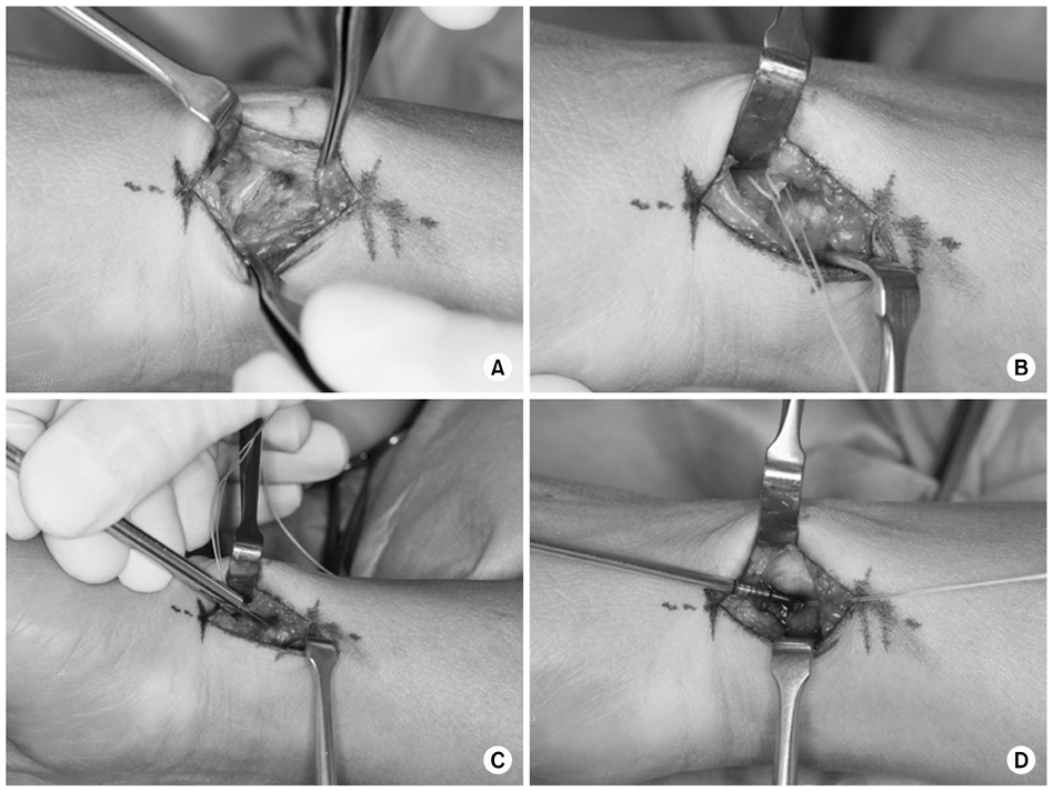

Fig. 1
Watson's scaphoid shift test. The examiners grasp the wrist with their thumb over the scaphoid tubercle and they will feel a clunk when the patient's wrist is moved from ulnar to radial deviation.
Fig. 2
Radiologic findings of the scapholunate dissociation. A wide scapholunate gap, cortical ring sign in the posteroanterior view, and dorsal intercalated segmental instability pattern in the lateral view can be seen.
Fig. 3
Brunelli's scapholunate ligament reconstruction using flexor carpi radialis (FCR) tendon. (A) Visible gap between scaphoid and lunate. (B) Harvesting the half-slip of FCR tendon. (C) Tendon passed through the scaphoid bone tunnel. (D) Tendon sutured to the lunate and dorsal intercapal ligament.
Fig. 4
Magnetic resonance imaging findings of the triangular fibrocartilage complex (TFCC) tear. (A, B) Subluxation of the ulnar head and flexor carpi ulnaris tendon. (C, D) Foveal tear of the TFCC.
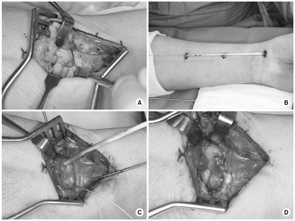
Fig. 5
Open repair for a triangular fibrocartilage complex foveal tear. (A) Exposing the distal radioulnar ligament. (B) Masson-Allen type suture to the ligament. (C) Making a bone hole to the fovea. (D) Inserting a suture anchor.
Fig. 6
Arthroscopic repair of the triangular fibrocartilage complex using double Fiberwire® suture.
Fig. 1
Fig. 2
Fig. 3
Fig. 4
Fig. 5
Fig. 6
Diagnosis and Management of Ligament Injuries of the Wrist

