 E-submission
 TOTA
TOTA TOTS
TOTS
Articles
- Page Path
- HOME > J Musculoskelet Trauma > Volume 30(3); 2017 > Article
-
Case Report
- Oncogenic Osteomalacia with Multiple Insufficiency Fractures: A Case Report
-
Young-Chang Park, M.D.
 , Joon-Oh Seo, M.D., Kyu-Hyun Yang, M.D., Ph.D.
 -
Journal of the Korean Fracture Society 2017;30(3):146-150.
DOI: https://doi.org/10.12671/jkfs.2017.30.3.146
Published online: July 21, 2017
Department of Orthopaedic Surgery, Gangnam Severance Hospital, Yonsei University College of Medicine, Seoul, Korea.
- Correspondence to: Kyu-Hyun Yang, M.D., Ph.D. Department of Orthopaedic Surgery, Gangnam Severance Hospital, Yonsei University College of Medicine, 211 Eonju-ro, Gangnam-gu, Seoul 06273, Korea. Tel: +82-2-2019-3414, Fax: +82-2-573-5393, kyang@yuhs.ac
• Received: May 2, 2017 • Revised: May 27, 2017 • Accepted: May 27, 2017
Copyright © 2017 The Korean Fracture Society. All rights reserved.
This is an Open Access article distributed under the terms of the Creative Commons Attribution Non-Commercial License (http://creativecommons.org/licenses/by-nc/4.0) which permits unrestricted non-commercial use, distribution, and reproduction in any medium, provided the original work is properly cited.
- 1,111 Views
- 2 Download
- 1 Crossref
Abstract
- Oncogenic osteomalacia is a rare paraneoplastic syndrome, characterized by hypophosphatemia, renal phosphate wasting, osteomalacia, and multiple insufficiency fractures, as a result of the tumor. A wide excision of the causative tumor is considered as the treatment of choice, following which, a dramatic recovery is expected. Authors report a case in which the symptoms and bone mineral density were dramatically recovered after an excision of the causative tumor around the tibialis posterior muscle in oncogenic osteomalacia.
- 1. McCance RA. Osteomalacia with Looser's nodes (Milkman's syndrome) due to a raised resistance to vitamin D acquired about the age of 15 years. Q J Med, 1947;16:33-46.PubMed
- 2. Kumar R, Folpe AL, Mullan BP. Tumor-induced osteomalacia. Transl Endocrinol Metab, 2015;7:1871.
- 3. Ledford CK, Zelenski NA, Cardona DM, Brigman BE, Eward WC. The phosphaturic mesenchymal tumor: why is definitive diagnosis and curative surgery often delayed? Clin Orthop Relat Res, 2013;471:3618-3625.ArticlePubMedPMC
- 4. Jan de Beur SM. Tumor-induced osteomalacia. JAMA, 2005;294:1260-1267.ArticlePubMed
- 5. Jiang Y, Xia WB, Xing XP, et al. Tumor-induced osteomalacia: an important cause of adult-onset hypophosphatemic osteomalacia in China: report of 39 cases and review of the literature. J Bone Miner Res, 2012;27:1967-1975.ArticlePDF
- 6. Chong WH, Andreopoulou P, Chen CC, et al. Tumor localization and biochemical response to cure in tumor-induced osteomalacia. J Bone Miner Res, 2013;28:1386-1398.ArticlePDF
- 7. Zhang J, Zhu Z, Zhong D, et al. 68Ga DOTATATE PET/CT is an accurate imaging modality in the detection of culprit tumors causing osteomalacia. Clin Nucl Med, 2015;40:642-646.Article
- 8. Andreopoulou P, Dumitrescu CE, Kelly MH, et al. Selective venous catheterization for the localization of phosphaturic mesenchymal tumors. J Bone Miner Res, 2011;26:1295-1302.ArticlePubMedPMCPDF
- 9. Umphrey LG, Whitaker MD, Bosch EP, Cook CB. Clinical and bone density outcomes of tumor-induced osteomalacia after treatment. Endocr Pract, 2007;13:458-462.Article
REFERENCES
Fig. 1
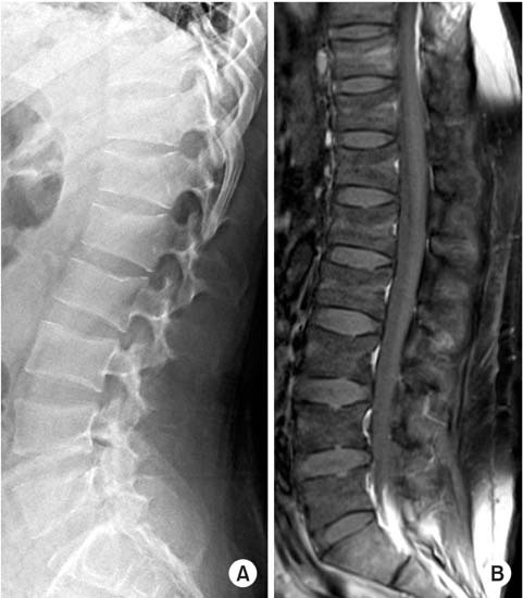
Spine simple x-ray lateral view shows that vertebral height of T10, 11, 12, L1, and 2 decreased slightly (A) and magnetic resonance imaging T1-weighted enhanced image shows multiple compression fracture (B).
Fig. 2

Whole body bone scan shows multifocal bony uptakes in the bilateral ribs, pelvis, femoral neck, knee, and ankle. RT: right, LT: left.
Fig. 3
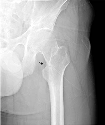
Insufficiency fracture of the femoral neck. A lucent line (arrow) is called a Looser's zone or pseudofracture.
Fig. 4

(A, B) Tumor localization. Causative tumor (arrows) was found on the left lower leg by Octreoscan. (C) Magnetic resonance imaging T2-weighted image shows 4.5×2.0×2.0-cm-sized soft tissue tumor located at the tibialis posterior muscle belly. Tumor had a round, well-defined margin with heterogeneous signal intensity. RT: right.

Fig. 6

Bone mineral density (BMD) change. Compared to the preoperative status, BMD and Z-score at 1 year and six months follow-up after surgery showed good recovery. BMC: bone mineral content.
Table 1
![jkfs-30-146-i001.jpg]()
Laboratory Findings in Oncogenic Osteomalcia
Figure & Data
REFERENCES
Citations
Citations to this article as recorded by 
- Multiple Insufficiency Fractures Caused by Tumor-induced Osteomalacia: A Case Report
Youngkwan Moon, Youngrak Choi
Journal of Korean Foot and Ankle Society.2025; 29(4): 177. CrossRef
 Cite
CiteOncogenic Osteomalacia with Multiple Insufficiency Fractures: A Case Report

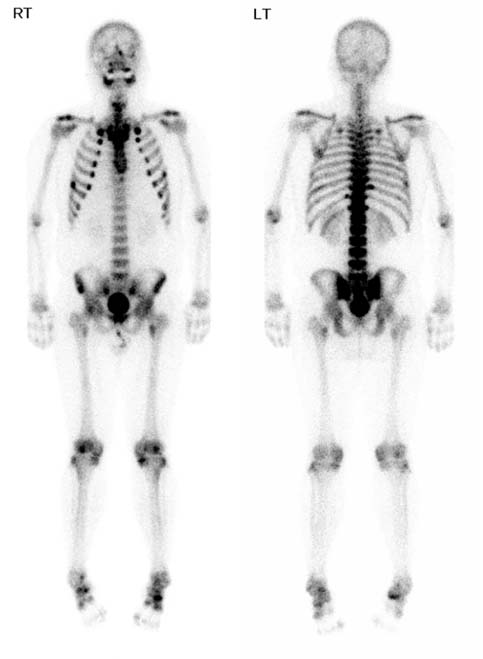

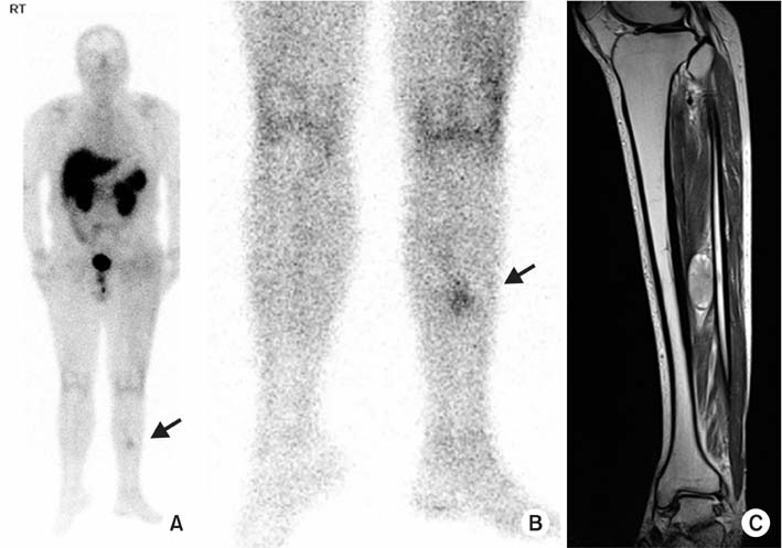
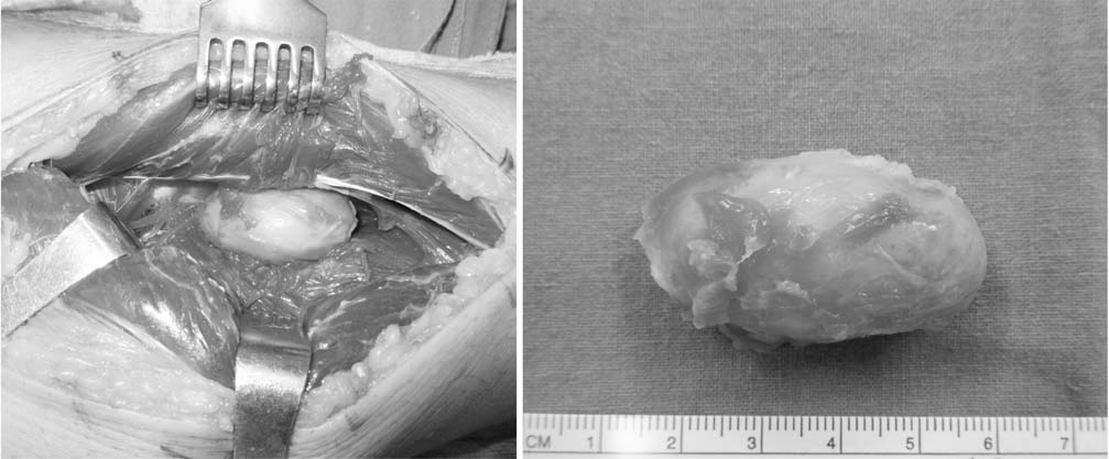
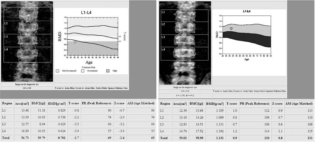
Fig. 1
Spine simple x-ray lateral view shows that vertebral height of T10, 11, 12, L1, and 2 decreased slightly (A) and magnetic resonance imaging T1-weighted enhanced image shows multiple compression fracture (B).
Fig. 2
Whole body bone scan shows multifocal bony uptakes in the bilateral ribs, pelvis, femoral neck, knee, and ankle. RT: right, LT: left.
Fig. 3
Insufficiency fracture of the femoral neck. A lucent line (arrow) is called a Looser's zone or pseudofracture.
Fig. 4
(A, B) Tumor localization. Causative tumor (arrows) was found on the left lower leg by Octreoscan. (C) Magnetic resonance imaging T2-weighted image shows 4.5×2.0×2.0-cm-sized soft tissue tumor located at the tibialis posterior muscle belly. Tumor had a round, well-defined margin with heterogeneous signal intensity. RT: right.
Fig. 5
Intraoperative image. Tumor was excised completely.
Fig. 6
Bone mineral density (BMD) change. Compared to the preoperative status, BMD and Z-score at 1 year and six months follow-up after surgery showed good recovery. BMC: bone mineral content.
Fig. 1
Fig. 2
Fig. 3
Fig. 4
Fig. 5
Fig. 6
Oncogenic Osteomalacia with Multiple Insufficiency Fractures: A Case Report
Laboratory Findings in Oncogenic Osteomalcia
| Variable | Oncogenic osteomalacia | Case | Reference range | |||
|---|---|---|---|---|---|---|
| Initial visit | Preoperative | POD 1 day | POD 1 week† | |||
| Phosphorus (mg/dl) | ↓ | 1.7* | 1.9* | 3.5 | 3.7 | 2.9-4.6 |
| 25(OH)D (ng/ml) | NL or ↓ | 11.1* | 12.1* | - | 16.5* | 30-100 |
| 1,25(OH)2D (pg/ml) | ↓ or NL | - | 18.79 | - | 278.62* | 19.6-54.3 |
| Alkaline phosphatase (IU/L) | ↑ | 172* | 215* | 201* | 220* | 44-99 |
| PTH (pg/ml) | NL or ↑ | 118.3* | 48 | - | - | 15-65 |
| Calcium (mg/dl) | NL | 8.8 | 8.9 | 8.6 | 8.8 | 8.5-10.1 |
| FGF-23 (RU/ml) | ↑ | - | 608* | - | 72 | <180 |
*Abnormal laboratory findings. †Serum phosphate and FGF-23 levels were normalized 1 week after excision of phosphaturic mesenchymal tumor. POD: postoperative day, PTH: parathyroid hormone, FGF-23: fibroblast growth factor-23, NL: normal.
Table 1
Laboratory Findings in Oncogenic Osteomalcia
*Abnormal laboratory findings. †Serum phosphate and FGF-23 levels were normalized 1 week after excision of phosphaturic mesenchymal tumor. POD: postoperative day, PTH: parathyroid hormone, FGF-23: fibroblast growth factor-23, NL: normal.

