 E-submission
 TOTA
TOTA TOTS
TOTS
Articles
- Page Path
- HOME > J Musculoskelet Trauma > Volume 21(3); 2008 > Article
-
Original Article
- Treatment of Intra-articular Calcaneal Fractures Using Minimally Invasive Sinus Tarsi Approach in Diabetic Patients
- Hong-Moon Sohn, M.D., Sang-Ho Ha, M.D., Sang-Hong Lee, M.D., Jun-Young Lee, M.D., Jeong-Ho Kim, M.D., Sang-Jun Lee, M.D.
-
Journal of the Korean Fracture Society 2008;21(3):195-199.
DOI: https://doi.org/10.12671/jkfs.2008.21.3.195
Published online: July 31, 2008
Department of Orthopaedic Surgery, College of Medicine, Chosun University, Gwangju, Korea.
- Address reprint requests to: Jun-Young Lee, M.D. Department of Orthopaedic Surgery, Chosun University Hospital, 588, Seosuk-dong, Dong-gu, Gwangju 501-717, Korea. Tel: 82-62-220-3147, Fax: 82-62-226-3379, leejy88@chosun.ac.kr
Copyright © 2008 The Korean Fracture Society. All rights reserved.
This is an Open Access article distributed under the terms of the Creative Commons Attribution Non-Commercial License (http://creativecommons.org/licenses/by-nc/3.0/) which permits unrestricted non-commercial use, distribution, and reproduction in any medium, provided the original work is properly cited.
- 1,003 Views
- 2 Download
- 1 Crossref
Abstract
-
Purpose
- Wound problems occur in 5~30% of intra-articular calcaneal fractures following operation. Diabetes mellitus, large incisions and abundant dissection can increase the risk of wound problems that may require skin graft or other additional care. The authors used minimally invasive technique to treat intra-articular calcaneal fractures in diabetic patients and evaluated the results and complications of this method.
-
Materials and Methods
- Between January 2002 and July 2005, 12 patients with intra-articular calcaneal fractures who had underlying diabetes mellitus were treated using minimally invasive technique with a modified sinus tarsi approach. The patients had an average age of 47 years (39~67) and were followed an average of 19 months (13~32). The mean period between injury and operation was 8 days (5~14). Crutch assisted partial weight bearing was advised for an average of 7.3 weeks (6~9) and full weight bearing was allowed after average of 9.3 weeks (7~11).
-
Results
- According to AOFAS scale for ankle and hindfoot, patients had the following results: excellent - 1 patient (8%), good - 9 patients (75%), fair - 1 patient (8%), unsatisfied - 1 patient (8%). Bone union was achieved in all cases and there were no events of deep infection or skin necrosis.
-
Conclusion
- Treating intra-articular calcaneal fractures by minimally invasive technique is an excellent operative method for patients with diabetes mellitus, as this method can minimize soft tissue incision and resulting deep infection and skin necrosis.
- 1. Abidi NA, Dhawan S, Gruen GS, Vogt MT, Conti SF. Wound-healing risk factors after open reduction and internal fixation of calcaneal fractures. Foot Ankle Int, 1998;19:856-861.ArticlePubMedPDF
- 2. Allon SM, Mears DC. Three dimensional analysis of calcaneal fractures. Foot Ankle, 1991;11:254-263.ArticlePubMedPDF
- 3. Byun YS, Cho YH, Park JW, Lee JS, Kim JH. Early postoperative complications of calcaneal fractures following operative treatment by lateral extensile approach. J Korean Fract Soc, 2004;17:323-327.Article
- 4. Catani F, Benedetti MG, Simoncini L, Leardin A, Giannini S. Analysis of function after intra-articular fracture of the os calcis. Foot Ankle Int, 1999;20:417-421.ArticlePDF
- 5. Cho SD, Cho YS, Kim BS, Prak TW, Kim GB, Kim KY. Result of surgical treatment of calcaneal fractures using extensile lateral approach. J Korean Fracture Soc, 1999;12:320-327.Article
- 6. Ebraheim NA, Elgafy H, Sabry FF, Freih M, Abou-Chakra IS. Sinus tarsi approach with trans-articular fixation for displaced intra-articular fractures of the calcaneus. Foot Ankle Int, 2000;21:105-113.ArticlePubMedPDF
- 7. Essex-Lopresti P. The mechanism, reduction technique, and results in fractures of the os calcis. Br J Surg, 1952;39:395-419.ArticlePubMedPDF
- 8. Fernandez DL, Koella C. Combined percutaneous and "minimal" internal fixation for displaced articular fractures of the calcaneus. Clin Orthop Relat Res, 1993;290:108-116.Article
- 9. Folk JW, Starr AJ, Early JS. Early wound complications of operative treatment of calcaneus fractures: analysis of 190 fractures. J Orthop Trauma, 1999;13:369-372.ArticlePubMed
- 10. Harvey EJ, Grujic L, Early JS, Benirschke SK, Sangeorzan BJ. Morbidity associated with ORIF of intra-articular calcaneus fractures using a lateral approach. Foot Ankle Int, 2001;22:868-873.ArticlePubMedPDF
- 11. Kim CW, Chung MY, Jung KT. Comparison of the conservative and operative treatment of the intraarticular calcaneal fractures. J Korean Soc Fract, 1999;12:335-343.Article
- 12. Kitaoka HB, Alexander IJ, Adelaar RS, Nunley JA, Myerson MS, Sanders M. Clinical rating systems for the ankle-hindfoot, midfoot, hallux, and lesser toes. Foot Ankle Int, 1994;14:349-353.ArticlePDF
- 13. Kundel K, Funk E, Brutscher M, Bickel R. Calcaneal fractures: operative versus nonoperative treatment. J Trauma, 1996;41:839-845.PubMed
- 14. Loder RT. The influence of diabetes mellitus on the healing of closed fractures. Clin Orthop Relat Res, 1988;232:210-216.Article
- 15. O'Brien J, Buckley R, McCormack R, et al. Personal gait satisfaction after displaced intra-articular calcaneal fractures: a 2-8 year followup. Foot Ankle Int, 2004;25:657-665.ArticlePubMedPDF
- 16. Paley D, Hall H. Intra-articular fractures of the calcaneus. A critical analysis of results and prognostic factors. J Bone Joint Surg Am, 1993;75:342-354.ArticlePubMed
- 17. Randle JA, Kreder HJ, Stephen D, Williams J, Jaglal S, Hu R. Should calcaneal fractures be treated surgically? A meta-analysis. Clin Orthop Relat Res, 2000;377:217-227.
- 18. Sanders R. Displaced intra-articular fractures of the calcaneus. J Bone Joint Surg Am, 2000;82:225-250.ArticlePubMed
- 19. Sanders R, Fortin P, DiPasquale T, Walling A. Operative treatment in 120 displaced intra-articular calcaneal fractures. Results using a prognostic computed tomography scan classification. Clin Orthop Relat Res, 1993;290:87-95.
REFERENCES
Fig. 1
(B) Intra-operative photograph demonstrating posterior facet reduced with traction bow (left), and fixated with temporary 1.4 mm K-wires (right).
(C) Intra-operative lateral C-arm image demonstrating posterior facet reduced and stabilized with K-wire.
(D) At 12 months after operation, good alignment and union was achieved.
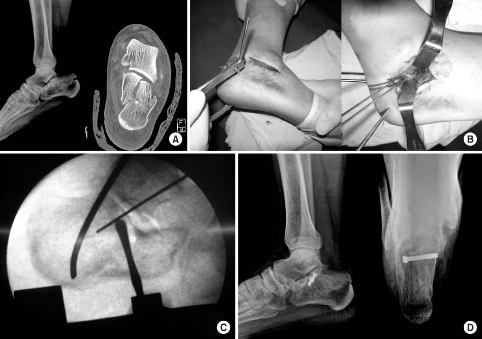
(A) Initial ankle lateral X-ray and semicoronal CT scan of a 39 year-old man with inatraarticular calcaneal fracture demonstrating displaced lateral posterior facet fragment.
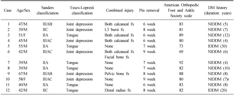
Figure & Data
REFERENCES
Citations
Citations to this article as recorded by 
- Ankle and foot fracture management in patients with diabetes: an evidence-based review
Caroline E Plant, Arham Farukh Qureshi, Anna Chapman
Orthopaedics and Trauma.2026; 40(2): 105. CrossRef
 Cite
CiteTreatment of Intra-articular Calcaneal Fractures Using Minimally Invasive Sinus Tarsi Approach in Diabetic Patients

Fig. 1
(A) Initial ankle lateral X-ray and semicoronal CT scan of a 39 year-old man with inatraarticular calcaneal fracture demonstrating displaced lateral posterior facet fragment.
(B) Intra-operative photograph demonstrating posterior facet reduced with traction bow (left), and fixated with temporary 1.4 mm K-wires (right).
(C) Intra-operative lateral C-arm image demonstrating posterior facet reduced and stabilized with K-wire.
(D) At 12 months after operation, good alignment and union was achieved.
Fig. 1
Treatment of Intra-articular Calcaneal Fractures Using Minimally Invasive Sinus Tarsi Approach in Diabetic Patients
Patients data
Table 1
Patients data

