 E-submission
 TOTA
TOTA TOTS
TOTS
Articles
- Page Path
- HOME > J Musculoskelet Trauma > Volume 23(4); 2010 > Article
-
Case Report
- Removal of a Broken Intramedullary Nail with a Narrow Hollow Using a Bulb-tipped Guide Wire and Kirschner Wire: A Case Report
- Moses Lee, M.D., Kyu-Hyun Yang, M.D.
-
Journal of the Korean Fracture Society 2010;23(4):377-381.
DOI: https://doi.org/10.12671/jkfs.2010.23.4.377
Published online: October 31, 2010
Department of Orthopedic Surgery, Yonsei University College of Medicine, Seoul, Korea.
- Address reprint requests to: Kyu-Hyun Yang, M.D. Department of Orthopedic Surgery, Gangnam Severance Hospital, College of Medicine, Yonsei University, 146-92, Dokok-dong, Gannam-gu, Seoul 135-720, Korea. Tel: 82-2-2019-3414, Fax: 82-2-573-5393, kyang@yuhs.ac
• Received: June 9, 2010 • Accepted: July 23, 2010
Copyright © 2010 The Korean Fracture Society
- 1,385 Views
- 5 Download
- 4 Crossref
Abstract
- To report the unusual failure of proximal femoral nail antirotation (PFNA) at the level of lag screw hole and introduce a simple technique for removal of a broken intramedullary nail with a narrow hollow using a bulb-tipped guide wire and Kirschner wire.
CASE REPORT
DISCUSSION
- 1. Brewster NT, Ashcroft GP, Scotland TR. Extraction of broken intramedullary nails--an improvement in technique. Injury, 1995;26:286. Article
- 2. Georgilas I, Mouzopoulos G, Neila C, Morakis E, Tzurbakis M. Removal of broken distal intramedullary nail with a simple method: a case report. Arch Orthop Trauma Surg, 2009;129:203-205.ArticlePDF
- 3. Karladani AH. Removal of a broken nail using a guide wire and a screw. Acta Orthop, 2006;77:986-988.Article
- 4. Levine JW, Georgiadis GM. Removal of a broken cannulated tibial nail: a simple intramedullary technique. J Orthop Trauma, 2004;18:247-249.
- 5. Magu NK, Sharma AK, Singh R. Extraction of the broken intramedullary femoral nail--an innovative technique. Injury, 2004;35:1322-1323.Article
- 6. Park SY, Yang KH, Yoo JH. Removal of a broken intramedullary nail with a narrow hollow. J Orthop Trauma, 2006;20:492-494.Article
- 7. Poehling GG, Webb LX. Retrieval and replacement of a broken Küntscher rod by a closed technique. Technical note. J Bone Joint Surg Am, 1982;64:1389-1390.Article
- 8. Winquist RA. Broken interlocking nails and screws. Tech Orthop, 1998;13:15-26.Article
REFERENCES
Fig. 1
(A) Preoperative radiograph shows subtrochanteric fracture of left femur: Hypertrophy of the lateral cortex and transverse fracture with medial beak were noticed.
(B) Postoperative radiograph after the initial operation: inadequate reduction status with flexion deformity at the fracture site.
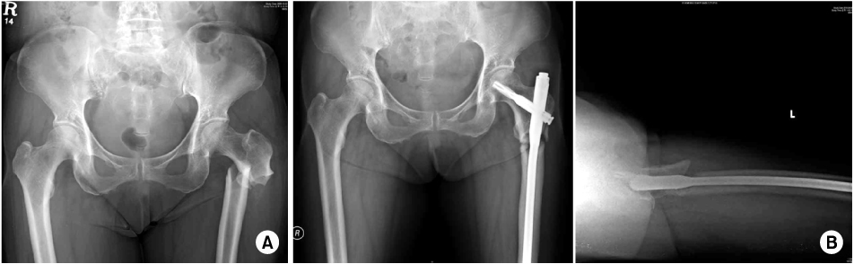
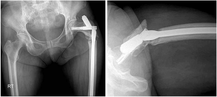
Fig. 3Intraoperative radiographs from image intensifier; after removing of the proximal portion of broken nail, bulb-tipped guide wire was introduced (arrow).

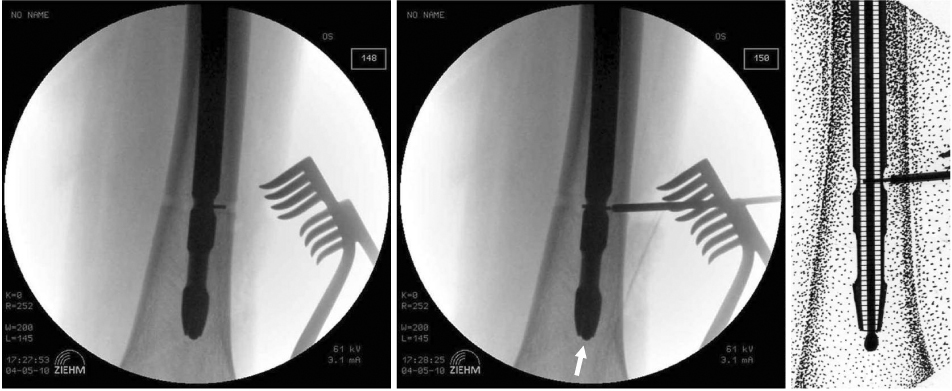
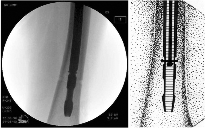
Figure & Data
REFERENCES
Citations
Citations to this article as recorded by 
- Novel Method for Removal of Broken Intramedullary Interlocking Nail with a Subtrochanteric Fracture
Ashwani Soni, Anmol Sharma, Rajeev Kumar Kansay, Deepam Vashist
JBJS Case Connector.2019; 9(4): e0182. CrossRef - METHOD FOR REMOVING BROKEN PROXIMAL FEMORAL NAILS USING EXISTING SCREW HOLE
CHO HONG MAN, MIN WOONG BAE
Acta Ortopédica Brasileira.2018; 26(1): 72. CrossRef - A blocking-wire technique for removal of a broken hollow intramedullary nail
Xuan-Lin Zheng, Young-Chang Park, Dong-Hyun Kang, Sang-Ok Seok, Yeo-Kwon Yoon, Kyu-Hyun Yang
Injury.2016; 47(7): 1601. CrossRef - Removal Methods for Broken Proximal Femoral Nails Using Ball Tip Guide Wire: Technical Note and Two Cases Report
Bong-Ju Park, Hong-Man Cho
Journal of the Korean Fracture Society.2014; 27(4): 315. CrossRef
 Cite
CiteRemoval of a Broken Intramedullary Nail with a Narrow Hollow Using a Bulb-tipped Guide Wire and Kirschner Wire: A Case Report


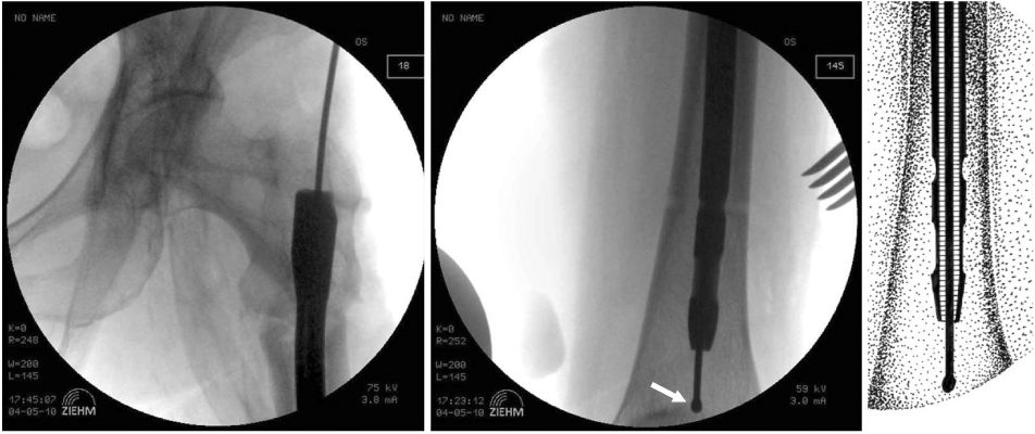


Fig. 1
(A) Preoperative radiograph shows subtrochanteric fracture of left femur: Hypertrophy of the lateral cortex and transverse fracture with medial beak were noticed.
(B) Postoperative radiograph after the initial operation: inadequate reduction status with flexion deformity at the fracture site.
Fig. 2
Nonunion and hardware failure developed at seven months after the operation.
Fig. 3
Intraoperative radiographs from image intensifier; after removing of the proximal portion of broken nail, bulb-tipped guide wire was introduced (arrow).
Fig. 4
Intraoperative radiographs from image intensifier; after removing distal interlocking screws a 10 mm length no. 3 Kirschiner wire was introduced into the distal interlocking hole and further pushed with a no 3. Steinmann pin. An arrow indicates the tip of a bulb-tipped guide wire.
Fig. 5
Intraoperative radiographs from image intensifier; bulb-tipped guide wire was gently pulled out until the bulb abutted and locked with the K-wire at the distal interlocking hole, then exchange IM nailing was done.
Fig. 1
Fig. 2
Fig. 3
Fig. 4
Fig. 5
Removal of a Broken Intramedullary Nail with a Narrow Hollow Using a Bulb-tipped Guide Wire and Kirschner Wire: A Case Report

