 E-submission
 TOTA
TOTA TOTS
TOTS
Articles
- Page Path
- HOME > J Musculoskelet Trauma > Volume 23(3); 2010 > Article
-
Original Article
- Staged Minimally Invasive Plate Osteosynthesis of Distal Tibial Fractures
-
Sung-Ki Park, M.D., Chang-Wug Oh, M.D., Jong-Keon Oh, M.D., Kyung-Hoon Kim, M.D., Woo-Kie Min, M.D.
 , Byung-Chul Park, M.D., Won-Ju Jeong, M.D., Joo-Chul Ihn, M.D.
 -
Journal of the Korean Fracture Society 2010;23(3):289-295.
DOI: https://doi.org/10.12671/jkfs.2010.23.3.289
Published online: July 31, 2010
Department of Orthopedic Surgery, School of Medicine, Kyungpook National University, Daegu, Korea.
*Department of Orthopedic Surgery, School of Medicine, Korea University, Seoul, Korea.
†Department of Orthopedic Surgery, Daegu Veterans Hospital, Daegu, Korea.
- Address reprint requests to: Chang-Wug Oh, M.D. Department of Orthopedic Surgery, School of Medicine, Kyungpook National University, 101, Dongin-dong 2-ga, Jung-gu, Deagu 700-422, Korea. Tel: 82-53-420-5630, Fax: 82-53-422-6605, cwoh@knu.ac.kr
• Received: October 17, 2009 • Revised: April 28, 2010 • Accepted: May 12, 2010
Copyright © 2010 The Korean Fracture Society
- 1,418 Views
- 6 Download
- 2 Crossref
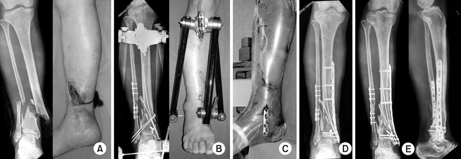
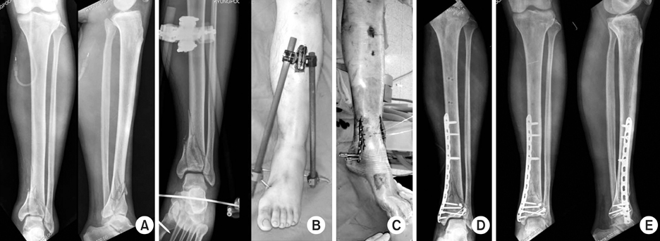
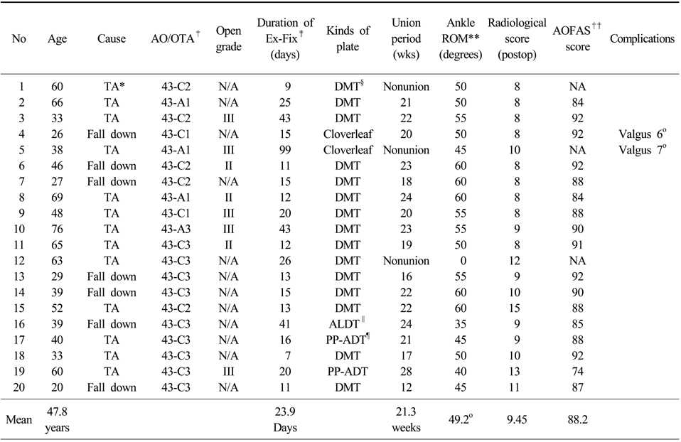
Figure & Data
REFERENCES
Citations
Citations to this article as recorded by 
- Anterolateral Minimally Invasive Plate Osteosynthesis of Distal Tibial Fractures Using an Anterolateral Locking Plate
Dongwhan Suh, Hwan Hee Lee, Young Hoon Han, Jae Jung Jeong
Journal of Korean Foot and Ankle Society.2020; 24(1): 19. CrossRef - Minimally Invasive Osteosynthesis with Locking Compression Plate for Distal Tibia Fractures
Sung-Kyu Kim, Keun-Bae Lee, Keun-Young Lim, Eun-Sun Moon
Journal of the Korean Fracture Society.2011; 24(1): 33. CrossRef
 Cite
CiteStaged Minimally Invasive Plate Osteosynthesis of Distal Tibial Fractures


Fig. 1
(A) A 48-year old man sustained the distal tibial fracture with grade IIIA open wound.
(B) After fixation of fibula fracture, bridging external fixation was done with wound debridement and closure.
(C, D) After the improvement of soft tissue condition, medial plating was done with minimally invasive technique.
(E) At 6 month, a successful union was achieved with the good alignment.
Fig. 2
(A) An AO-OTA 43-C2 fracture.
(B) A bridging external fixation was applied.
(C, D) After the improvement of soft tissue condition, medial MIPO was done with joint reconstruction.
(E) A successful healing was achieved after 8 months.
Fig. 1
Fig. 2
Staged Minimally Invasive Plate Osteosynthesis of Distal Tibial Fractures
Distal tibial fractures treated with staged MIPO technique
*TA: Traffic accident, †AO/OTA: Arbeitsgemeinschaft für Osteosynthesefragen and orthopedic trauma association, ‡Ex-Fix: External fixator, §DMT: LCP distal medial tibia, ∥ALDT: LCP Anterolateral distal tibia plate, ¶PP-ADT: Periarticular anterolateral distal tibia plate, **ROM: Range of motion, ††AOFAS: American orthopedic foot and ankle society.
Table 1
Distal tibial fractures treated with staged MIPO technique
*TA: Traffic accident, †AO/OTA: Arbeitsgemeinschaft für Osteosynthesefragen and orthopedic trauma association, ‡Ex-Fix: External fixator, §DMT: LCP distal medial tibia, ∥ALDT: LCP Anterolateral distal tibia plate, ¶PP-ADT: Periarticular anterolateral distal tibia plate, **ROM: Range of motion, ††AOFAS: American orthopedic foot and ankle society.

