 E-submission
 TOTA
TOTA TOTS
TOTS
Articles
- Page Path
- HOME > J Musculoskelet Trauma > Volume 23(2); 2010 > Article
-
Case Report
- Bursting Fracture of the Proximal Femur during Insertion of Unreamed Femoral Nail for Femur Shaft Fracture: A Case Report
- Ji Wan Kim, M.D., Seong-Eun Byun, M.D., Won-Hyuk Oh, M.D., Jung Jae Kim, M.D.
-
Journal of the Korean Fracture Society 2010;23(2):227-231.
DOI: https://doi.org/10.12671/jkfs.2010.23.2.227
Published online: April 30, 2010
Department of Orthopaedic Surgery, Asan Medical Center, College of Medicine, University of Ulsan, Seoul, Korea.
*Department of Orthopaedic Surgery, Inje University, Haeundae Paik Hospital, Busan, Korea.
†Samchunpo Seoul Hospital, Samchunpo, Korea.
- Address reprint requests to: Jung Jae Kim, M.D. Department of Orthopaedic Surgery, Asan Medical Center, College of Medicine, University of Ulsan, 86, Asanbyeongwon-gil, Songpa-gu, Seoul 138-736, Korea. Tel: 82-2-3010-3530, Fax: 82-2-488-7877, jjkim2@amc.seoul.kr
• Received: November 11, 2009 • Revised: December 22, 2009 • Accepted: January 28, 2010
Copyright © 2010 The Korean Fracture Society
- 1,378 Views
- 4 Download
- 3 Crossref
Abstract
- When treating femur shaft fracture in adults, undreamed nail can be an option in order to avoid systemic complications. To appropriately insert unreamed intramedullary nail, an accurate entry point and sufficient reaming of the entry portal is essential. The intramedullary canal of the proximal femur must be reamed over than the diameter of the proximal end of the nail. If the proximal reaming is not sufficient, complications such as bursting fracture of proximal femur can occur. We present two cases of bursting fracture of proximal femur following insertion of undreamed intramedullary nail as well as a literature review.
- 1. Abbas D, Faisal M, Butt MS. Unreamed femoral nailing. Injury, 2000;31:711-717.Article
- 2. Alho A, Strømsøe K, Ekeland A. Locked intramedullary nailing of femoral shaft fractures. J Trauma, 1991;31:49-59.Article
- 3. Brumback RJ, Virkus WW. Intramedullary nailing of the femur: reamed versus nonreamed. J Am Acad Orthop Surg, 2000;8:83-90.Article
- 4. Canadian Orthopaedic Trauma Society. Reamed versus unreamed intramedullary nailing of the femur: comparison of the rate of ARDS in multiple injured patients. J Orthop Trauma, 2006;20:384-387.Article
- 5. Forster MC, Aster AS, Ahmed S. Reaming during anterograde femoral nailing: is it worth it? Injury, 2005;36:445-449.Article
- 6. Johnson KD, Tencer AF, Sherman MC. Biomechanical factors affecting fracture stability and femoral bursting in closed intramedullary nailing of femoral shaft fractures, with illustrative case presentations. J Orthop Trauma, 1987;1:1-11.Article
- 7. Papadakis SA, Zalavras C, Mirzayan R, Shepherd L. Undetected iatrogenic lesions of the anterior femoral shaft during intramedullary nailing: a cadaveric study. J Orthop Surg Res, 2008;3:30. ArticlePDF
- 8. Shepherd LE, Shean CJ, Gelalis ID, Lee J, Carter VS. Prospective randomized study of reamed versus unreamed femoral intramedullary nailing: an assessment of procedures. J Orthop Trauma, 2001;15:28-32.Article
- 9. Tencer AF, Sherman MC, Johnson KD. Biomechanical factors affecting fracture stability and femoral bursting in closed intramedullary rod fixation of femur fractures. J Biomech Eng, 1985;107:104-111.ArticlePDF
- 10. Yoon HK, Jeon KP, Kang KH, Kim JI, Kim DS, Koh YK. A Clinical Comparative Study of Reamed and Unreamed Nail of Femoral Shaft Fracture. J Korean Soc Fract, 1998;11:495-500.Article
REFERENCES
Figure 1
(A) Initial AP view shows AO type A3 femoral shaft fracture and contralateral femoral shaft.
(B) Intraoperative C-arm image shows bursting fracture and (C) Bursting fracture is fixed by additional interlocking screw.
(D) Postoperative AP and (E) lateral x-rays show union of femoral shaft fracture and bursting fracture.
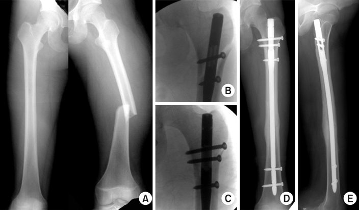
Figure 2
(A) Initial AP view shows AO type B1 femoral shaft fracture.
(B) Postoperative AP view, (C) lateral view, (D) preoperative 3D CT image and (E) postoperative 3D CT image show bursting fracture of proximal femur.

Figure 3
(A) Postoperative CT shows the appropriate entry point of the nail.
(B) Preoperative CT shows 9.8 mm of diameter of the isthmus of the femur.
(C) The x-ray shows the level of the following axial images (D~I).


Figure & Data
REFERENCES
Citations
Citations to this article as recorded by 
- Risk Factors Associated with Intraoperative Iatrogenic Fracture in Patients Undergoing Intramedullary Nailing for Atypical Femoral Fractures with Marked Anterior and Lateral Bowing
Yong Bum Joo, Yoo Sun Jeon, Woo Yong Lee, Hyung Jin Chung
Medicina.2023; 59(4): 735. CrossRef - Results of Intramedullary Nailing of Femoral Shaft Fracture - Trochanteric Entry Portal (Sirus Nail) versus Piriformis Entry Portal (M/DN Nail) -
Sang Ho Ha, Woong-Hee Kim, Gwang Chul Lee
Journal of the Korean Fracture Society.2014; 27(1): 50. CrossRef - Iatrogenic Femur Proximal Shaft Fracture during Nailing Using Lateral Entry Portal on Femur Shaft Fracture
Hong Moon Sohn, Gwang Chul Lee, Chae Won Lim
Journal of the Korean Orthopaedic Association.2014; 49(4): 272. CrossRef
 Cite
CiteBursting Fracture of the Proximal Femur during Insertion of Unreamed Femoral Nail for Femur Shaft Fracture: A Case Report

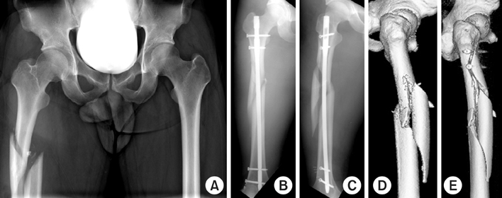

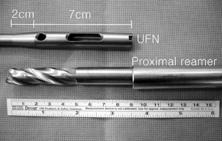
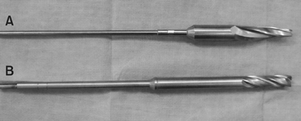
Figure 1
(A) Initial AP view shows AO type A3 femoral shaft fracture and contralateral femoral shaft.
(B) Intraoperative C-arm image shows bursting fracture and (C) Bursting fracture is fixed by additional interlocking screw.
(D) Postoperative AP and (E) lateral x-rays show union of femoral shaft fracture and bursting fracture.
Figure 2
(A) Initial AP view shows AO type B1 femoral shaft fracture.
(B) Postoperative AP view, (C) lateral view, (D) preoperative 3D CT image and (E) postoperative 3D CT image show bursting fracture of proximal femur.
Figure 3
(A) Postoperative CT shows the appropriate entry point of the nail.
(B) Preoperative CT shows 9.8 mm of diameter of the isthmus of the femur.
(C) The x-ray shows the level of the following axial images (D~I).
Figure 4
The diameter of the most proximal part of the UFN is 13 mm, the diameter of the following portion becomes slender and the diameter of the other part is 9~12 mm.
Figure 5
The comparison of proximal reamer for UFN and PFNA. (A) is the proximal reamer for PFNA and the end of PFNA proximal reamer is tapered, and (B) is the proximal reamer for UFN.
Figure 1
Figure 2
Figure 3
Figure 4
Figure 5
Bursting Fracture of the Proximal Femur during Insertion of Unreamed Femoral Nail for Femur Shaft Fracture: A Case Report

