 E-submission
 TOTA
TOTA TOTS
TOTS
Articles
- Page Path
- HOME > J Musculoskelet Trauma > Volume 23(2); 2010 > Article
-
Case Report
- Fracture-Dislocation of S1 in 3-Year-Old Boy: A Case Report
- Sang-Bong Ko, M.D., Sang-Wook Lee, M.D.
-
Journal of the Korean Fracture Society 2010;23(2):232-235.
DOI: https://doi.org/10.12671/jkfs.2010.23.2.232
Published online: April 30, 2010
Department of Orthopedic Surgery, Daegu Catholic University Medical Center, Daegu, Korea.
- Address reprint requests to: Sang-Bong Ko, M.D. Department of Orthopedic Surgery, Daegu Catholic University Medical Center, 3056-6, Daemyung 4-dong, Nam-gu, Daegu 705-825, Korea. Tel: 82-53-650-4283, Fax: 82-53-626-4272, bong@cu.ac.kr
• Received: November 4, 2009 • Revised: February 24, 2010 • Accepted: February 27, 2010
Copyright © 2010 The Korean Fracture Society
- 620 Views
- 1 Download
Abstract
- Fracture-dislocation of the sacrum that has not yet fully developed is common in the distal sacrococcygeal joint of children, but this injury is rarely seen in 1st Sacrum. Most of these patients have a severe neurological deficit, so this injury generally requires surgical decompression. We managed a three year old patient who had a S1 fracture-dislocation without a neurological deficit, and the patient was treated with simple skin traction and bed rest without surgery. The child had a satisfactory result, so we report on this case with reviewing the relevant literatures.
- 1. Beguiristain J, Schweitzer D, Mora G, Pombo V. Traumatic lumbosacral dislocation in a 5-year-old boy with eight years follow-up. Spine (Phila Pa 1976), 1995;20:362-366.Article
- 2. Bucknill TM, Blackburne JS. Fracture-Dislocations of the sacrum. Report of three cases. J Bone Joint Surg Br, 1976;58-B:476-470.ArticlePDF
- 3. Gibbons KJ, Solonluk DS, Razack N. Neurological injury and patterns of sacral fractures. J Neurosurg, 1990;72:889-893.Article
- 4. Lyon RM. Frymoyer JW, Wiesel SW. Pediatric Spine Injuries. In: The Adult & Pediatric Spine, 2004;3rd ed. Philadelphia, Lippincott Williams & Wilkins. 440.
- 5. Mann DC, Dodds JA. Spinal injuries in 57 patients 17 years or younger. Orthopedics, 1993;16:159-164.Article
- 6. Novkov HV, Tanchev PJ, Gyorev IS. Severe fracture-dislocation of S1 in a 12-year-old boy. A case report. . Spine (Phila Pa 1976), 1996;21:2500-2503.
- 7. Rodriguez-Fuentes AE. Traumatic sacrolisthesis S1-2. Report of a case. Spine Phila Pa 1976, 1993;18:768-771.
- 8. Roy-Camille R, Saillant G, Gagna G, Mazel C. Transverse fractures of the upper sacrum. Suicidal jumper's fracture. Spine (Phila Pa 1976), 1985;10:838-845.
- 9. Strange-Vognsen HH, Lebech A. An unusual type of fracture in the upper sacrum. J Orthop Trauma, 1991;5:200-203.Article
REFERENCES

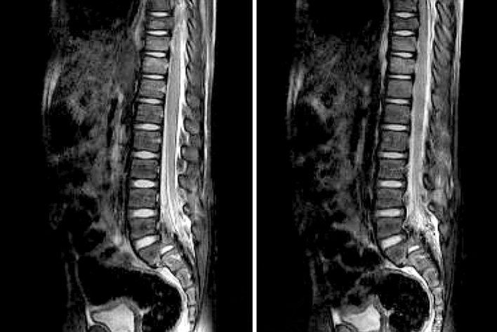

Figure & Data
REFERENCES
Citations
Citations to this article as recorded by 
 Cite
CiteFracture-Dislocation of S1 in 3-Year-Old Boy: A Case Report
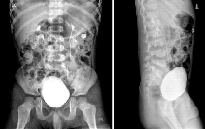

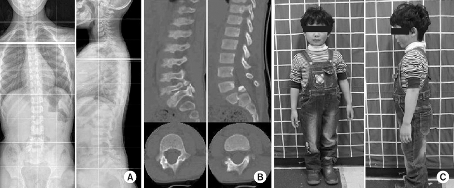
Figure 1
Initial radiographs shows fracture-dislocation of 1st Sacrum in L-spine AP and Lateral views.
Figure 2
Fracture dislocation of 1st Sacrum is seen in sagittal section of MRI.
Figure 3
At 2 year later follow up.
(A) Simple whole spine radiographs show good sagittal and frontal alignment.
(B) CT show union of each pedicle, elongation of pedicle and remodeling state of 1st sacrum.
(C) Gross photographs show good sagittal and frontal alignment.
Figure 1
Figure 2
Figure 3
Fracture-Dislocation of S1 in 3-Year-Old Boy: A Case Report

