 E-submission
 TOTA
TOTA TOTS
TOTS
Articles
- Page Path
- HOME > J Musculoskelet Trauma > Volume 26(2); 2013 > Article
-
Original Article
- Minimally Invasive Plate Osteosynthesis for Femoral Mid-Diaphyseal Fractures
- Hyoung-Keun Oh, M.D., Suk-Kyoo Choo, M.D., Jong-In Kim, M.D., Sung-Jong Woo, M.D.
-
Journal of the Korean Fracture Society 2013;26(2):140-146.
DOI: https://doi.org/10.12671/jkfs.2013.26.2.140
Published online: April 22, 2013
Department of Orthopedic Surgery, Inje University Ilsan Paik Hospital, Goyang, Korea.
- Address reprint requests to: Hyoung-Keun Oh, M.D. Department of Orthopedic Surgery, Inje University Ilsan Paik Hospital, 170 Juhwa-ro, Ilsanseo-gu, Goyang 411-706, Korea. Tel: 82-31-910-7968, Fax: 82-31-910-7967, osd11@paik.ac.kr
• Received: October 23, 2012 • Revised: January 14, 2013 • Accepted: February 12, 2013
Copyright © 2013 The Korean Fracture Society
- 629 Views
- 4 Download
Abstract
-
Purpose
- To investigate the surgical outcomes of patients with femoral mid-diaphyseal fractures treated with minimally invasive plate osteosynthesis (MIPO), which were difficult to intramedullary nailing.
-
Materials and Methods
- We evaluated 11 patients with femoral mid-diaphyseal fractures who were treated with MIPO. There were 7 males and 4 females and the mean age was 47 years (20-85 years). According to AO/OTA classification, there were 1 type of A1, 5 types of A3, 1 of B2 and 4 of B3. The reason of plate fixation instead of intramedullary nailing is as follows: femoral vessel and severe soft tissue injuries-2 cases, polytrauma patients with chest injury-6 cases, and narrow medullary canal diameter-3 cases. Six out of 11 cases were treated with initial external fixation as a damage control orthopedics.
-
Results
- The mean union time of 6 cases was 3.7 months (3-5 months). There were 5 cases (45%) of nonunion, which should be treated with autogenous bone graft. All cases of nonunion resulted from severe soft tissue damage and polytrauma, which needed initial external fixation. There was no case of malalignment and implant-related complication.
-
Conclusion
- In cases of difficult intramedullary nailing for the femoral mid-diaphyseal fractures, MIPO could be an alternative surgical option, but concurrent soft tissue injuries and multiple trauma may increase the risk of nonunion in spite of biological fixation.
- 1. Apivatthakakul T, Chiewcharntanakit S. Minimally invasive plate osteosynthesis (MIPO) in the treatment of the femoral shaft fracture where intramedullary nailing is not indicated. Int Orthop, 2009;33:1119-1126.ArticlePubMedPDF
- 2. Bosse MJ, MacKenzie EJ, Riemer BL, et al. Adult respiratory distress syndrome, pneumonia, and mortality following thoracic injury and a femoral fracture treated either with intramedullary nailing with reaming or with a plate. A comparative study. J Bone Joint Surg Am, 1997;79:799-809.ArticlePubMed
- 3. Celebi L, Can M, Muratli HH, Yagmurlu MF, Yuksel HY, Bicimoğlu A. Indirect reduction and biological internal fixation of comminuted subtrochanteric fractures of the femur. Injury, 2006;37:740-750.ArticlePubMed
- 4. Farouk O, Krettek C, Miclau T, Schandelmaier P, Guy P, Tscherne H. Minimally invasive plate osteosynthesis: does percutaneous plating disrupt femoral blood supply less than the traditional technique? J Orthop Trauma, 1999;13:401-406.ArticlePubMed
- 5. Geissler WB, Powell TE, Blickenstaff KR, Savoie FH. Compression plating of acute femoral shaft fractures. Orthopedics, 1995;18:655-660.ArticlePubMed
- 6. Giannoudis PV, Smith RM, Bellamy MC, Morrison JF, Dickson RA, Guillou PJ. Stimulation of the inflammatory system by reamed and unreamed nailing of femoral fractures. An analysis of the second hit. J Bone Joint Surg Br, 1999;81:356-361.ArticlePubMed
- 7. Han SB, Choi IC, Lee SH, Suh DH, Cho HJ. Minimal invasive plate osteosynthesis for distal femoral fracture. J Korean Fract Soc, 2006;19:11-16.Article
- 8. Huang HT, Huang PJ, Su JY, Lin SY. Indirect reduction and bridge plating of supracondylar fractures of the femur. Injury, 2003;34:135-140.ArticlePubMed
- 9. Kesemenli C, Subasi M, Necmioglu S, Kapukaya A. Treatment of multifragmentary fractures of the femur by indirect reduction (biological) and plate fixation. Injury, 2002;33:691-699.ArticlePubMed
- 10. Krettek C, Müller M, Miclau T. Evolution of minimally invasive plate osteosynthesis (MIPO) in the femur. Injury, 2001;32:Suppl 3. SC14-SC23.ArticlePubMed
- 11. Krettek C, Schandelmaier P, Miclau T, Tscherne H. Minimally invasive percutaneous plate osteosynthesis (MIPPO) using the DCS in proximal and distal femoral fractures. Injury, 1997;28:Suppl 1. A20-A30.ArticlePubMed
- 12. Oh CW, Oh JK, Kim SJ, et al. Minimally invasive plate osteosynthesis for comminuted subtrochanteric fracture of the femur. J Korean Fract Soc, 2006;19:407-411.Article
- 13. Pape HC, Hildebrand F, Pertschy S, et al. Changes in the management of femoral shaft fractures in polytrauma patients: from early total care to damage control orthopedic surgery. J Trauma, 2002;53:452-461.ArticlePubMed
- 14. Thompson F, O'Beirne J, Gallagher J, Sheehan J, Quinlan W. Fractures of the femoral shaft treated by plating. Injury, 1985;16:535-538.ArticlePubMed
- 15. Tornetta P 3rd, Tiburzi D. Antegrade or retrograde reamed femoral nailing. A prospective, randomised trial. J Bone Joint Surg Br, 2000;82:652-654.PubMed
- 16. Tuttle MS, Smith WR, Williams AE, et al. Safety and efficacy of damage control external fixation versus early definitive stabilization for femoral shaft fractures in the multiple-injured patient. J Trauma, 2009;67:602-605.ArticlePubMed
- 17. Winquist RA, Hansen ST Jr, Clawson DK. Closed intramedullary nailing of femoral fractures. A report of fire hundred and twenty cases. J Bone Joint Surg Am, 1984;66:529-539.PubMed
- 18. Zhiquan A, Bingfang Z, Yeming W, Chi Z, Peiyan H. Minimally invasive plating osteosynthesis (MIPO) of middle and distal third humeral shaft fractures. J Orthop Trauma, 2007;21:628-633.ArticlePubMed
- 19. Zlowodzki M, Vogt D, Cole PA, Kregor PJ. Plating of femoral shaft fractures: open reduction and internal fixation versus submuscular fixation. J Trauma, 2007;63:1061-1065.ArticlePubMed
REFERENCES
Fig. 1
(A) A case of 20 years old male. Plain radiographs shows B3 femoral mid-diaphyseal fracture. Intamedullary nailing was contraindicated because of narrow medullay canal.
(B) (a) Postoperative radiograph shows good reduction and alignment of fracture after minimally invasive plate osteosynthesis. (b) Three months follow-up radiograph shows bridging callus. (c) Twelve months follow-up radiographs show solid bony union.

Fig. 2
(A) A case of 32 years old male. Plain radiographs and angiography show B3 femoral mid-diaphyseal fracture with total occlusion of the femoral artery (arrow).
(B) (a) There was severe soft tissue injury on the lateral thigh. (b) Bypass femoral artery reconstruction (arrow) was carried out through the medial open wound. (c) Femoral fracture was stabilized by temporary external fixation.
(C) (a) External fixator was converted to interal fixator using minimally invasive plate osteosynthesis technique after 18 days of the initial injury. (b) Previous external fixator and prevented locking compression plate could be used as a reduction tool. (c) Postoperative radiographs show good alignment of the fracture.
(D) (a) Plain radiographs show nonunion of femoral mid-diaphyseal fracture after 8 months of plate fixation. (b) After autogenous bone graft and supplementary plate fixation, solid bony union could be obtained.
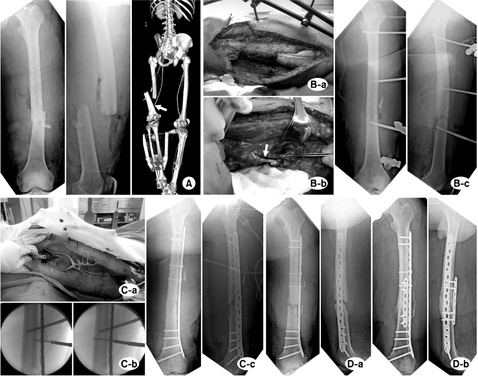
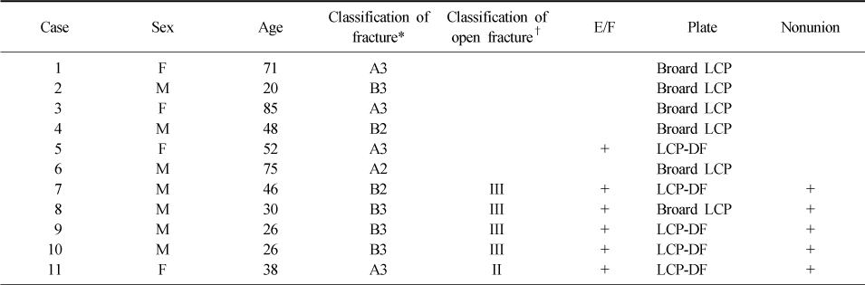
Figure & Data
REFERENCES
Citations
Citations to this article as recorded by 
 Cite
CiteMinimally Invasive Plate Osteosynthesis for Femoral Mid-Diaphyseal Fractures
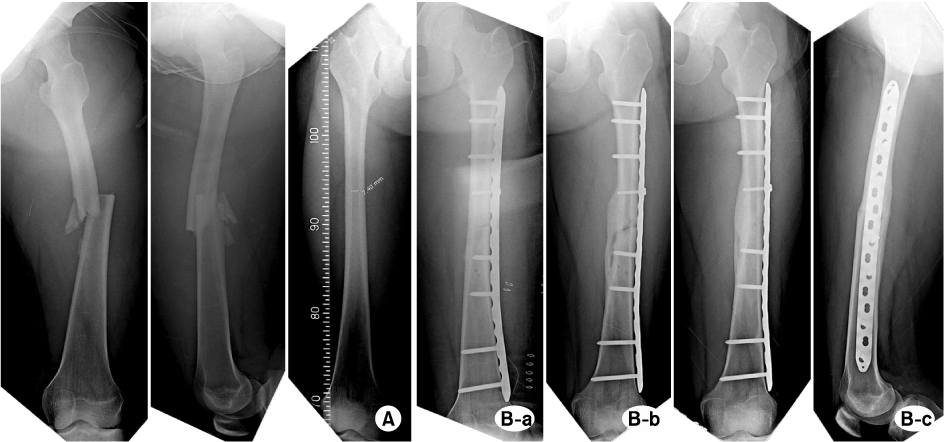

Fig. 1
(A) A case of 20 years old male. Plain radiographs shows B3 femoral mid-diaphyseal fracture. Intamedullary nailing was contraindicated because of narrow medullay canal.
(B) (a) Postoperative radiograph shows good reduction and alignment of fracture after minimally invasive plate osteosynthesis. (b) Three months follow-up radiograph shows bridging callus. (c) Twelve months follow-up radiographs show solid bony union.
Fig. 2
(A) A case of 32 years old male. Plain radiographs and angiography show B3 femoral mid-diaphyseal fracture with total occlusion of the femoral artery (arrow).
(B) (a) There was severe soft tissue injury on the lateral thigh. (b) Bypass femoral artery reconstruction (arrow) was carried out through the medial open wound. (c) Femoral fracture was stabilized by temporary external fixation.
(C) (a) External fixator was converted to interal fixator using minimally invasive plate osteosynthesis technique after 18 days of the initial injury. (b) Previous external fixator and prevented locking compression plate could be used as a reduction tool. (c) Postoperative radiographs show good alignment of the fracture.
(D) (a) Plain radiographs show nonunion of femoral mid-diaphyseal fracture after 8 months of plate fixation. (b) After autogenous bone graft and supplementary plate fixation, solid bony union could be obtained.
Fig. 1
Fig. 2
Minimally Invasive Plate Osteosynthesis for Femoral Mid-Diaphyseal Fractures
Demographic Data and Surgical Outcomes
E/F: External fixation, F: Female, M: Male, LCP: Locking compression plate, LCP-DF: Locking compression plate in distal femur.
*AO/OTA classification, †Gustillo-Anderson classification.
Table 1
Demographic Data and Surgical Outcomes
E/F: External fixation, F: Female, M: Male, LCP: Locking compression plate, LCP-DF: Locking compression plate in distal femur. *AO/OTA classification, †Gustillo-Anderson classification.

