 E-submission
 TOTA
TOTA TOTS
TOTS
Articles
- Page Path
- HOME > J Musculoskelet Trauma > Volume 28(3); 2015 > Article
-
Original Article
- Outcomes of Severe Comminuted Distal Radius Fractures with Pronator Preserving Approach
- Seung Hyun Cho, M.D., Hong Gi Park, M.D., Deuk Soo Jun, M.D., Jae Ang Sim, M.D., Ph.D, Young Hak Roh, M.D., Yong-Cheol Yoon, M.D., Jong-Ryoon Baek, M.D., Ph.D
-
Journal of the Korean Fracture Society 2015;28(3):178-185.
DOI: https://doi.org/10.12671/jkfs.2015.28.3.178
Published online: July 22, 2015
Department of Orthopedic Surgery, Gachon University Gil Medical Center, Incheon, Korea.
- Address reprint requests to: Jong-Ryoon Baek, M.D., Ph.D. Department of Orthopedic Surgery, Gachon University Gil Medical Center, 21 Namdong-daero 774beon-gil, Namdong-gu, Incheon 405-760, Korea. Tel: 82-32-460-3384, Fax: 82-32-468-5437, baekjr@gilhospital.com
• Received: June 14, 2015 • Revised: June 21, 2015 • Accepted: June 22, 2015
Copyright © 2015 The Korean Fracture Society. All rights reserved.
This is an Open Access article distributed under the terms of the Creative Commons Attribution Non-Commercial License (http://creativecommons.org/licenses/by-nc/4.0/) which permits unrestricted non-commercial use, distribution, and reproduction in any medium, provided the original work is properly cited.
- 1,362 Views
- 2 Download
- 1 Crossref
Abstract
-
Purpose
- We investigate the outcomes of treatment of patients with severe comminuted distal radius fractures with volar plate fixation using a pronator-preserving approach.
-
Materials and Methods
- Fourteen patients with severe comminution of the distal radius fractures for whom anatomical reduction of the fractures was deemed difficult to achieve with traditional approaches were enrolled. The gender ratio was 8 males to 6 females, and the average age of the patients was 64.9 years. According to the AO/OTA classification of fractures, 2 patients had 23-A3 fractures, 7 patients had 23-C2, and 5 patients had 23-C3. Radial length, radial inclination, and volar tilt were measured for radiologic evaluation. Modified Mayo wrist score (MMWS) was used for clinical outcome.
-
Results
- Bony union was achieved in all 14 patients without signs of complications. The average time-to-union was 4.3 months (3-6 months). The radiological findings at the final follow-up were as follows: the average radial inclination was 20.5°; the average volar tilt, 7.57°; and the average radial length, 11.8 mm. At the final follow-up, the results of the MMWS were 'Fair' in 1 patient, 'Good' in 4, and 'Excellent' in 9.
-
Conclusion
- We propose that a pronator-preserving approach is an effective treatment for severe comminuted distal radius fracture.
- 1. Helfet DL, Shonnard PY, Levine D, Borrelli J Jr. Minimally invasive plate osteosynthesis of distal fractures of the tibia. Injury, 1997;28:Suppl 1. A42-A47. discussion A47-A48. Article
- 2. Apivatthakakul T, Chiewcharntanakit S. Minimally invasive plate osteosynthesis (MIPO) in the treatment of the femoral shaft fracture where intramedullary nailing is not indicated. Int Orthop, 2009;33:1119-1126.ArticlePDF
- 3. Kim JW, Oh CW, Jung WJ, Kim JS. Minimally invasive plate osteosynthesis for open fractures of the proximal tibia. Clin Orthop Surg, 2012;4:313-320.Article
- 4. Aksu N, Karaca S, Kara AN, Işiklar ZU. Minimally invasive plate osteosynthesis (MIPO) in diaphyseal humerus and proximal humerus fractures. Acta Orthop Traumatol Turc, 2012;46:154-160.Article
- 5. Kapoor H, Agarwal A, Dhaon BK. Displaced intra-articular fractures of distal radius: a comparative evaluation of results following closed reduction, external fixation and open reduction with internal fixation. Injury, 2000;31:75-79.Article
- 6. Kreder HJ, Hanel DP, Agel J, et al. Indirect reduction and percutaneous fixation versus open reduction and internal fixation for displaced intra-articular fractures of the distal radius: a randomised, controlled trial. J Bone Joint Surg Br, 2005;87:829-836.
- 7. Harley BJ, Scharfenberger A, Beaupre LA, Jomha N, Weber DW. Augmented external fixation versus percutaneous pinning and casting for unstable fractures of the distal radius--a prospective randomized trial. J Hand Surg Am, 2004;29:815-824.Article
- 8. Gruber G, Bernhardt GA, Köhler G, Gruber K. Surgical treatment of distal radius fractures with an angle fixed bar palmar plating system: a single center study of 102 patients over a 2-year period. Arch Orthop Trauma Surg, 2006;126:680-685.ArticlePDF
- 9. Figl M, Weninger P, Liska M, Hofbauer M, Leixnering M. Volar fixed-angle plate osteosynthesis of unstable distal radius fractures: 12 months results. Arch Orthop Trauma Surg, 2009;129:661-669.ArticlePDF
- 10. Chung KC, Watt AJ, Kotsis SV, Margaliot Z, Haase SC, Kim HM. Treatment of unstable distal radial fractures with the volar locking plating system. J Bone Joint Surg Am, 2006;88:2687-2694.Article
- 11. Downing ND, Karantana A. A revolution in the management of fractures of the distal radius? J Bone Joint Surg Br, 2008;90:1271-1275.ArticlePDF
- 12. Imatani J, Noda T, Morito Y, Sato T, Hashizume H, Inoue H. Minimally invasive plate osteosynthesis for comminuted fractures of the metaphysis of the radius. J Hand Surg Br, 2005;30:220-225.ArticlePDF
- 13. Farouk O, Krettek C, Miclau T, Schandelmaier P, Guy P, Tscherne H. Minimally invasive plate osteosynthesis: does percutaneous plating disrupt femoral blood supply less than the traditional technique? J Orthop Trauma, 1999;13:401-406.Article
- 14. Ganz R, Mast J, Weber B, Perren S. Clinical aspects of biological plating. Injury, 1991;22:4-5.
- 15. Takada N, Otsuka T. Anatomical features of the pronator quadratus muscle related to minimally invasive plate osteosynthesis of distal radial fractures with a volar locking plate: a cadaver study. Eur Orthop Traumatol, 2011;2:133-136.ArticlePDF
- 16. Lo HY, Cheng HY. Clinical study of the pronator quadratus muscle: anatomical features and feasibility of pronator-sparing surgery. BMC Musculoskelet Disord, 2014;15:136. ArticlePDF
- 17. Stuart PR. Pronator quadratus revisited. J Hand Surg Br, 1996;21:714-722.ArticlePDF
- 18. Heidari N, Clement H, Kosuge D, Grechenig W, Tesch NP, Weinberg AM. Is sparing the pronator quadratus muscle possible in volar plating of the distal radius? J Hand Surg Eur Vol, 2012;37:402-406.ArticlePDF
- 19. Rath S, Hung LK, Leung PC. Vascular anatomy of the pronator quadratus muscle-bone flap: a justification for its use with a distally based blood supply. J Hand Surg Am, 1990;15:630-636.Article
- 20. Lee JC, Lim J, Chacha PB. The anatomical basis of the vascularized pronator quadratus pedicled bone graft. J Hand Surg Br, 1997;22:644-646.ArticlePDF
- 21. Lamas C, Llusà M, Méndez A, Proubasta I, Carrera A, Forcada P. Intraosseous vascularity of the distal radius: anatomy and clinical implications in distal radius fractures. Hand (N Y), 2009;4:418-423.ArticlePDF
- 22. Wei XM, Sun ZZ, Rui YJ, Song XJ. Minimally invasive plate osteosynthesis for distal radius fractures. Indian J Orthop, 2014;48:20-24.ArticlePDF
- 23. Sen MK, Strauss N, Harvey EJ. Minimally invasive plate osteosynthesis of distal radius fractures using a pronator sparing approach. Tech Hand Up Extrem Surg, 2008;12:2-6.Article
- 24. Rey PB, Rochet S, Loisel F, Obert L. Technical note: How to spare the pronator quadratus during MIPO of distal radius fractures by using a mini-volar plate. Chir Main, 2014;33:95-99.Article
- 25. Jung GH, Cho CH, Kim JD. Distractive bridge plating in metaphyseal comminuted fractures of the distal radius through a pronator sparing approach. Eur J Orthop Surg Traumatol, 2012;22:613-619.ArticlePDF
REFERENCES
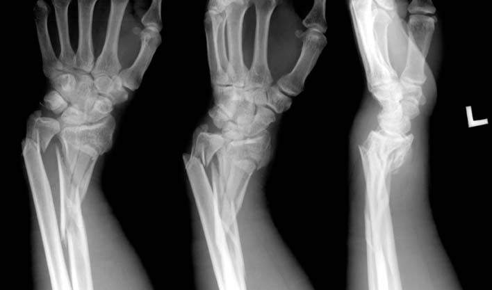
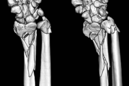
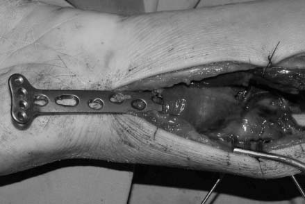


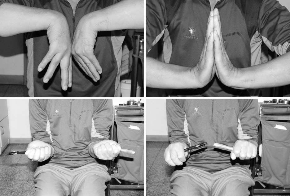
Figure & Data
REFERENCES
Citations
Citations to this article as recorded by 
- Use of Miniplate for Severe Comminuted Metadiaphyseal Fractures of the Distal Radius
Jong-Ryoon Baek, Yong-Cheol Yoon, Seung Hyun Baek
Journal of the Korean Fracture Society.2019; 32(4): 204. CrossRef
 Cite
CiteOutcomes of Severe Comminuted Distal Radius Fractures with Pronator Preserving Approach


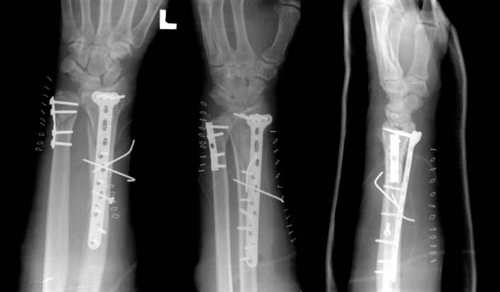
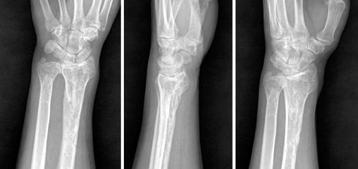

Fig. 1
A 55-year-old male patient presented with a severely comminuted fracture of the distal radius.
Fig. 2
Preoperative 3-dimensional computed tomography image.
Fig. 3
A pronator-preserving approach was used as the surgical intervention.
Fig. 4
The immediate postoperative photograph shows a successful reduction of the fracture.
Fig. 5
A radiographic image showing a successful bony union at the final follow-up.
Fig. 6
The range of motion of the wrist was normal and the modified Mayo wrist score score was 'excellent'.
Fig. 1
Fig. 2
Fig. 3
Fig. 4
Fig. 5
Fig. 6
Outcomes of Severe Comminuted Distal Radius Fractures with Pronator Preserving Approach

