 E-submission
 TOTA
TOTA TOTS
TOTS
Articles
- Page Path
- HOME > J Musculoskelet Trauma > Volume 20(3); 2007 > Article
-
Case Report
- Vertically Unstable Fracture of the Pelvis Combined with Anterior Dislocation of the Hip Joint: A Case Report
- Kap Jung Kim, M.D., Ha Yong Kim, M.D., Dae Suk Yang, M.D., Won Sik Choy, M.D.
-
Journal of the Korean Fracture Society 2007;20(3):272-276.
DOI: https://doi.org/10.12671/jkfs.2007.20.3.272
Published online: June 14, 2016
Department of Orthopaedic Surgery, School of Medicine, Eulji University, Daejeon, Korea.
- Address reprint requests to: Kap Jung Kim, M.D. Department of Orthopaedic Surgery, Eulji University Hospital, 1306, Dunsan-dong, Seo-gu, Daejeon 302-799, Korea. Tel: 82-42-611-3279, Fax:82-42-259-1289, oskkj@eulji.ac.kr
Copyright © The Korean Fracture Society. All rights reserved
- 772 Views
- 1 Download
Abstract
- Pelvic fractures result from high energy trauma and often associated with concomitant injuries. But, vertically unstable pelvic fractures combined with anterior dislocation of the hip is far less common. The traumatic dislocation of the hip is a true orthopedic emergency and it should be considered that a femoral head can be exposed to deteriorized vascularity. We report a case of vertically unstable pelvic fractures combined with traumatic anterior dislocation of the hip joint with the review of the literature.
- 1. Brav EA. Traumatic dislocation of the hip. Army experience and results over a twelve-year period. J Bone Joint Surg Am, 1962;44:1115-1134.
- 2. Duwelius PJ, Van Allen M, Bray TJ, Nelson D. Computed tomography-guided fixation of unstable posterior pelvic ring disruptions. J Orthop Trauma, 1992;6:420-426.Article
- 3. Epstein HC. Posterior fracture-dislocations of the hip; long-term follow-up. J Bone Joint Surg Am, 1974;56:1103-1127.PubMed
- 4. Lyddon DW, Hartman JT. Traumatic dislocation of the hip with ipsilateral femoral fracture. A case report. J Bone Joint Surg Am, 1971;53:1012-1016.PubMed
- 5. Pietrafesa CA, Hoffman JR. Traumatic dislocation of the hip. JAMA, 1983;249:3342-3346.ArticlePubMed
- 6. Reigstad A. Traumatic dislocation of the hip. J Trauma, 1980;20:603-606.ArticlePubMed
- 7. Tile M. Acute pelvic fractures: I. Causation and classification. J Am Acad Orthop Surg, 1996;4:143-151.ArticlePubMed
- 8. Young JW, Burgess AR, Brumback RJ, Poka A. Pelvic fractures: value of plain radiography in early assessment and management. Radiology, 1986;160:445-451.ArticlePubMed
REFERENCES
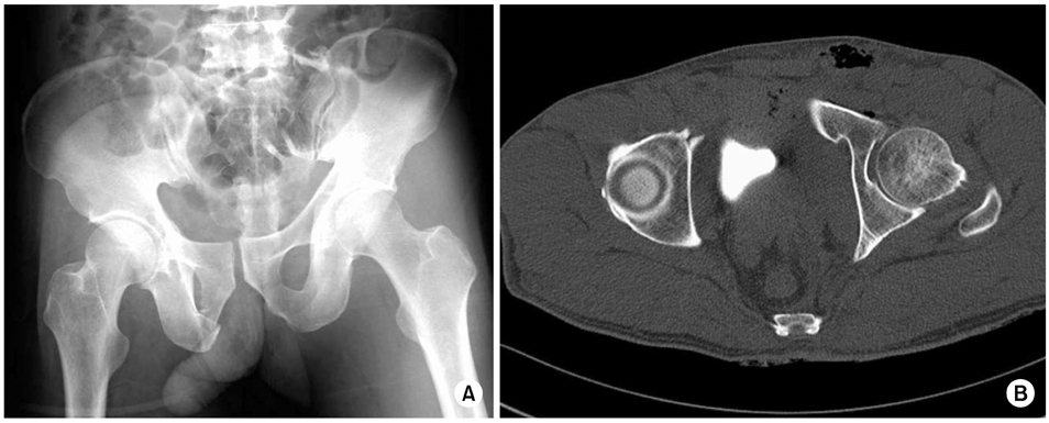
Fig. 1

Initial pelvis AP (A) and inlet view (B) show anteroinferior dislocation of left femoral head, right pubic fracture and sacral fracture.
Fig. 2

Pelvis CT shows sacral fracture (Type I) (A), anteroinferior dislocation of left femoral head and right pubic fractures (B,C).
Fig. 3
Post reduction pelvis AP radiograph and pelvis CT show concentric reduction of left femoral head (A, B). Concentric reduction was maintained by skeletal traction (A).
Fig. 4
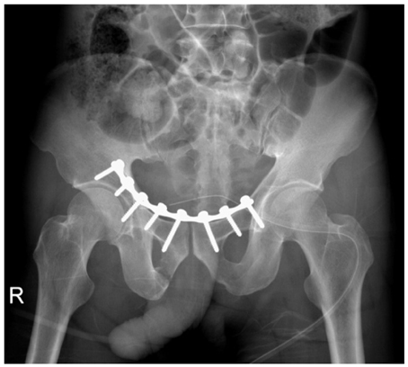
The first postoperative radiograph shows open reduction and internal fixation of the anterior ring of the pelvis by modified Stoppa approach.
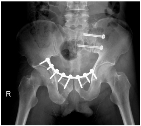
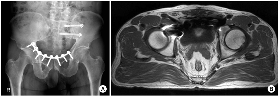
Figure & Data
REFERENCES
Citations
Citations to this article as recorded by 
 Cite
CiteVertically Unstable Fracture of the Pelvis Combined with Anterior Dislocation of the Hip Joint: A Case Report
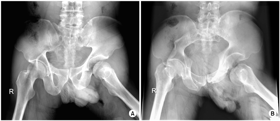
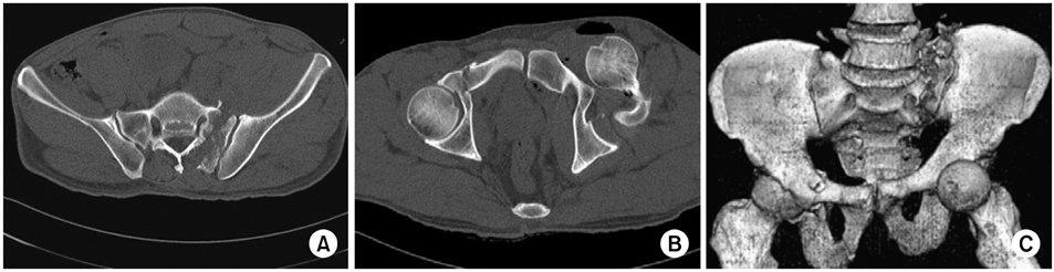


Fig. 1
Initial pelvis AP (A) and inlet view (B) show anteroinferior dislocation of left femoral head, right pubic fracture and sacral fracture.
Fig. 2
Pelvis CT shows sacral fracture (Type I) (A), anteroinferior dislocation of left femoral head and right pubic fractures (B,C).
Fig. 3
Post reduction pelvis AP radiograph and pelvis CT show concentric reduction of left femoral head (A, B). Concentric reduction was maintained by skeletal traction (A).
Fig. 4
The first postoperative radiograph shows open reduction and internal fixation of the anterior ring of the pelvis by modified Stoppa approach.
Fig. 5
The second postoperative radiograph shows percutaneous iliosacral screw fixation under the CT guided for sacral fracture.
Fig. 6
Complete union of pelvic fractures (A)and no visible evidence of avascular necrosis of left femoral head (B) at the time of final follow-up, postoperative 2 years.
Fig. 1
Fig. 2
Fig. 3
Fig. 4
Fig. 5
Fig. 6
Vertically Unstable Fracture of the Pelvis Combined with Anterior Dislocation of the Hip Joint: A Case Report

