 E-submission
 TOTA
TOTA TOTS
TOTS
Articles
- Page Path
- HOME > J Musculoskelet Trauma > Volume 35(4); 2022 > Article
- Original Article Intra-Articular Alterations after Suprapatellar Nailing in Tibial Shaft Fractures: An Arthroscopic Evaluation
- GwangChul Lee, Sung Hun Yang, Sung Min Jo, Jeong Min Kook
-
Journal of Musculoskeletal Trauma 2022;35(4):129-134.
DOI: https://doi.org/10.12671/jkfs.2022.35.4.129
Published online: October 31, 2022
Department of Orthopaedic Surgery, College of Medicine, Chosun University, Gwangju, Korea
- 868 Views
- 11 Download
- 0 Crossref
- 0 Scopus
Abstract
Purpose
This study attempted to study the intra-articular changes due to intramedullary nailing through the suprapatellar approach by evaluating the joint cartilage damage and presence of foreign bodies through a comparison of the pre- and post-operative status evaluated by knee arthroscopy.
Materials and Methods
This retrospective study analyzed fifteen patients who underwent intramedullary nailing through the suprapatellar approach for proximal tibial shaft fracture from January 2017 to March 2020. The condition of the joint cartilage and the presence of foreign substances in the patellofemoral joint were evaluated. The cartilage status of the patellofemoral joint was evaluated using the International Cartilage Repair Society (ICRS) grading system. Data from the ICRS grading and the visual analogue scale (VAS) scores of the femoral and patellar cartilage were compared to each independent variable surveyed.
Results
All the intra-articular structures before nailing were normal. In all cases after nailing, articular cartilage damage of the patellofemoral joint and intra-articular debris were observed. The average VAS score was 0.6 (0-1) before surgery and 2.27 (0-4) after surgery. There were no statistically significant differences except for the correlation in the diameter of the tibia nail and femoral ICRS grade (p=0.001) and the damage to the cartilage was greater in the femoral cartilage than that to the patella (p=0.001).
Conclusion
Intra-articular damage appears to be unavoidable in suprapatellar nailing. Further research is needed on the long-term effects of intra-articular damage and on methods to reduce this damage.
J Korean Fract Soc. 2022 Oct;35(4):129-134. Korean.
Published online Oct 20, 2022.
https://doi.org/10.12671/jkfs.2022.35.4.129
Published online Oct 20, 2022.
https://doi.org/10.12671/jkfs.2022.35.4.129
Copyright © 2022 The Korean Fracture Society.
Original Article
슬개 상부 접근법을 통한 금속정 고정술 후 관절내의 변화: 관절경적 관찰

Intra-Articular Alterations after Suprapatellar Nailing in Tibial Shaft Fractures: An Arthroscopic Evaluation
초록
목적
경골 근위 간부 골절에 대해 슬개 상부 접근법을 통한 금속정 고정술 후 관절경을 통한 관절내 변화와 요인을 알아보고자 하였다.
대상 및 방법
2017년 1월부터 2020년 3월까지 본원에서 슬개 상부 접근법을 통해 골수강내 금속정 고정술을 시행한 환자를 대상으로 하였다. 술 전, 후 관절경을 시행하여 관절내 변화를 관찰하였다. 관절 연골의 변화를 ICRS grade로 평가하였고, 임상적 평가로 VAS 점수를 이용하였다. 관련된 요인들과 ICRS grade, VAS 점수와의 연관성에 대해 통계적으로 분석하였다.
결과
술 전 관절내 구조물은 모두 정상이었다. 술 후 전 예에서 대퇴골 및 슬개골의 관절 연골 손상 및 관절내 유리체가 관찰되었다. VAS 점수는 술 전 평균 0.6점(범위 0-1점), 술 후 평균 2.27점(범위 0-4점)이었다. 금속정 크기와 대퇴골 연골 손상 정도의 관계는 통계적으로 유의하였고(p=0.001) 대퇴골 연골의 손상이 슬개골보다 심하였으며(p=0.001) 그 외 인자들 간의 유의한 상관관계는 없었다.
결론
슬개 상부 접근법을 통한 금속정 고정술은 관절내 손상을 야기한다. 향후 지속적인 연구를 통한 관절내 손상의 장기적인 영향과 손상을 줄일 수 있는 방법들에 대해 추가적인 연구가 필요하다.
Abstract
Purpose
This study attempted to study the intra-articular changes due to intramedullary nailing through the suprapatellar approach by evaluating the joint cartilage damage and presence of foreign bodies through a comparison of the pre- and post-operative status evaluated by knee arthroscopy.
Materials and Methods
This retrospective study analyzed fifteen patients who underwent intramedullary nailing through the suprapatellar approach for proximal tibial shaft fracture from January 2017 to March 2020. The condition of the joint cartilage and the presence of foreign substances in the patellofemoral joint were evaluated. The cartilage status of the patellofemoral joint was evaluated using the International Cartilage Repair Society (ICRS) grading system. Data from the ICRS grading and the visual analogue scale (VAS) scores of the femoral and patellar cartilage were compared to each independent variable surveyed.
Results
All the intra-articular structures before nailing were normal. In all cases after nailing, articular cartilage damage of the patellofemoral joint and intra-articular debris were observed. The average VAS score was 0.6 (0-1) before surgery and 2.27 (0-4) after surgery. There were no statistically significant differences except for the correlation in the diameter of the tibia nail and femoral ICRS grade (p=0.001) and the damage to the cartilage was greater in the femoral caritlage than that to the patella (p=0.001).
Conclusion
Intra-articular damage appears to be unavoidable in suprapatellar nailing. Further research is needed on the long-term effects of intra-articular damage and on methods to reduce this damage.
Keywords
Tibial fracture, Proximal, Patellofemoral joint, Suprapatellar, Intramedullary nailing
경골 골절, 근위부, 대퇴-슬개 관절, 슬개상부, 골수강내 금속정 고정술
서론
경골 간부 골절에서 골수강내 금속정 고정술은 금속판 고정술에 비해 생역학적인 장점과 연부조직의 손상을 줄일 수 있어 비교적 좋은 결과를 보인다.1) 그러나 근위 경골 간부 골절의 경우 일반적인 슬개 하부 접근법을 이용한 금속정 고정술 시 슬관절 굴곡으로 인해 발생하는 근위 골편의 부정정렬로 인해 불유합, 부정유합 등의 합병증이 발생할 수 있는 것으로 알려져 있다.2)
최근에 시행되고 있는 슬개 상부 접근법을 이용한 금속정 고정술은 슬개 상부의 대퇴 사두건 부위 삽입구로 관절강 내부를 통과해 금속정의 삽입구를 만들게 됨으로써 경골 근위부 골절에서 사용될 경우 반신전 상태로 수술을 시행하기 때문에 슬관절 굴곡 각도를 줄여 골절부의 전위가 상대적으로 적게 일어난다.3,4) 따라서 골절의 정복을 용이하게 유지시킬 수 있을 뿐만 아니라 부정정렬을 줄일 수 있다.5)
그러나 슬개 상부 접근법은 삽입을 위한 기구 조작 및 삽입 시에 대퇴-슬개 관절에 압력이 가해지고 확공이나 금속정을 삽입할 때 발생하는 골 파편 등으로 인해 슬관절 내 활액막염 등이 발생할 수 있으며, 금속정 삽입 과정에서 대퇴-슬개 관절의 의인성 연골 손상이 일어날 수 있는 것으로 보고되고 있다.6)
이에 본 연구에서는 슬개 상부 접근법을 이용하여 골수강내 금속정 고정술 시 관절내 변화가 일어날 수 있다는 가정하에 관절경을 통한 관절내 변화를 관찰하였다. 수술과 관련된 인자들이 관절내 변화에 어떠한 영향을 주는지 알아보고자 하였으며, 임상적, 방사선학적 평가를 통해 관절내 변화와의 연관성에 대해 평가하였다.
대상 및 방법
1. 연구 대상
본 연구는 조선대학교병원 의학연구윤리심의위원회(Institutional Review Board, IRB)의 승인을 받아 진행되었으며(IRB No. 2020-12-056), 후향적 의무기록 연구로 환자 서면 동의서는 IRB에 의해 면제받았다.
본원 정형외과에서 2017년 1월부터 2020년 3월까지 경골 근위 간부 골절에 대하여 슬개 상부 접근법을 이용한 골수강내 금속정 삽입술을 시행한 환자를 대상으로 하였다. 골절선이 관절면을 침범하지 않으면서 전체 경골 길이의 근위 1/3 이내에 위치하고 협부 이상에 골절선이 있는 경우를 경골 근위 간부 골절로 정의하였다. 단순 방사선 소견상 대퇴-슬개 관절의 관절염이 있거나 동반된 슬관절 병변이 있는 환자, 과거 슬관절부에 수술적 치료를 시행 받았던 환자를 제외한 15예를 대상으로 하였다. 남자가 11예, 여자가 4예였으며 평균 연령은 47.5세(범위 18-79세)였다. 골절의 분류는 AO/OTA 분류를 이용하였고 골절의 개방성 유무 및 다발성 손상 여부에 대해 평가하였다(Table 1).7)
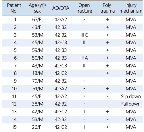
Table 1
Demographic Characteristics
2. 수술 방법 및 연구 방법
전 예에서 전신마취하에 앙와위로 눕히고, 환측 하퇴부 아래 적당한 두께의 포를 개어 넣어 슬부를 약 15도에서 30도 정도 구부린 반신전(semiextended) 상태로 유지한 상태에서 견인술을 통한 비관혈적 정복술을 시행하고, 영상증폭기 영상을 통해 정복 상태를 확인하였다. 우선적으로 슬개골을 내-외측으로 밀어 유동성을 평가한 후 슬개 상부 접근이 가능함을 확인하고, 슬개 상부 대퇴사두건 부위를 3-5 cm 절개하여 관절을 노출시켰다. 이후 절개부를 통한 관절경 검사를 시행하여 금속정 삽입 전 대퇴-슬개 관절의 연골 상태, 관절내 이물 등의 관절 상태를 평가하였다(Fig. 1B). 유도핀 삽입기를 넣기 전 검지 손가락을 대퇴-슬개 관절에 넣어서 금속정 삽입을 위한 충분한 공간이 있음을 확인하였다(Fig. 1A). 유도핀 삽입기를 통하여 삽입된 유도핀이 영상증폭기상 경골의 간부 전방과 평행한 것을 확인한 뒤 확공술을 시행하였다(Fig. 1C). 삽입기를 유지한 상태에서 적절하게 확공술을 시행한 후 금속정의 크기 및 길이를 결정하였다. 금속정을 삽입할 때는 슬개 상부 접근법에 사용되는 금속정 결착 기구를 사용하였고 삽입기를 제거한 상태에서 금속정을 삽입하였다. 금속정은 모든 예에서 Zimmer® Natural Nail® System-Tibia (Zimmer Biomet, Warsaw IN, USA)를 사용하였다.
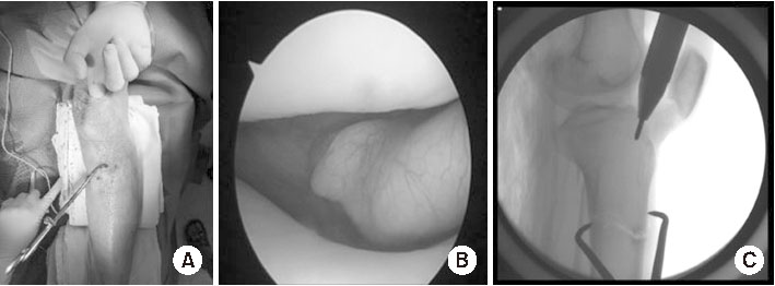
Fig. 1
(A) A 3-5 cm incision was made in the quadriceps tendon to expose the joint. By inserting a finger into the patellofemoral joint, it was confirmed that there was sufficient space for the protector to enter. (B) Diagnostic arthroscopic image of the patellofemoral joint before nail insertion. (C) Lateral C-arm image just before inserting the guide pin.
금속정을 삽입한 이후 결착 기구를 제거하고 기존 슬개 상부 절개부를 통하여 관절경 검사를 시행하였고, 대퇴-슬개 관절의 관절 연골 상태 및 관절내 이물의 유무 등을 평가하였다.8) 대퇴-슬개 관절의 연골 평가는 International Cartilage Repair Society (ICRS) grading system을 이용하였다(Fig. 2).9)
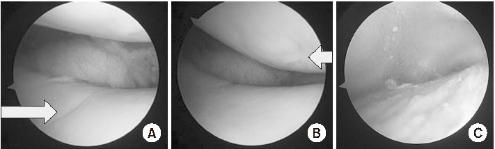
Fig. 2
Arthroscopic images after nail insertion with a suprapatellar approach. ICRS (International Cartilage Repair Society) grade II forms of femoral damage (A, arrow) and patellar damage (B, arrow), bony and joint debris (C) are observed.
단순 방사선 추시를 통해 술 전, 후 대퇴-슬개 관절의 변화를 평가하였고 임상적으로는 술 전과 술 후 일주일째 patella gliding test를 통한 통증 정도에 대해 visual analogue scale (VAS) 점수를 이용하여 평가하였다.10)
조사한 각각의 독립변수와 수술 전과 후를 비교한 대퇴골 및 슬개골 연골의 ICRS grade 및 VAS 점수에 대하여 상관분석 및 Mann–Whitney 검정을 이용하여 통계적으로 분석하였고, 유의 확률이 0.05 이하인 경우를 통계적으로 의미가 있는 것으로 하였다. 모든 통계 분석은 IBM SPSS 프로그램(ver. 20.0; IBM, Armonk, NY, USA)을 사용하였다(Table 2).
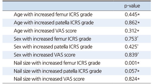
Table 2
Results of the Analysis of Age, Sex, and Nail Size of the Patients
결과
금속정 삽입 전 관절경 검사상 대퇴 슬개 관절 및 관절내 상태에서 이상 소견을 보인 경우는 없었다. 금속정 삽입 후 관절경 검사상 관절 연골의 상태는 대퇴골의 경우 ICRS grade I은 0예, II는 10예, III는 5예였고, 슬개골의 경우 I은 5예, II는 10예로, 모든 예에서 grade I 이상의 관절 연골 손상 및 관절내 유리체(bony debris)가 관찰되었다. 금속정 삽입 전, 후 방사선적 평가상 대퇴-슬개 관절의 변화는 관찰되지 않았다. 수술 전, 후 VAS 점수의 평균은 0.6점(범위 0-1점), 2.27점(범위 0-4점)이었다(Table 3).
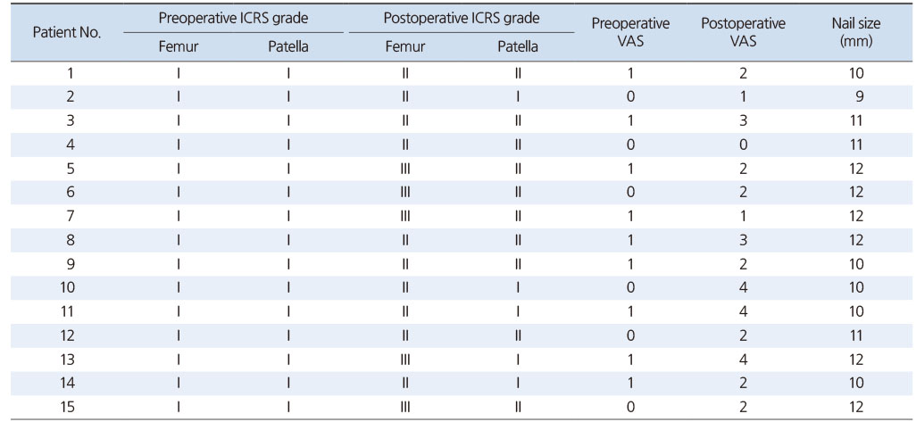
Table 3
ICRS Grade and VAS Score before and after Surgery according to the Size of the Nail
나이, 성별, 금속정 크기와 술 후 관절내 손상 정도, 임상적 결과와의 상관관계 분석 결과 금속정 크기와 대퇴골 연골 ICRS grade 간에 유의미함을 보였으나(p=0.001), 다른 요소들 간에는 통계적인 유의함이 없었다(Table 2).
수술 후 슬개골과 대퇴골의 연골에 대한 ICRS grade는 수술 전에 비해 모두 증가하였다. 독립표본 t-검정을 통해 수술 전과 후 슬개골 연골의 ICRS grade 변화와 대퇴골 연골의 ICRS grade 변화를 비교하였을 때, 대퇴골 연골의 변화가 통계적으로 유의하였다(p=0.001; Table 4).
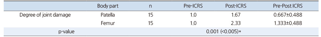
Table 4
Differences in the Preoperative-Postoperative Patella and Femur ICRS Grade Scores
고찰
본 연구에서 슬개 상부 접근법을 이용한 골수강내 금속정 고정술 시 관절내 손상은 전 예에서 관찰되었고, 특히 대퇴골 연골부 손상이 더 많았으며, 금속정 직경이 클수록 더 심한 연골 손상을 보였다. 하지만 관절내 손상과 임상적, 방사선적 결과와의 연관성은 없었다. 이러한 결과를 바탕으로 보다 추가적인 연구와 장기적인 추시가 필요할 것으로 보인다.
경골 간부의 골절에서 슬개 하부 접근법을 통한 골수강내 금속정 삽입술이 표준치료로 쓰여왔지만 슬부 전방부 통증의 발생률은 34%-71%까지 보고될 정도로 높다.11,12,13) Morandi 등14)에 의하면, 슬개 하부 접근법으로 골수강내 금속정을 삽입한 후에 발생하는 슬부 통증은 연골 손상, 슬개 인대 손상, 슬개 하부 신경의 의인성 손상 및 경골 천정부의 금속정의 돌출로 인하여 발생한다고 보고하였다.
이에, 슬개하부 접근법에서 발생하는 통증과 근위경골 골절 정복의 어려움을 보완하기 위해 Tornetta와 Collins15)는 슬관절 내측 관절낭을 절개해 15도 정도의 슬관절 굴곡 상태에서 금속정을 삽입하는 반신전 상태의 수술법을 이용하여 슬관절을 과굴곡시키지 않고 금속정을 삽입하는 방법을 소개하였다. 이어 슬개 상부 접근법을 위한 기구들이 개발되면서 Wang 등16)은 슬개 상부 접근법으로 골수강내 금속정을 삽입하는 것이 슬개 하부 접근법보다 전방 슬부 통증 및 슬개 관절의 통증(VAS)을 감소시키고, 방사선 조사 시간, 대퇴-슬개 관절의 임상적 예후를 개선시킨다고 하였다.
하지만 슬개 상부 접근법은 슬관절 내부를 손상시킬 수 있기 때문에 이러한 변화가 장기적으로 어떠한 양상으로 변화할 것인지, 임상적으로 어떠한 영향을 일으킬 것인지에 대해서는 추가적인 연구가 필요하다. Garnavos17)는 슬개 상부 접근법으로 골수강내 금속정 삽입을 시행한 17명 중 2명에게 합병증이 발생했다고 보고했는데, 1명은 관절면의 불만족스러운 정복으로 재수술을 시행하였으며, 다른 1명은 체중부하 시 슬개건염이 발생하였다. 이에 반해 Zamora 등18)과 Gaines 등19)은 카데바 연구에서 슬개 상부와 슬개 하부 접근법으로 골수강내 금속정을 삽입 시 슬개 상부 접근법이 관절내 구조물을 손상시키는 빈도가 더 적었다고 보고했다.
저자들의 연구에서는 수술적 처치 시 사용한 골수강내 금속정의 내강에 따른 대퇴 관절의 연골 손상 정도가 증가하는 것이 통계적으로 유의한 결과를 보였다(p=0.001). 이는 슬개 상부 접근법 시행 시 삽입기를 이용하여 관절면에 손상이 가지 않도록 보호하고는 있으나 금속정 내강이 클수록 내강 확공술을 시행하는 빈도수나 총 시간이 늘어나, 대퇴-슬개 관절에 압박력을 가하는 시간이 늘어 손상이 발생했을 수 있다고 판단하였다. 또한 금속정 삽입 시에는 삽입기를 제거한 상태로 삽입을 해야 했기 때문에 금속정의 내강이 클수록 대퇴 슬개 관절의 손상을 더 유발할 수 있다고 보았다. 저자들은 내강 확공술이 완료될 때까지 걸린 시간을 측정하지 못하였고 이에 관한 보고도 없어, 이와 관련한 추가적인 연구가 필요할 것으로 생각된다.
Loening 등20)의 소(bovine)를 대상으로 시행한 연구에 의하면, 관절 연골의 경우 25 MPa을 넘는 압력에서 손상이 발생하게 되고 연골세포의 경우 4.5 MPa 이상에서 세포자멸사가 발생한다고 말하고 있다. Gelbke 등6)의 카데바 연구에서는 접점압이 슬개 상부 접근법의 경우 슬개골에서 평균 1.84 MPa, 대퇴골에서 평균 2.13 MPa로, 슬개 하부 접근법에서는 평균 0.90 MPa로 슬개 상부 접근법이 슬개 하부 접근법에 비해 많은 접점압이 발생하지만, 이는 Loening 등20)이 말한 관절연골의 통합성(integrity)이나 연골세포의 손상을 일으킬 만한 수치에 비해서는 낮은 수치로 연골의 손상이나 연골세포의 세포자멸사 조장에 크게 영향을 끼치지 않을 것으로 보인다.
본 연구에서 슬개골의 연골 손상에 비해 대퇴골의 연골 손상이 통계적으로 유의하게 심한 양상을 확인할 수 있었다(p=0.001). 이는 Gelbke 등6)의 카데바 연구에서 금속정 삽입술 시 접점압이 슬개골보다 대퇴골에 더 크게 발생한 점과 관련하여 생각해 볼 수 있다. 그리고 근위 골편의 전위를 줄이기 위해 경골 전방 피질골의 방향에 평행하게 금속정 삽입을 하려 했던 노력도 대퇴골 연골 손상에 더 많은 영향을 준 것으로 판단된다.
또한 저자들은 금속정 삽입술 전, 후에 대한 관절경 소견과 방사선학적 변화만을 확인하여 비교하였다. Sanders 등21)의 연구에서는 관절경 검사상 약 5.4%에서 grade II 연골연 화증이 관찰되었으나 1년 후 추시상의 자기공명영상 검사 및 임상적 평가와 큰 연관성이 없다고 말하였다. 따라서 장기 추시 시에 대한 추가적인 연구가 필요할 것으로 생각된다.
추가적인 한계점으로는 증례수가 적으며, 술자의 숙련도에 따른 효과를 고려하지 못했다. 본 연구는 수술 직후 나타나는 연골 변화에 대한 기술에 대한 것으로 이에 대하여 전향적 연구 및 장기 추시 연구와 추가적인 증례 수집이 필요할 것으로 생각된다.
결론
슬개 상부 접근법을 통한 골수강내 금속정을 시행한 경우 관절내 손상은 피할 수 없는 것으로 보인다. 특히 대퇴부의 관절 연골 손상이 더 많았고 금속정의 직경이 클수록 더 심한 관절 연골 손상을 보였다.
지속적인 연구를 통해 이러한 관절내 손상이 장기적으로 어떠한 변화를 일으킬지에 대한 관찰이 필요하며 손상을 줄일 수 있는 방법들에 대해서도 추가적인 연구가 필요할 것으로 생각된다.
Notes
Financial support:This study was supported by research fund from Chosun University Hospital, 2018.
Conflict of interests:None.
References
-
Larsen P, Lund H, Laessoe U, Graven-Nielsen T, Rasmussen S. Restrictions in quality of life after intramedullary nailing of tibial shaft fracture: a retrospective follow-up study of 223 cases. J Orthop Trauma 2014;28:507–512.
-
-
Freedman EL, Johnson EE. Radiographic analysis of tibial fracture malalignment following intramedullary nailing. Clin Orthop Relat Res 1995;(315):25–33.
-
-
Zelle BA, Boni G. Safe surgical technique: intramedullary nail fixation of tibial shaft fractures. Patient Saf Surg 2015;9:40
-
-
Hessmann MH, Buhl M, Finkemeier C, Khoury A, Mosheiff R, Blauth M. Suprapatellar nailing of fractures of the tibia. Oper Orthop Traumatol 2020;32:440–454.
-
-
Williamson M, Iliopoulos E, Williams R, Trompeter A. Intra-operative fluoroscopy time and radiation dose during suprapatellar tibial nailing versus infrapatellar tibial nailing. Injury 2018;49:1891–1894.
-
-
Gelbke MK, Coombs D, Powell S, DiPasquale TG. Suprapatellar versus infra-patellar intramedullary nail insertion of the tibia: a cadaveric model for comparison of patellofemoral contact pressures and forces. J Orthop Trauma 2010;24:665–671.
-
-
Müller ME, Nazarian S, Koch P, Schatzker J. In: The comprehensive classification of fractures of long bones. Berlin: Springer; 1990.
-
-
Jakma T, Reynders-Frederix P, Rajmohan R. Insertion of intramedullary nails from the suprapatellar pouch for proximal tibial shaft fractures. A technical note. Acta Orthop Belg 2011;77:834–837.
-
-
Brittberg M, Winalski CS. Evaluation of cartilage injuries and repair. J Bone Joint Surg Am 2003;85-A Suppl 2:58–69.
-
-
Yang L, Sun Y, Li G. Comparison of suprapatellar and infrapatellar intramedullary nailing for tibial shaft fractures: a systematic review and meta-analysis. J Orthop Surg Res 2018;13:146
-
-
Toivanen JA, Väistö O, Kannus P, Latvala K, Honkonen SE, Järvinen MJ. Anterior knee pain after intramedullary nailing of fractures of the tibial shaft. A prospective, randomized study comparing two different nail-insertion techniques. J Bone Joint Surg Am 2002;84:580–585.
-
-
Lefaivre KA, Guy P, Chan H, Blachut PA. Long-term follow-up of tibial shaft fractures treated with intramedullary nailing. J Orthop Trauma 2008;22:525–529.
-
-
Habernek H, Kwasny O, Schmid L, Ortner F. Complications of interlocking nailing for lower leg fractures: a 3-year follow up of 102 cases. J Trauma 1992;33:863–869.
-
-
Morandi M, Banka T, Gaiarsa GP, et al. Intramedullary nailing of tibial fractures: review of surgical techniques and description of a percutaneous lateral suprapatellar approach. Orthopedics 2010;33:172–179.
-
-
Tornetta P 3rd, Collins E. Semiextended position of intramedullary nailing of the proximal tibia. Clin Orthop Relat Res 1996;(328):185–189.
-
-
Wang C, Chen E, Ye C, Pan Z. Suprapatellar versus infrapatellar approach for tibia intramedullary nailing: a meta-analysis. Int J Surg 2018;51:133–139.
-
-
Garnavos C. Intramedullary nailing with a suprapatellar approach and condylar bolts for the treatment of bicondylar fractures of the tibial plateau. JB JS Open Access 2017;2:e0017
-
-
Zamora R, Wright C, Short A, Seligson D. Comparison between suprapatellar and parapatellar approaches for intramedullary nailing of the tibia. Cadaveric study. Injury 2016;47:2087–2090.
-
-
Gaines RJ, Rockwood J, Garland J, Ellingson C, Demaio M. Comparison of insertional trauma between suprapatellar and infrapatellar portals for tibial nailing. Orthopedics 2013;36:e1155–e1158.
-
-
Loening AM, James IE, Levenston ME, et al. Injurious mechanical compression of bovine articular cartilage induces chondrocyte apoptosis. Arch Biochem Biophys 2000;381:205–212.
-
-
Sanders RW, DiPasquale TG, Jordan CJ, Arrington JA, Sagi HC. radiographic results and clinical outcomes at a minimum of 12 months follow-up. J Orthop Trauma 2014;28:245–255.
-
 Cite
Cite
