 E-submission
 TOTA
TOTA TOTS
TOTS
Articles
- Page Path
- HOME > J Musculoskelet Trauma > Volume 26(3); 2013 > Article
-
Original Article
- Arthroscopic Assisted Intra-Articular Reduction and Internal Fixation of Tibia Plateau Fracture
- Dong Hwi Kim, M.D., Gwang Chul Lee, M.D., Kwi Youn Choi, M.D., Sung Won Cho, M.D., Sang Ho Ha, M.D.
-
Journal of the Korean Fracture Society 2013;26(3):191-198.
DOI: https://doi.org/10.12671/jkfs.2013.26.3.191
Published online: July 15, 2013
Department of Orthopaedic Surgery, School of Medicine, Chosun University, Gwangju, Korea.
- Address reprint requests to: Gwang Chul Lee, M.D. Department of Orthopaedic Surgery, Chosun University Hospital, 365 Pilmun-daero, Dong-gu, Gwangju 501-717, Korea. Tel: 82-62-220-3147, Fax: 82-62-226-3379, leekci@chosun.ac.kr
• Received: January 29, 2013 • Revised: March 12, 2013 • Accepted: April 25, 2013
Copyright © 2013 The Korean Fracture Society. All rights reserved.
This is anOpen Access article distributed under the terms of the Creative Commons Attribution Non-Commercial License (http://creativecommons.org/licenses/by-nc/3.0/) which permits unrestricted non-commercial use, distribution, and reproduction in any medium, provided the original work is properly cited.
- 1,064 Views
- 4 Download
- 1 Crossref
Abstract
-
Purpose
- We evaluated the results of arthroscopic intra-articular reduction and internal fixation of tibial plateau fractures without cortical window along with any additional bone grafts.
-
Materials and Methods
- From March 2006 to March 2009, twelve patients with arthroscopic intra-articular reduction and internal fixation of tibial plateau fractures over 5 mm in depression and displacement on the articular surface in computed tomography (CT) were enrolled in this study. We reduced or removed the depressed fracture fragment using freer without making a cortical window. Then, we accomplished internal fixation by a cannulated screw. All cases have not received bone graft. Both the postoperative clinical and radiological results were evaluated by the Rasmussen system.
-
Results
- The fractures were healed completely in an average of 9 (range from 7 to 12) weeks. According to Rasmussen classification, we obtained satisfactory clinical results as excellent in 8 cases, good in 3 cases, and fair in 1 case; and radiological results were excellent in 7 cases and good in 5 cases.
-
Conclusion
- We consider that arthroscopic intra-articular reduction and internal fixation of tibial plateau fractures without cortical window and any additional bone grafts is are a useful methods for attaining satisfactory results.
- 1. Asik M, Cetik O, Talu U, Sozen YV. Arthroscopy-assisted operative management of tibial plateau fractures. Knee Surg Sports Traumatol Arthrosc, 2002;10:364-370.ArticlePubMedPDF
- 2. Bernfeld B, Kligman M, Roffman M. Arthroscopic assistance for unselected tibial plateau fractures. Arthroscopy, 1996;12:598-602.ArticlePDF
- 3. Buchko GM, Johnson DH. Arthroscopy assisted operative management of tibial plateau fractures. Clin Orthop Relat Res, 1996;(332):29-36.Article
- 4. Caspari RB, Hutton PM, Whipple TL, Meyers JF. The role of arthroscopy in the management of tibial plateau fractures. Arthroscopy, 1985;1:76-82.ArticlePubMedPDF
- 5. Chan YS, Chiu CH, Lo YP, et al. Arthroscopy-assisted surgery for tibial plateau fractures: 2- to 10-year follow-up results. Arthroscopy, 2008;24:760-768.ArticlePubMedPDF
- 6. Delamarter R, Hohl M. The cast brace and tibial plateau fractures. Clin Orthop Relat Res, 1989;(242):26-31.Article
- 7. Fowble CD, Zimmer JW, Schepsis AA. The role of arthroscopy in the assessment and treatment of tibial plateau fractures. Arthroscopy, 1993;9:584-590.ArticlePDF
- 8. Gausewitz S, Hohl M. The significance of early motion in the treatment of tibial plateau fractures. Clin Orthop Relat Res, 1986;(202):135-138.Article
- 9. Guanche CA, Markman AW. Arthroscopic management of tibial plateau fractures. Arthroscopy, 1993;9:467-471.ArticlePubMedPDF
- 10. Holzach P, Matter P, Minter J. Arthroscopically assisted treatment of lateral tibial plateau fractures in skiers: use of a cannulated reduction system. J Orthop Trauma, 1994;8:273-281.Article
- 11. Honkonen SE. Indications for surgical treatment of tibial condyle fractures. Clin Orthop Relat Res, 1994;(302):199-205.Article
- 12. Honkonen SE. Degenerative arthritis after tibial plateau fractures. J Orthop Trauma, 1995;9:273-277.Article
- 13. Jennings JE. Arthroscopic management of tibial plateau fractures. Arthroscopy, 1985;1:160-168.ArticlePubMedPDF
- 14. Keogh P, Kelly C, Cashman WF, McGuinness AJ, O'Rourke SK. Percutaneous screw fixation of tibial plateau fractures. Injury, 1992;23:387-389.Article
- 15. Lansinger O, Bergman B, Körner L, Andersson GB. Tibial condylar fractures. A twenty-year follow-up. J Bone Joint Surg Am, 1986;68:13-19.ArticlePubMed
- 16. Lee KW, Lee HH, Yang DH, Choy WS. Arthroscopic reduction and internal fixation of intra-articular fractures of lateral tibial plateau. J Korean Arthrosc Soc, 2006;10:53-60.
- 17. Lemon RA, Bartlett DH. Arthroscopic assisted internal fixation of certain fractures about the knee. J Trauma, 1985;25:355-358.Article
- 18. Lubowitz JH, Elson WS, Guttmann D. Part I: arthroscopic management of tibial plateau fractures. Arthroscopy, 2004;20:1063-1070.ArticlePubMedPDF
- 19. Marsh JL, Smith ST, Do TT. External fixation and limited internal fixation for complex fractures of the tibial plateau. J Bone Joint Surg Am, 1995;77:661-673.Article
- 20. McCarthy JJ, Parker RD. Arthroscopic reduction and internal fixation of a displaced intraarticular lateral femoral condyle fracture of the knee. Arthroscopy, 1996;12:224-227.ArticlePubMedPDF
- 21. McLennan JG. The role of arthroscopic surgery in the treatment of fractures of the intercondylar eminence of the tibia. J Bone Joint Surg Br, 1982;64:477-480.ArticlePDF
- 22. Moore TM, Patzakis MJ, Harvey JP. Tibial plateau fractures: definition, demographics, treatment rationale, and long-term results of closed traction management or operative reduction. J Orthop Trauma, 1987;1:97-119.PubMed
- 23. O'Dwyer KJ, Bobic VR. Arthroscopic management of tibial plateau fractures. Injury, 1992;23:261-264.Article
- 24. O'Keeffe RM Jr, Riemer BL, Butterfield SL. Harvesting of autogenous cancellous bone graft from the proximal tibial metaphysis. A review of 230 cases. J Orthop Trauma, 1991;5:469-474.PubMed
- 25. Park IH, Lee KB, Park MR, Lee JY, Rhee DY. Arthroscopic management of the tibial condylar fracture. J Korean Soc Fract, 1990;25:1323-1332.
- 26. Perez Carro L. Arthroscopic management of tibial plateau fractures: special techniques. Arthroscopy, 1997;13:265-267.ArticlePubMedPDF
- 27. Rasmussen PS. Tibial condylar fractures. Impairment of knee joint stability as an indication for surgical treatment. J Bone Joint Surg Am, 1973;55:1331-1350.PubMed
- 28. Roerdink WH, Oskam J, Vierhout PA. Arthroscopically assisted osteosynthesis of tibial plateau fractures in patients older than 55 years. Arthroscopy, 2001;17:826-831.ArticlePDF
- 29. Schatzker J, McBroom R, Bruce D. The tibial plateau fracture. The Toronto experience 1968--1975. Clin Orthop Relat Res, 1979;(138):94-104.
- 30. Scheerlinck T, Ng CS, Handelberg F, Casteleyn PP. Medium-term results of percutaneous, arthroscopically-assisted osteosynthesis of fractures of the tibial plateau. J Bone Joint Surg Br, 1998;80:959-964.ArticlePDF
- 31. Tscherne H, Lobenhoffer P. Tibial plateau fractures. Management and expected results. Clin Orthop Relat Res, 1993;(292):87-100.
- 32. Volpin G, Dowd GS, Stein H, Bentley G. Degenerative arthritis after intra-articular fractures of the knee. Long-term results. J Bone Joint Surg Br, 1990;72:634-638.ArticlePubMedPDF
- 33. Waddell JP, Johnston DW, Neidre A. Fractures of the tibial plateau: a review of ninety-five patients and comparison of treatment methods. J Trauma, 1981;21:376-381.Article
- 34. Watson JT. High-energy fractures of the tibial plateau. Orthop Clin North Am, 1994;25:723-752.ArticlePubMed
- 35. Young MJ, Barrack RL. Complications of internal fixation of tibial plateau fractures. Orthop Rev, 1994;23:149-154.PubMed
REFERENCES
Fig. 1A fragment interfering with the reduction is called a key fragment (short arrows). The landmark of the screw insertion site was fibular head and Gerdy's tubercle. Long arrows indicate direction of screw insertion.
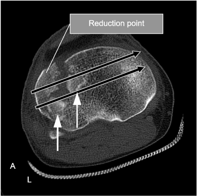
Fig. 2Arthroscopic findings See the depressed fracture fragment (A) and depressed fragment reduced by freer (B).

Fig. 3Preoperative radiologic findings. Antero-posterior (AP) view (A) and computed tomography images (B) of the right knee of a 28-year-old male patient shows a depressed lateral tibial plateau fracture (Schatzker type II). Both AP and lateral view (C, D) of the right knee shows a complete fracture reduction and internal fixation using a cannulated screw without bone graft.

Fig. 4Preoperative radiologic findings. Antero-posterior (AP) view (A) and computed tomography (CT) image (B) of the left knee of a 60-year-old male patient shows a tibia plateau fracture (Schatzker type V). Postoperative radiographic findings. AP view (C) and CT image (D) of the left knee shows an additional fixation by locking compression plate-proximal lateral tibia minimally invasive plate osteosynthesis (LCP-PLT) (MIPO technique).

Fig. 5
(A) Ten months after the operation, computed tomography images show a complete bone union with congruency of the articular surface. (B) Second look arthroscopy shows a healed articular surface as well as lateral meniscus.

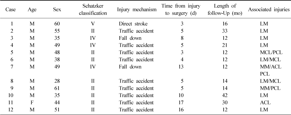
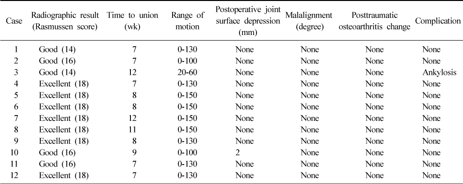
Figure & Data
REFERENCES
Citations
Citations to this article as recorded by 
- Current Concepts in Management of Tibia Plateau Fracture
Sang Hak Lee, Kang-Il Kim
Journal of the Korean Fracture Society.2014; 27(3): 245. CrossRef
 Cite
CiteArthroscopic Assisted Intra-Articular Reduction and Internal Fixation of Tibia Plateau Fracture

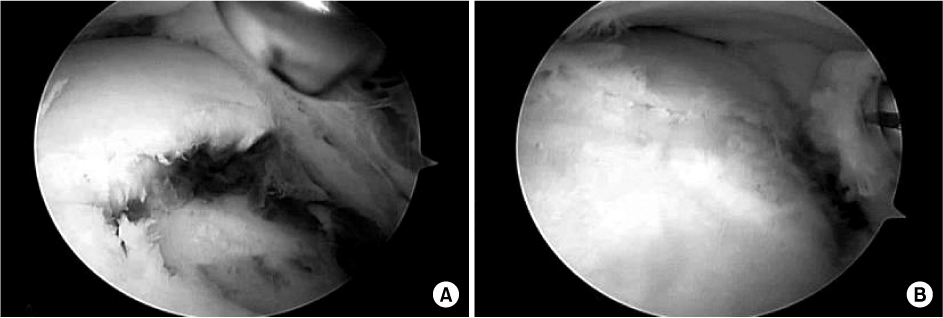
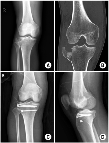
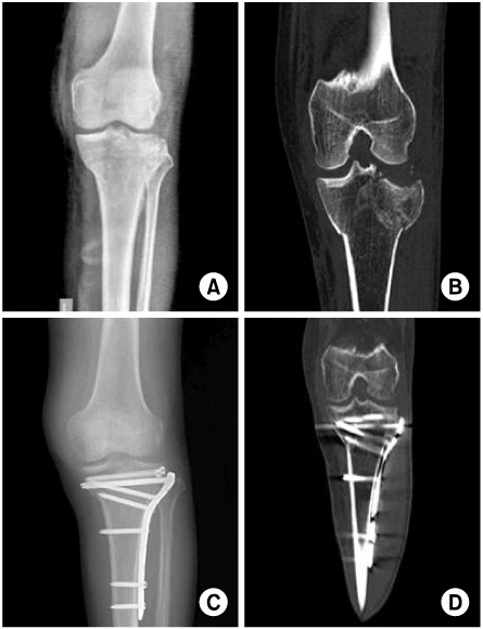
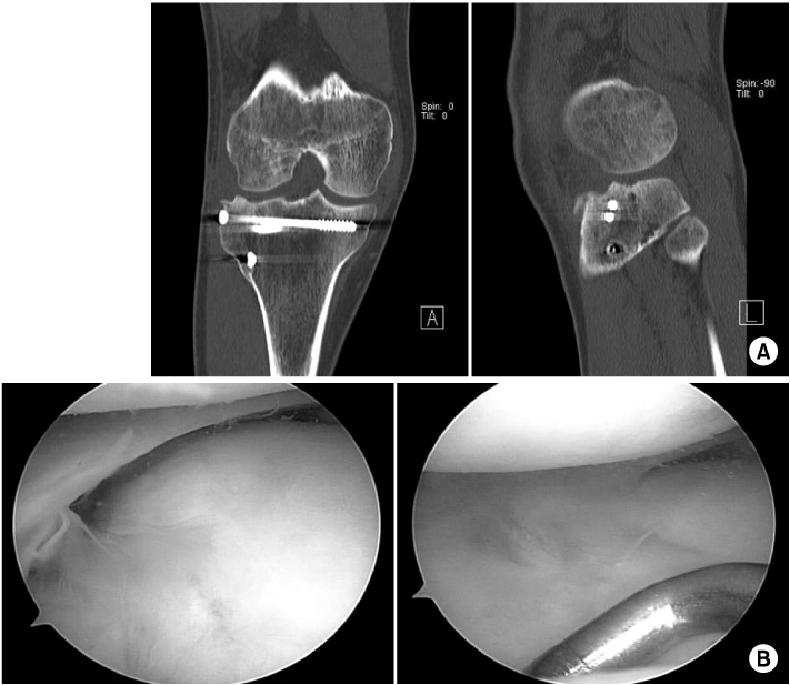
Fig. 1
A fragment interfering with the reduction is called a key fragment (short arrows). The landmark of the screw insertion site was fibular head and Gerdy's tubercle. Long arrows indicate direction of screw insertion.
Fig. 2
Arthroscopic findings See the depressed fracture fragment (A) and depressed fragment reduced by freer (B).
Fig. 3
Preoperative radiologic findings. Antero-posterior (AP) view (A) and computed tomography images (B) of the right knee of a 28-year-old male patient shows a depressed lateral tibial plateau fracture (Schatzker type II). Both AP and lateral view (C, D) of the right knee shows a complete fracture reduction and internal fixation using a cannulated screw without bone graft.
Fig. 4
Preoperative radiologic findings. Antero-posterior (AP) view (A) and computed tomography (CT) image (B) of the left knee of a 60-year-old male patient shows a tibia plateau fracture (Schatzker type V). Postoperative radiographic findings. AP view (C) and CT image (D) of the left knee shows an additional fixation by locking compression plate-proximal lateral tibia minimally invasive plate osteosynthesis (LCP-PLT) (MIPO technique).
Fig. 5
(A) Ten months after the operation, computed tomography images show a complete bone union with congruency of the articular surface. (B) Second look arthroscopy shows a healed articular surface as well as lateral meniscus.
Fig. 1
Fig. 2
Fig. 3
Fig. 4
Fig. 5
Arthroscopic Assisted Intra-Articular Reduction and Internal Fixation of Tibia Plateau Fracture
Patient Demographics (N=12)
M: Male, F: Female, LM: Lateral meniscus, MCL: Medial collateral ligament, PCL: Posterior cruciate ligament, MM: Medial meniscus, ACL: Anterior cruciate ligament.
Results (N=12)
Table 1
Patient Demographics (N=12)
M: Male, F: Female, LM: Lateral meniscus, MCL: Medial collateral ligament, PCL: Posterior cruciate ligament, MM: Medial meniscus, ACL: Anterior cruciate ligament.
Table 2
Results (N=12)

