 E-submission
 TOTA
TOTA TOTS
TOTS
Articles
- Page Path
- HOME > J Musculoskelet Trauma > Volume 26(3); 2013 > Article
-
Original Article
- The Character of Reverse Obliquity Intertrochanteric Fractures in Elderly Patients
- Ji Wan Kim, M.D., Jae-Suk Chang, M.D., Jung Hwan Sung, M.D., Jung Jae Kim, M.D.
-
Journal of the Korean Fracture Society 2013;26(3):173-177.
DOI: https://doi.org/10.12671/jkfs.2013.26.3.173
Published online: July 15, 2013
Department of Orthopaedic Surgery, Asan Medical Center, College of Medicine, University of Ulsan, Seoul, Korea.
*Department of Orthopaedic Surgery, Haeundae Paik Hospital, College of Medicine, Inje University, Busan, Korea.
- Address reprint requests to: Jae-Suk Chang, M.D. Department of Orthopaedic Surgery, Asan Medical Center, 86 Asanbyeongwon-gil, Songpa-gu, Seoul 138-736, Korea. Tel: 82-2-3010-3530·Fax: 82-2-488-7877, jschang@amc.seoul.kr
• Received: March 25, 2013 • Revised: May 12, 2013 • Accepted: June 11, 2013
Copyright © 2013 The Korean Fracture Society. All rights reserved.
This is anOpen Access article distributed under the terms of the Creative Commons Attribution Non-Commercial License (http://creativecommons.org/licenses/by-nc/3.0/) which permits unrestricted non-commercial use, distribution, and reproduction in any medium, provided the original work is properly cited.
- 1,428 Views
- 15 Download
- 1 Crossref
Abstract
-
Purpose
- To discriminate the characteristics between reverse obliquity fractures in the elderly and that of young adults using three-dimensional computed tomography (3D CT).
-
Materials and Methods
- Eighteen patients who had reverse obliquity intertrochanteric fractures were enrolled from January 2007 to March 2012. The fracture pattern was analyzed using the 3D CT. The area showing low density (bone defect) of trochanter and femoral neck region was measured. Patients were divided into two groups: Group I, less than 65 years old and Group 2, 65 years and over.
-
Results
- In all 9 cases of group 1, the proximal fragment had a 'V' shape with an average of 5.6 cm below the vastus ridge; however, the fracture of 8 cases (88.97%) in group 2 had a 'Λ' shape of the distal fragment at the level of vastus ridge and an additional fracture line extending to the greater trochanter tip. The bone defect volume of the trochanter and femoral neck region was larger significantly in group 2 than in group 1.
-
Conclusion
- Reverse obliquity intertrochanteric fracture in the elderly demonstrated a pattern of bursting fracture with 4 parts, which had different patterns from that of young patients. We believe that the larger volume of bone defects resulted in the difference of fracture patterns between the two groups.
- 1. Bergman GD, Winquist RA, Mayo KA, Hansen ST Jr. Subtrochanteric fracture of the femur. Fixation using the Zickel nail. J Bone Joint Surg Am, 1987;69:1032-1040.Article
- 2. Gnudi S, Ripamonti C, Lisi L, Fini M, Giardino R, Giavaresi G. Proximal femur geometry to detect and distinguish femoral neck fractures from trochanteric fractures in postmenopausal women. Osteoporos Int, 2002;13:69-73.ArticlePDF
- 3. Haidukewych GJ, Israel TA, Berry DJ. Reverse obliquity fractures of the intertrochanteric region of the femur. J Bone Joint Surg Am, 2001;83:643-650.ArticlePubMed
- 4. Hansen S, Jensen JE, Ahrberg F, Hauge EM, Brixen K. The combination of structural parameters and areal bone mineral density improves relation to proximal femur strength: an in vitro study with high-resolution peripheral quantitative computed tomography. Calcif Tissue Int, 2011;89:335-346.ArticlePubMedPDF
- 5. Holzer G, von Skrbensky G, Holzer LA, Pichl W. Hip fractures and the contribution of cortical versus trabecular bone to femoral neck strength. J Bone Miner Res, 2009;24:468-474.ArticlePDF
- 6. Im GI, Lim MJ. Proximal hip geometry and hip fracture risk assessment in a Korean population. Osteoporos Int, 2011;22:803-807.ArticlePDF
- 7. Ito M, Wakao N, Hida T, et al. Analysis of hip geometry by clinical CT for the assessment of hip fracture risk in elderly Japanese women. Bone, 2010;46:453-457.Article
- 8. Kim JW, Chang JS, Lee H, Bae JY, Kim JJ. Clinical results of femoral subtrochanteric fractures. J Korean Hip Soc, 2010;22:222-226.Article
- 9. Madsen JE, Naess L, Aune AK, Alho A, Ekeland A, Strømsøe K. Dynamic hip screw with trochanteric stabilizing plate in the treatment of unstable proximal femoral fractures: a comparative study with the Gamma nail and compression hip screw. J Orthop Trauma, 1998;12:241-248.Article
- 10. Orthopaedic Trauma Association. Fracture and dislocation compendium. Orthopaedic Trauma Association Committee for Coding and Classification. J Orthop Trauma, 1996;10:suppl 1. v-ix. 1-154.
- 11. Szulc P, Duboeuf F, Schott AM, et al. Structural determinants of hip fracture in elderly women: re-analysis of the data from the EPIDOS study. Osteoporos Int, 2006;17:231-236.ArticlePDF
- 12. Willoughby R. Dynamic hip screw in the management of reverse obliquity intertrochanteric neck of femur fractures. Injury, 2005;36:105-109.Article
REFERENCES
Fig. 1
(A) Region of interest in trochanter from the greater trochanter tip to the upper part of lesser trochanter.
(B) Measurement of bone defect of the trochanter and femoral neck region.
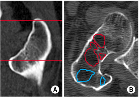
Fig. 2
Three-dimensional image of reverse obliquity fracture in the young group (group 1).
(A) Anterior-posterior view showing a typical reverse obliquity pattern.
(B) Lateral view showing a 'V' shape apex of proximal fragment.
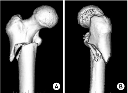
Fig. 3
Three-dimensional image of reverse obliquity fracture in the elderly group (group 2).
(A) Anterior-posterior view showing a typical reverse obliquity pattern with additional fracture line along the trochanteric line.
(B) Lateral view showing a 'Λ' shape apex of distal fragment and additional vertical splitting of the greater trochanter.

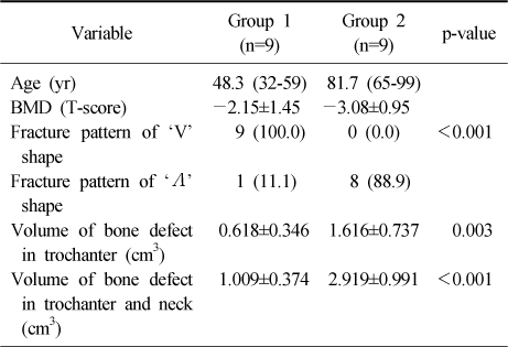
Figure & Data
REFERENCES
Citations
Citations to this article as recorded by 
- A Comparison of Internal Fixation and Bipolar Hemiarthroplasty for the Treatment of Reverse Oblique Intertrochanteric Femoral Fractures in Elderly Patients
Bong-Ju Park, Hong-Man Cho, Woong-Bae Min
Hip & Pelvis.2015; 27(3): 152. CrossRef
 Cite
CiteThe Character of Reverse Obliquity Intertrochanteric Fractures in Elderly Patients


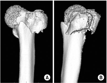
Fig. 1
(A) Region of interest in trochanter from the greater trochanter tip to the upper part of lesser trochanter.
(B) Measurement of bone defect of the trochanter and femoral neck region.
Fig. 2
Three-dimensional image of reverse obliquity fracture in the young group (group 1).
(A) Anterior-posterior view showing a typical reverse obliquity pattern.
(B) Lateral view showing a 'V' shape apex of proximal fragment.
Fig. 3
Three-dimensional image of reverse obliquity fracture in the elderly group (group 2).
(A) Anterior-posterior view showing a typical reverse obliquity pattern with additional fracture line along the trochanteric line.
(B) Lateral view showing a 'Λ' shape apex of distal fragment and additional vertical splitting of the greater trochanter.
Fig. 1
Fig. 2
Fig. 3
The Character of Reverse Obliquity Intertrochanteric Fractures in Elderly Patients
Clinical Data between the Two Groups
Values are presented as average (range) or mean±standard deviation or average (%). BMD: Bone mineral density.
Table 1
Clinical Data between the Two Groups
Values are presented as average (range) or mean±standard deviation or average (%). BMD: Bone mineral density.

