 E-submission
 TOTA
TOTA TOTS
TOTS
Articles
- Page Path
- HOME > J Musculoskelet Trauma > Volume 29(1); 2016 > Article
-
Case Report
- Calcified Anterior Tibial Artery Entrapment in Distal Third Tibial Fracture: A Case Report
- Kyu-Hyun Yang, M.D., Ph.D., Yougun Won, M.D., Sang Bum Kim, M.D., Ph.D., Won Kuen Park, M.D., You Sun Jung, M.D.
-
Journal of the Korean Fracture Society 2016;29(1):68-72.
DOI: https://doi.org/10.12671/jkfs.2016.29.1.68
Published online: January 19, 2016
Department of Orthopaedic Surgery, Yonsei University College of Medicine, Seoul, Korea.
*Department of Orthopaedic Surgery, College of Medicine, Konyang University, Daejeon, Korea.
- Address reprint requests to: Yougun Won, M.D. Department of Orthopaedic Surgery, Konyang University Hospital, 158 Gwanjeodong-ro, Seo-gu, Daejeon 35365, Korea. Tel: 82-42-600-9862, Fax: 82-42-545-2373, yougunwon@gmail.com
• Received: September 30, 2015 • Revised: October 29, 2015 • Accepted: December 21, 2015
Copyright © 2016 The Korean Fracture Society. All rights reserved.
This is an Open Access article distributed under the terms of the Creative Commons Attribution Non-Commercial License (http://creativecommons.org/licenses/by-nc/4.0) which permits unrestricted non-commercial use, distribution, and reproduction in any medium, provided the original work is properly cited.
- 1,208 Views
- 5 Download
- 1 Crossref
Abstract
- In the distal third of the tibia, the anterior tibial artery runs close to the anterolateral surface of the tibial cortex. In a clinical situation, without vascular evaluation, injury or entrapment of the anterior tibial artery is difficult to detect. Because, an intact dorsalis pedis pulse is supplied with the collateral vessels of the posterior tibial artery. An entrapped anterior tibial artery can be injured during closed reduction in an emergency room or open reduction and internal fixation in the operating room. Care must be taken to prevent iatrogenic anterior tibial artery. In this case, an entrapped anterior tibial artery was observed in a simple radiograph and computed tomograph without contrast media for the vessel. We report on a rare case of calcified anterior tibial artery entrapment in a distal tibial fracture.
- 1. Brinker MR, Bailey DE Jr. Fracture healing in tibia fractures with an associated vascular injury. J Trauma, 1997;42:11-19.Article
- 2. Segal D, Brenner M, Gorczyca J. Tibial fractures with infrapopliteal arterial injuries. J Orthop Trauma, 1987;1:160-169.Article
- 3. Tan ET, Tan TJ, Poon KB. Entrapment of the deep peroneal nerve and anterior tibial vessels by a spiral tibial fracture causing partial non-union: a case report. Skeletal Radiol, 2015;[epub].ArticlePDF
- 4. Court-Brown CM, McBirnie J. The epidemiology of tibial fractures. J Bone Joint Surg Br, 1995;77:417-421.ArticlePDF
- 5. Labler L, Wedler V, Mica L, Trentz O. Entrapment of the anterior tibial artery in a distal tibial fracture after intramedullary nailing. Unfallchirurg, 2006;109:156-159.ArticlePDF
- 6. Miki RA, Lawrence JP, Gillon TJ, Lawrence BD, Zell RA. Anterior tibial artery and deep peroneal nerve entrapment in spiral distal third tibia fracture. Orthopedics, 2008;31:cited 2008 Dec. [Internet]. Available from: http://www.healio.com/orthopedics/trauma/journals/ortho/2008-12-31-12/%7B75123d28-e2b6-42a5-95b9-07ee2cb7b83f%7D/anterior-tibial-artery-and-deep-peroneal-nerve-entrapmentin-spiral-distal-third-tibia-fractureArticle
- 7. Sanders RJ, Alston GK. Variations and anomalies of the popliteal and tibial arteries. Am J Surg, 1986;152:531-534.Article
- 8. Ebraheim NA, Lu J, Hao Y, Biyani A, Yeasting RA. Anterior tibial artery and its actual projection on the lateral aspect of the tibia: a cadaveric study. Surg Radiol Anat, 1998;20:259-262.ArticlePDF
- 9. Borrelli J Jr, Prickett W, Song E, Becker D, Ricci W. Extraosseous blood supply of the tibia and the effects of different plating techniques: a human cadaveric study. J Orthop Trauma, 2002;16:691-695.Article
REFERENCES
Fig. 1

An 80-year-old female patient with a spiral distal tibial fracture. (A) Antero-posterior view. (B) Lateral view. (C) Oblique view. Entrapped vessel is shown at the fracture site in the plain radiograph (arrow).

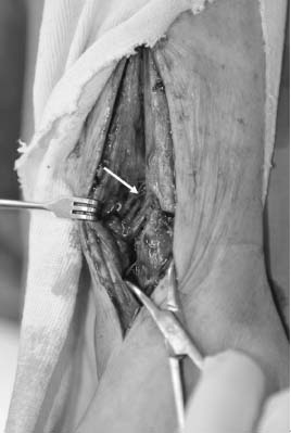

Figure & Data
REFERENCES
Citations
Citations to this article as recorded by 
- Comparison of Time to Operation and Efficacies of Ultrasound-Guided Nerve Block and General Anesthesia in Emergency External Fixation of Lower Leg Fractures (AO 42, 43, 44)
Chan Kang, Sang-Bum Kim, Youn-Moo Heo, You-Gun Won, Byung-Hak Oh, June-Bum Jun, Gi-Soo Lee
The Journal of Foot and Ankle Surgery.2017; 56(5): 1019. CrossRef
 Cite
CiteCalcified Anterior Tibial Artery Entrapment in Distal Third Tibial Fracture: A Case Report
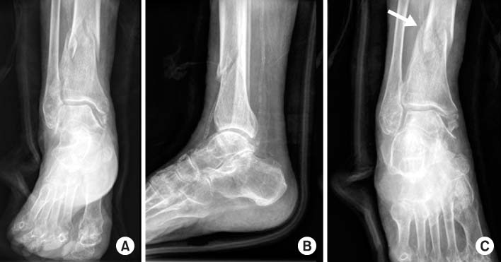
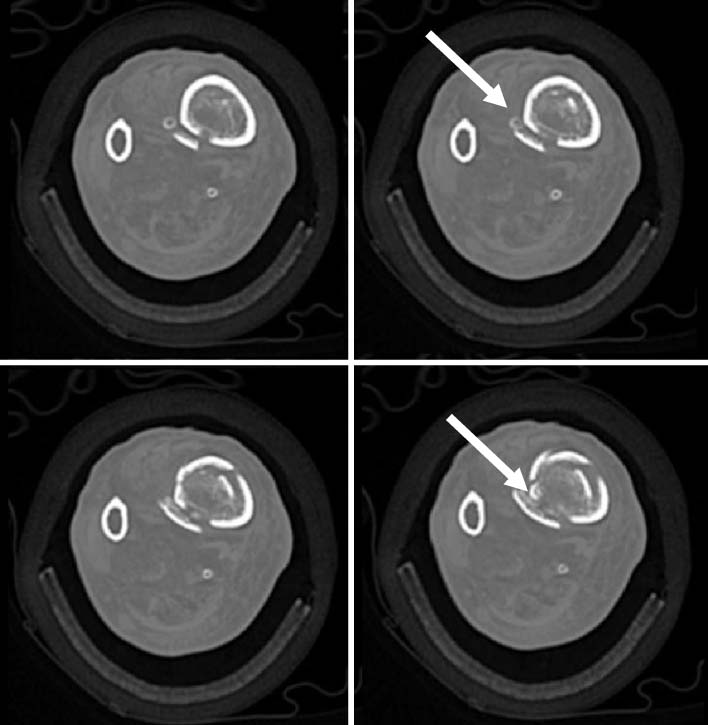

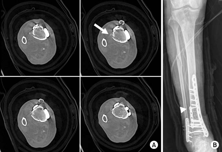
Fig. 1
An 80-year-old female patient with a spiral distal tibial fracture. (A) Antero-posterior view. (B) Lateral view. (C) Oblique view. Entrapped vessel is shown at the fracture site in the plain radiograph (arrow).
Fig. 2
Preoperative computed tomography axial images show the calcified anterior tibial artery (white arrows) at the fracture site.
Fig. 3
Intraoperative photograph shows entrapment of the anterior tibial artery (arrow).
Fig. 4
(A) Postoperative computed tomography axial images show the reduction of calcified anterior tibial artery (arrow) in the fracture site and the fixation of fracture with locking compression plate. (B) Postoperative plain radiograph shows intact anterior tibial artery (arrow head).
Fig. 1
Fig. 2
Fig. 3
Fig. 4
Calcified Anterior Tibial Artery Entrapment in Distal Third Tibial Fracture: A Case Report

