 E-submission
 TOTA
TOTA TOTS
TOTS
Articles
- Page Path
- HOME > J Musculoskelet Trauma > Volume 25(1); 2012 > Article
-
Review Article
- The Current Concepts in the Treatment of Proximal Humerus Fracture
- Joo Han Oh, M.D., Yeun Ho Kim, M.D.
-
Journal of the Korean Fracture Society 2012;25(1):94-104.
DOI: https://doi.org/10.12671/jkfs.2012.25.1.94
Published online: January 31, 2012
Department of Orthopaedic Surgery and Joint Reconstruction Center, Seoul National University Bundang Hospital, Seongnam, Korea.
- Address reprint requests to: Yeun Ho Kim, M.D. Department of Orthopaedic Surgery and Joint Reconstruction Center, Seoul National University Bundang Hospital, 166, Gumi-ro, Bundang-gu, Seongnam 463-707, Korea. Tel: 82-31-787-7197, Fax: 82-31-787-4056, younhk@hotmail.com
Copyright © 2012 The Korean Fracture Society
- 1,807 Views
- 42 Download
- 8 Crossref
- 1. Abu-Rajab RB, Stansfield BW, Nunn T, Nicol AC, Kelly IG. Re-attachment of the tuberosities of the humerus following hemiarthroplasty for four-part fracture. J Bone Joint Surg Br, 2006;88:1539-1544.ArticlePDF
- 2. Bae JH, Oh JK, Chon CS, Oh CW, Hwang JH, Yoon YC. The biomechanical performance of locking plate fixation with intramedullary fibular strut graft augmentation in the treatment of unstable fractures of the proximal humerus. J Bone Joint Surg Br, 2011;93:937-941.ArticlePDF
- 3. Bernard J, Charalambides C, Aderinto J, Mok D. Early failure of intramedullary nailing for proximal humeral fractures. Injury, 2000;31:789-792.Article
- 4. Connor PM, Flatow EL. Complications of internal fixation of proximal humeral fractures. Instr Course Lect, 1997;46:25-37.
- 5. Coste JS, Reig S, Trojani C, Berg M, Walch G, Boileau P. The management of infection in arthroplasty of the shoulder. J Bone Joint Surg Br, 2004;86:65-69.ArticlePDF
- 6. Court-Brown CM, Caesar B. Epidemiology of adult fractures: a review. Injury, 2006;37:691-697.Article
- 7. Court-Brown CM, Cattermole B, McQueen MM. Impacted valgus fractures (B1.1) of the proximal humerus. The results of non-operative treatment. J Bone Joint Surg Br, 2002;84:504-508.
- 8. Court-Brown CM, Garg A, McQueen MM. The translated two-part fracture of the proximal humerus. Epidemiology and outcome in the older patient. J Bone Joint Surg Br, 2001;83:799-804.
- 9. Court-Brown CM, McQueen MM. The impacted varus (A2.2) proximal humeral fracture: prediction of outcome and results of nonoperative treatment in 99 patients. Acta Orthop Scand, 2004;75:736-740.Article
- 10. DePalma AF, Cautilli RA. Fractures of the upper end of the humerus. Clin Orthop, 1961;20:73-93.
- 11. Dhar SA, Butt MF, Mir MR, Ali MF, Kawoosa AA. Use of the Ilizarov apparatus to improve alignment in proximal humeral fractures treated initially by a unilateral external fixator. Strategies Trauma Limb Reconstr, 2008;3:119-122.ArticlePDF
- 12. Edelson G, Kelly I, Vigder F, Reis ND. A three-dimensional classification for fractures of the proximal humerus. J Bone Joint Surg Br, 2004;86:413-425.ArticlePDF
- 13. Gardner MJ, Boraiah S, Helfet DL, Lorich DG. Indirect medial reduction and strut support of proximal humerus fractures using an endosteal implant. J Orthop Trauma, 2008;22:195-200.Article
- 14. Gardner MJ, Boraiah S, Helfet DL, Lorich DG. The anterolateral acromial approach for fractures of the proximal humerus. J Orthop Trauma, 2008;22:132-137.Article
- 15. Gardner MJ, Weil Y, Barker JU, Kelly BT, Helfet DL, Lorich DG. The importance of medial support in locked plating of proximal humerus fractures. J Orthop Trauma, 2007;21:185-191.Article
- 16. Gerber C, Lambert SM, Hoogewoud HM. Absence of avascular necrosis of the humeral head after post-traumatic rupture of the anterior and posterior humeral circumflex arteries. A case report. J Bone Joint Surg Am, 1996;78:1256-1259.Article
- 17. Gerber C, Schneeberger AG, Vinh TS. The arterial vascularization of the humeral head. An anatomical study. J Bone Joint Surg Am, 1990;72:1486-1494.Article
- 18. Gerber C, Werner CM, Vienne P. Internal fixation of complex fractures of the proximal humerus. J Bone Joint Surg Br, 2004;86:848-855.ArticlePDF
- 19. Hanson B, Neidenbach P, de Boer P, Stengel D. Functional outcomes after nonoperative management of fractures of the proximal humerus. J Shoulder Elbow Surg, 2009;18:612-621.Article
- 20. Healy WL, Jupiter JB, Kristiansen TK, White RR. Nonunion of the proximal humerus. A review of 25 cases. J Orthop Trauma, 1990;4:424-431.
- 21. Hertel R, Hempfing A, Stiehler M, Leunig M. Predictors of humeral head ischemia after intracapsular fracture of the proximal humerus. J Shoulder Elbow Surg, 2004;13:427-433.Article
- 22. Iannotti JP, Gabriel JP, Schneck SL, Evans BG, Misra S. The normal glenohumeral relationships. An anatomical study of one hundred and forty shoulders. J Bone Joint Surg Am, 1992;74:491-500.Article
- 23. Kamineni S, Ankem H, Sanghavi S. Anatomical considerations for percutaneous proximal humeral fracture fixation. Injury, 2004;35:1133-1136.Article
- 24. Klein M, Juschka M, Hinkenjann B, Scherger B, Ostermann PA. Treatment of comminuted fractures of the proximal humerus in elderly patients with the Delta III reverse shoulder prosthesis. J Orthop Trauma, 2008;22:698-704.Article
- 25. Koval KJ, Gallagher MA, Marsicano JG, Cuomo F, McShinawy A, Zuckerman JD. Functional outcome after minimally displaced fractures of the proximal part of the humerus. J Bone Joint Surg Am, 1997;79:203-207.Article
- 26. Krause FG, Huebschle L, Hertel R. Reattachment of the tuberosities with cable wires and bone graft in hemiarthroplasties done for proximal humeral fractures with cable wire and bone graft: 58 patients with a 22-month minimum follow-up. J Orthop Trauma, 2007;21:682-686.Article
- 27. Kristiansen B, Angermann P, Larsen TK. Functional results following fractures of the proximal humerus. A controlled clinical study comparing two periods of immobilization. Arch Orthop Trauma Surg, 1989;108:339-341.ArticlePDF
- 28. Leyshon RL. Closed treatment of fractures of the proximal humerus. Acta Orthop Scand, 1984;55:48-51.Article
- 29. Matsen FA 3rd, Rockwood CA Jr, Wirth MA, Lippitt SB. Rockwood CA, Matsen FA, Wirth MA, Lippitt SB. Glenohumeral arthritis and its management. In: The shoulder, 2004;3rd ed. Philadelphia, WB Saunders. 879-1008.Article
- 30. Mont MA, Maar DC, Urquhart MW, Lennox D, Hungerford DS. Avascular necrosis of the humeral head treated by core decompression. A retrospective review. J Bone Joint Surg Br, 1993;75:785-788.ArticlePDF
- 31. Murray IR, Amin AK, White TO, Robinson CM. Proximal humeral fractures: current concepts in classification, treatment and outcomes. J Bone Joint Surg Br, 2011;93:1-11.
- 32. Neer CS 2nd. Displaced proximal humeral fractures. I. Classification and evaluation. J Bone Joint Surg Am, 1970;52:1077-1089.
- 33. Neviaser AS, Hettrich CM, Beamer BS, Dines JS, Lorich DG. Endosteal strut augment reduces complications associated with proximal humeral locking plates. Clin Orthop Relat Res, 2011;469:3300-3306.Article
- 34. Palvanen M, Kannus P, Niemi S, Parkkari J. Update in the epidemiology of proximal humeral fractures. Clin Orthop Relat Res, 2006;442:87-92.Article
- 35. Plausinis D, Kwon YW, Zuckerman JD. Complications of humeral head replacement for proximal humeral fractures. Instr Course Lect, 2005;54:371-380.Article
- 36. Rajasekhar C, Ray PS, Bhamra MS. Fixation of proximal humeral fractures with the Polarus nail. J Shoulder Elbow Surg, 2001;10:7-10.Article
- 37. Resch H. Proximal humeral fractures: current controversies. J Shoulder Elbow Surg, 2011;20:827-832.Article
- 38. Robinson CM, Akhtar A, Mitchell M, Beavis C. Complex posterior fracture-dislocation of the shoulder. Epidemiology, injury patterns, and results of operative treatment. J Bone Joint Surg Am, 2007;89:1454-1466.
- 39. Robinson CM, Khan LA, Akhtar MA. Treatment of anterior fracture-dislocations of the proximal humerus by open reduction and internal fixation. J Bone Joint Surg Br, 2006;88:502-508.ArticlePDF
- 40. Robinson CM, Khan L, Akhtar A, Whittaker R. The extended deltoid-splitting approach to the proximal humerus. J Orthop Trauma, 2007;21:657-662.Article
- 41. Robinson CM, Page RS. Severely impacted valgus proximal humeral fractures. Results of operative treatment. J Bone Joint Surg Am, 2003;85:1647-1655.
- 42. Robinson CM, Wylie JR, Ray AG, et al. Proximal humeral fractures with a severe varus deformity treated by fixation with a locking plate. J Bone Joint Surg Br, 2010;92:672-678.ArticlePDF
- 43. Russo R, Visconti V, Lombardi LV, Ciccarelli M, Giudice G. The block-bridge system: a new concept and surgical technique to reconstruct articular surfaces and tuberosities in complex proximal humeral fractures. J Shoulder Elbow Surg, 2008;17:29-36.Article
- 44. Sidor ML, Zuckerman JD, Lyon T, Koval K, Cuomo F, Schoenberg N. The Neer classification system for proximal humeral fractures. An assessment of interobserver reliability and intraobserver reproducibility. J Bone Joint Surg Am, 1993;75:1745-1750.Article
- 45. Siebenrock KA, Gerber C. The reproducibility of classification of fractures of the proximal end of the humerus. J Bone Joint Surg Am, 1993;75:1751-1755.Article
- 46. Solberg BD, Moon CN, Franco DP, Paiement GD. Surgical treatment of three and four-part proximal humeral fractures. J Bone Joint Surg Am, 2009;91:1689-1697.Article
- 47. Volgas DA, Stannard JP, Alonso JE. Nonunions of the humerus. Clin Orthop Relat Res, 2004;419:46-50.Article
- 48. Walsh S, Reindl R, Harvey E, Berry G, Beckman L, Steffen T. Biomechanical comparison of a unique locking plate versus a standard plate for internal fixation of proximal humerus fractures in a cadaveric model. Clin Biomech (Bristol, Avon), 2006;21:1027-1031.Article
REFERENCES
Fig. 1Blood supply of the humeral head. 1: a.xillary artery, 2: posterior circumflex artery, 3: anterior circumflex artery, 4: anterolateral branch of the anterior circumflex artery, 5: greater tuberosity, 6: lesser tuberosity, 7: insertion of the subscapularis tendon, 8: constant site of entry of the anterolateral branch into bone, and 9: intertubercular groove17).
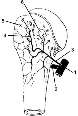

Fig. 3
Percutaneous pin fixation.
(A) Preoperative AP view.
(B) Preoperative axillary view.
(C) Postoperative AP view.
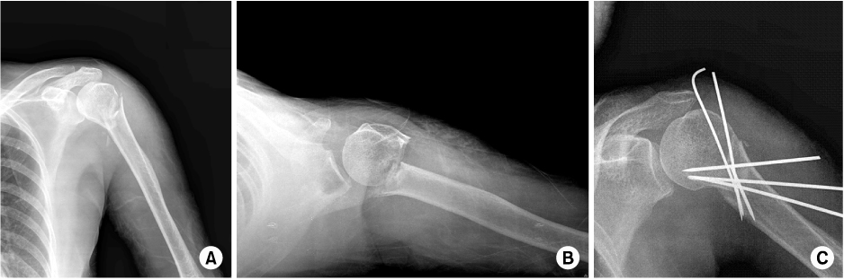


Figure & Data
REFERENCES
Citations
Citations to this article as recorded by 
- Tibiotalocalcaneal Arthrodesis Using a Lateral Distal Femoral Locking Plate in a Charcot Arthropathy Patient with Severe Varus Deformity and a Bone Defect: A Case Report
Jae-Wook Park, Seongwook Kim, Hyun-woo Park
Journal of Korean Foot and Ankle Society.2025; 29(3): 111. CrossRef - The application of a dual-lead locking screw could enhance the reduction and fixation stability of the proximal humerus fractures: a biomechanical evaluation
Eunju Lee, Hyeon Jang Jeong, Yeon Soo Lee, Joo Han Oh
Frontiers in Surgery.2024;[Epub] CrossRef - Effect of Calcar Screw in Locking Compression Plate System for Osteoporotic Proximal Humerus Fracture: A Finite Element Analysis Study
Jung-Soo Lee, Jong Hoon Kim, Kwang Gi Kim, Yong-Cheol Yoon, Piotr Gas
BioMed Research International.2022;[Epub] CrossRef - Medial and Lateral Dual Plate Fixation for Osteoporotic Proximal Humerus Comminuted Fracture: 2 Case Reports
Sam-Guk Park
Journal of the Korean Fracture Society.2016; 29(1): 61. CrossRef - Operative Treatment of Displaced Medial Epicondyle Fractures Using K-Wires Cross-Fixation
Youn Moo Heo, Sang-Bum Kim, Jin Woong Yi, Jae Ik Lee, Hyun Jin Yoo, Tae Gyun Kim
Journal of the Korean Orthopaedic Association.2015; 50(6): 513. CrossRef - The Analysis of the Treatment Outcomes of Proximal Humeral Fractures with Locking Plates
Kwang Won Lee, Yoon Sub Hwang, Choon Myeon Kim, Dae Suk Yang, Tae Soo Park
Clinics in Shoulder and Elbow.2014; 17(1): 10. CrossRef - Polarus Intramedullary Nail for Proximal Humeral and Humeral Shaft Fractures in Elderly Patients with Osteoporosis
Youn-Soo Hwang, Kwang-Yeol Kim, Hyung-Chun Kim, Su-Han Ahn, Dong-Eun Lee
Journal of the Korean Fracture Society.2013; 26(1): 14. CrossRef - The Result of Conservative Treatment of Proximal Humerus Fracture in Elderly Patients
Seung-Gil Baek, Chang-Wug Oh, Young-Soo Byun, Jong-Keon Oh, Joon-Woo Kim, Jong-Pil Yoon, Hyun-Joo Lee, Hyung-Sub Kim
Journal of the Korean Fracture Society.2013; 26(4): 292. CrossRef
 Cite
CiteThe Current Concepts in the Treatment of Proximal Humerus Fracture

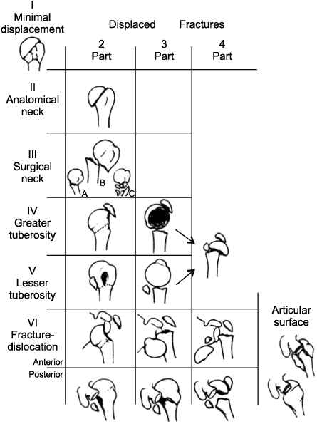


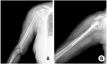
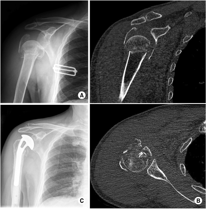
Fig. 1
Blood supply of the humeral head. 1: a.xillary artery, 2: posterior circumflex artery, 3: anterior circumflex artery, 4: anterolateral branch of the anterior circumflex artery, 5: greater tuberosity, 6: lesser tuberosity, 7: insertion of the subscapularis tendon, 8: constant site of entry of the anterolateral branch into bone, and 9: intertubercular groove17).
Fig. 2
Neer's classification of proximal humerus fractures32).
Fig. 3
Percutaneous pin fixation.
(A) Preoperative AP view.
(B) Preoperative axillary view.
(C) Postoperative AP view.
Fig. 4
Internal fixation with fibular allograft.
(A) Preoperative AP view.
(B) Preoperative 3D-CT image.
(C) Postoperative AP view.
Fig. 5
Internal fixation with intramedullary nail.
(A) Preoperative X-ray.
(B) Postoperative X-ray.
Fig. 6
Hemiarthroplasty in patient with 4-part proximal humeral fracture.
(A) Preoperative AP view.
(B) Preopetative CT image.
(C) Postoperative AP view.
Fig. 1
Fig. 2
Fig. 3
Fig. 4
Fig. 5
Fig. 6
The Current Concepts in the Treatment of Proximal Humerus Fracture

