 E-submission
 TOTA
TOTA TOTS
TOTS
Articles
- Page Path
- HOME > J Musculoskelet Trauma > Volume 25(4); 2012 > Article
-
Original Article
- The Treatment of Intertrochanteric Femoral Fracture with Proximal Femoral Nail Antirotation
- Jong Won Kim, M.D., Hyun Soo Park, M.D., Young Soo Jang, M.D., Jae Hyuk Choi, M.D., Sung Ju Bae, M.D., Chan Il Bae, M.D.
-
Journal of the Korean Fracture Society 2012;25(4):257-262.
DOI: https://doi.org/10.12671/jkfs.2012.25.4.257
Published online: October 19, 2012
Department of Orthopedic Surgery, KEPCO Medical Foundation, Hanil General Hospital, Seoul, Korea.
*Department of Orthopedic Surgery, Gangnam Himchan Hospital, Seoul, Korea.
- Address reprint requests to: Hyun Soo Park, M.D. Department of Orthopedic Surgery, Hanil General Hospital, 308, Uicheon-ro, Dobong-gu, Seoul 132-703, Korea. Tel: 82-2-901-3078, Fax: 82-2-900-1745, deanzang2@hanmail.net
• Received: May 5, 2012 • Revised: May 5, 2012 • Accepted: June 21, 2012
Copyright © 2012 The Korean Fracture Society
- 1,944 Views
- 17 Download
Abstract
-
Purpose
- This study was performed to evaluate the results of treating intertrochanteric fracture with proximal femoral nail antirotation (PFNA).
-
Materials and Methods
- We performed PFNA on 41 intertrochanteric femur fracture patients from May 2008, to August 2010. We analyzed the operation time, blood loss, recovery of ambulatory function, T-score, the tip apex distance (TAD), the sliding distance of the blade, neck-shaft angle, and complications.
-
Results
- The mean operation time was 51 minutes and the mean amount of blood loss was 350 ml. The time to ambulation averaged 7.2 days. Thirty-two cases (79%) recovered their previous walking status at 6 months after operation. The average T-score was 3.3 and TAD was 12.3 mm (8.6~27 mm). 35 cases (87%) achieved acceptable reduction. The average amount of PFNA blade sliding was 3.3 mm. The neck-shaft angle was changed 2.6 degrees varus displacement at the final follow-up. There was one case of nonunion due to tuberculosis infection.
-
Conclusion
- The findings from this study indicate that PFNA is a useful and reliable choice for the treatment of intertrochanteric fracture of the femur.
- 1. Banan H, Al-Sabti A, Jimulia T, Hart AJ. The treatment of unstable, extracapsular hip fractures with the AO/ASIF proximal femoral nail (PFN)--our first 60 cases. Injury, 2002;33:401-405.Article
- 2. Baumgaertner MR, Curtin SL, Lindskog DM, Keggi JM. The value of the tip-apex distance in predicting failure of fixation of peritrochanteric fractures of the hip. J Bone Joint Surg Am, 1995;77:1058-1064.Article
- 3. Boldin C, Seibert FJ, Fankhauser F, Peicha G, Grechenig W, Szyszkowitz R. The proximal femoral nail (PFN)-a minimal invasive treatment of unstable proximal femoral fractures: a prospective study of 55 patients with a follow-up of 15 months. Acta Orthop Scand, 2003;74:53-58.Article
- 4. Butt MS, Krikler SJ, Nafie S, Ali MS. Comparison of dynamic hip screw and gamma nail: a prospective, randomized, controlled trial. Injury, 1995;26:615-618.Article
- 5. Chevalley F, Gamba D. Gamma nailing of pertrochanteric and subtrochanteric fractures: clinical results of a series of 63 consecutive cases. J Orthop Trauma, 1997;11:412-415.Article
- 6. Cleveland M, Bosworth DM, Thompson FR, Wilson HJ Jr, Ishizuka T. A ten-year analysis of intertrochanteric fractures of the femur. J Bone Joint Surg Am, 1959;41-A:1399-1408.Article
- 7. Flores LA, Harrington IJ, Heller M. The stability of intertrochanteric fractures treated with a sliding screw-plate. J Bone Joint Surg Br, 1990;72:37-40.ArticlePDF
- 8. Friedl W, Clausen J. Experimental examination for optimized stabilisation of trochanteric femur fractures, intra- or extramedullary implant localisation and influence of femur neck component profile on cut-out risk. Chirurg, 2001;72:1344-1352.
- 9. Gotfried Y. The lateral trochanteric wall: a key element in the reconstruction of unstable pertrochanteric hip fractures. Clin Orthop Relat Res, 2004;(425):82-86.
- 10. Gundle R, Gargan MF, Simpson AH. How to minimize failures of fixation of unstable intertrochanteric fractures. Injury, 1995;26:611-614.Article
- 11. Halder SC. The Gamma nail for peritrochanteric fractures. J Bone Joint Surg Br, 1992;74:340-344.ArticlePDF
- 12. Hornby R, Evans JG, Vardon V. Operative or conservative treatment for trochanteric fractures of the femur. A randomised epidemiological trial in elderly patients. J Bone Joint Surg Br, 1989;71:619-623.ArticlePDF
- 13. Jacobs RR, McClain O, Armstrong HJ. Internal fixation of intertrochanteric hip fractures: a clinical and biomechanical study. Clin Orthop Relat Res, 1980;(146):62-70.
- 14. Jensen JS. Determinig factors for the mortality following hip fractures. Injury, 1984;15:411-414.Article
- 15. Kaufer H. Mechanics of the treatment of hip injuries. Clin Orthop Relat Res, 1980;(146):53-61.Article
- 16. Kim TH, Kim JO, Lee SY, Yun GU. Treatment of the unstable intertrochanteric fracture with proximal femoral nail antirotation: comparison with compression hip screw with trochanteric stabilizing plate. J Korean Fract Soc, 2010;23:353-359.Article
- 17. Koval KJ, Skovron ML, Aharonoff GB, Meadows SE, Zuckerman JD. Ambulatory ability after hip fracture. A prospective study in geriatric patients. Clin Orthop Relat Res, 1995;(310):150-159.
- 18. Koval KJ, Zuckerman JD. Hip fractures: I. overview and evaluation and treatment of femoral-neck fractures. J Am Acad Orthop Surg, 1994;2:141-149.Article
- 19. Lenich A, Mayr E, Rüter A, Möckl Ch, Füchtmeier B. First results with the trochanter fixation nail (TFN): a report on 120 cases. Arch Orthop Trauma Surg, 2006;126:706-712.ArticlePDF
- 20. Madsen JE, Naess L, Aune AK, Alho A, Ekeland A, Stromsoe K. Dynamic hip screw with trochanteric stabilizing plate in the treatment of unstable proximal femoral fractures: a comparative study with the Gamma nail and compression hip screw. J Orthop Trauma, 1998;12:241-248.Article
- 21. Pajarinen J, Lindahl J, Michelsson O, Savolainen V, Hirvensalo E. Pertrochanteric femoral fractures treated with a dynamic hip screw or a proximal femoral nail. A randomised study comparing post-operative rehabilitation. J Bone Joint Surg Br, 2005;87:76-81.
- 22. Papasimos S, Koutsojannis CM, Panagopoulos A, Megas P, Lambiris E. A randomised comparison of AMBI, TGN and PFN for treatment of unstable trochanteric fractures. Arch Orthop Trauma Surg, 2005;125:462-468.ArticlePDF
- 23. Park MS, Lim YJ, Kim YS, Kim KH, Cho HM. Treatment of the proximal femoral fractures with proximal femoral nail antirotation (PFNA). J Korean Fract Soc, 2009;22:91-97.Article
- 24. Parker MJ, Palmer CR. A new mobility score for predicting mortality after hip fracture. J Bone Joint Surg Br, 1993;75:797-798.ArticlePDF
- 25. Parker MJ, Pryor GA. Gamma versus DHS nailing for extracapsular femoral fractures. Meta-analysis of ten randomised trials. Int Orthop, 1996;20:163-168.
- 26. Pugh WL. A self-adjusting nail-plate for fractures about the hip joint. J Bone Joint Surg Am, 1955;37-A:1085-1093.Article
- 27. Rha JD, Kim YH, Yoon SI, Park TS, Lee MH. Factors affecting sliding of the lag screw in intertrochanteric fractures. Int Orthop, 1993;17:320-324.ArticlePDF
- 28. Richmond J, Aharonoff GB, Zuckerman JD, Koval KJ. Mortality risk after hip fractures. J Orthop Trauma, 2003;17:53-56.
- 29. Siegmeth AW, Gurusamy K, Parker MJ. Delay to surgery prolongs hospital stay in patients with fractures of the proximal femur. J Bone Joint Surg Br, 2005;87:1123-1126.ArticlePDF
- 30. Simpson AH, Varty K, Dodd CA. Sliding hip screws: modes of failure. Injury, 1989;20:227-231.Article
- 31. Sommers MB, Roth C, Hall H, et al. A laboratory model to evaluate cutout resistance of implants for pertrochanteric fracture fixation. J Orthop Trauma, 2004;18:361-368.Article
- 32. Steinberg GG, Desai SS, Kornwitz NA, Sullivan TJ. The intertrochanteric hip fracture. A retrospective analysis. Orthopedics, 1988;11:265-273.Article
- 33. Strauss E, Frank J, Lee J, Kummer FJ, Tejwani N. Helical blade versus sliding hip screw for treatment of unstable intertrochanteric hip fractures: a biomechanical evaluation. Injury, 2006;37:984-989.Article
REFERENCES
Fig. 1
A 87-year-old male who experienced intertrochanteric fracture from a fall.
(A) Preoperative radiograph showed intertrochanteric fracture.
(B) The fracture was reduced and fixed with proximal femoral nail antirotation.
(C) Radiologic findings 17 months postoperatively showed bony union of the fracture site.
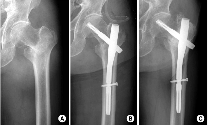
Fig. 2
A 85-year-old female who experienced intertrochanteric fracture from a fall.
(A) Preoperative radiograph showed intertrochanteric fracture.
(B) The fracture was reduced and fixed with proximal femoral nail antirotation (PFNA).
(C) Two weeks after PFNA removed, radiologic findings showed non-union.
(D) Because of the infected non-union, an antibiotic-loaded cement insertion was performed three weeks after PFNA removal.
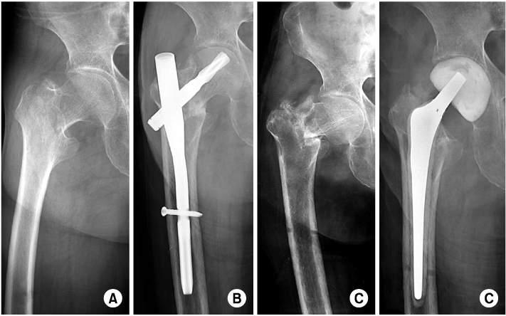
Fig. 3
A 67-year-old female who experienced intertrochanteric fracture from a fall.
(A) The preoperative radiograph showed intertrochanteric fracture.
(B) The fracture was reduced and fixed with proximal femoral nail antirotation.
(C) Radiologic findings 13 months postoperatively showed an impending cut-out.


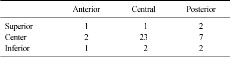
Figure & Data
REFERENCES
Citations
Citations to this article as recorded by 
 Cite
CiteThe Treatment of Intertrochanteric Femoral Fracture with Proximal Femoral Nail Antirotation


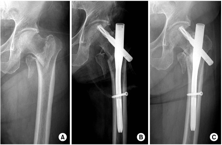
Fig. 1
A 87-year-old male who experienced intertrochanteric fracture from a fall.
(A) Preoperative radiograph showed intertrochanteric fracture.
(B) The fracture was reduced and fixed with proximal femoral nail antirotation.
(C) Radiologic findings 17 months postoperatively showed bony union of the fracture site.
Fig. 2
A 85-year-old female who experienced intertrochanteric fracture from a fall.
(A) Preoperative radiograph showed intertrochanteric fracture.
(B) The fracture was reduced and fixed with proximal femoral nail antirotation (PFNA).
(C) Two weeks after PFNA removed, radiologic findings showed non-union.
(D) Because of the infected non-union, an antibiotic-loaded cement insertion was performed three weeks after PFNA removal.
Fig. 3
A 67-year-old female who experienced intertrochanteric fracture from a fall.
(A) The preoperative radiograph showed intertrochanteric fracture.
(B) The fracture was reduced and fixed with proximal femoral nail antirotation.
(C) Radiologic findings 13 months postoperatively showed an impending cut-out.
Fig. 1
Fig. 2
Fig. 3
The Treatment of Intertrochanteric Femoral Fracture with Proximal Femoral Nail Antirotation
Blade Length
Position of Blade
Table 1
Blade Length
Table 2
Position of Blade

