 E-submission
 TOTA
TOTA TOTS
TOTS
Articles
- Page Path
- HOME > J Musculoskelet Trauma > Volume 29(3); 2016 > Article
-
Case Report
- Avulsion of the Femoral Attachment of Anterior Cruciate Ligament Associated with Ipsilateral Femoral Shaft Fracture in Skeletally Mature Patient: A Case Report
- Seong-Eun Byun, M.D., Taesup Kim, M.D., Bang-Hyun Kim, M.D., Jae-Hwa Kim, M.D., Ph.D, Soo-Hong Han, M.D., Ph.D, Wonchul Choi, M.D., Ph.D
-
Journal of the Korean Fracture Society 2016;29(3):200-205.
DOI: https://doi.org/10.12671/jkfs.2016.29.3.200
Published online: July 21, 2016
Department of Orthopaedic Surgery, CHA Bundang Medical Center, CHA University, Seongnam, Korea.
- Address reprint requests to: Wonchul Choi, M.D., Ph.D. Department of Orthopaedic Surgery, CHA Bundang Medical Center, CHA University, 59 Yatap-ro, Bundang-gu, Seongnam 13496, Korea. Tel: 82-31-780-5289, Fax: 82-31-708-3578, wcchoios@chamc.co.kr
• Received: February 24, 2016 • Revised: April 13, 2016 • Accepted: May 12, 2016
Copyright © 2016 The Korean Fracture Society. All rights reserved.
This is an Open Access article distributed under the terms of the Creative Commons Attribution Non-Commercial License (http://creativecommons.org/licenses/by-nc/4.0) which permits unrestricted non-commercial use, distribution, and reproduction in any medium, provided the original work is properly cited.
- 775 Views
- 2 Download
Abstract
- Avulsion fracture at the femoral attachment of the anterior cruciate ligament (ACL) is very rare and has been reported mostly in skeletally immature patients. Authors experienced a case of avulsion fracture at the femoral attachment of ACL in a skeletally mature, a 21-year-old male associated with ipsilateral femoral shaft fracture. Here, authors report on the case with a literature review. Care should be taken because an avulsion fracture at the femoral attachment of ACL can be accompanied by ipsilateral femoral shaft fracture in skeletally mature patients.
- 1. Bengtson H, Giangarra C. Osteochondral avulsion fracture of the anterior cruciate ligament femoral origin in a 10-year-old child: a case report. J Athl Train, 2011;46:451-455.ArticlePDF
- 2. Kawate K, Fujisawa Y, Yajima H, Sugimoto K, Tomita Y, Takakura Y. Avulsion of the cartilaginous femoral origin of the anterior cruciate ligament in a three-year-old child. A case report with a thirteen-year follow-up. J Bone Joint Surg Am, 2004;86:1787-1792.
- 3. Griffith JF, Antonio GE, Tong CW, Ming CK. Cruciate ligament avulsion fractures. Arthroscopy, 2004;20:803-812.Article
- 4. Song EK, Seon JK, Park SJ, Yoon TR. Clinical outcome of avulsion fracture of the anterior cruciate ligament between children and adults. J Pediatr Orthop B, 2009;18:335-338.Article
- 5. Nagarj R, Bali T, Kumar MN. Avulsion fracture of anterior cruciate ligament from femoral attachment in a skeletally mature patient: a case report. Southeast Asian J Case Rep Rev, 2015;4:1595-1600.
- 6. Matthews DE, Geissler WB. Arthroscopic suture fixation of displaced tibial eminence fractures. Arthroscopy, 1994;10:418-423.Article
- 7. Caldas MTL, Malheiros DS, Lazzaroni AP, Avelino EA, Santos AJ. Injury of the knee ligaments associated with ipsilateral femoral shaft fractures. Rev Bras Ortop, 2013;48:438-440.Article
- 8. van Raay JJ, Raaymakers EL, Dupree HW. Knee ligament injuries combined with ipsilateral tibial and femoral diaphyseal fractures: the "floating knee". Arch Orthop Trauma Surg, 1991;110:75-77.ArticlePDF
- 9. Trickey EL. Rupture of the posterior cruciate ligament of the knee. J Bone Joint Surg Br, 1968;50:334-341.ArticlePDF
- 10. Blacksin MF, Zurlo JV, Levy AS. Internal derangement of the knee after ipsilateral femoral shaft fracture: MR imaging findings. Skeletal Radiol, 1998;27:434-439.ArticlePDF
REFERENCES
Fig. 1
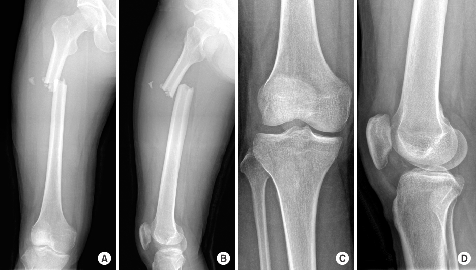
Initial femur anteroposterior (A) and lateral (B) image show a right femoral shaft fracture. And initial knee anteroposterior (C) and lateral (D) image without a sign of fracture.
Fig. 2
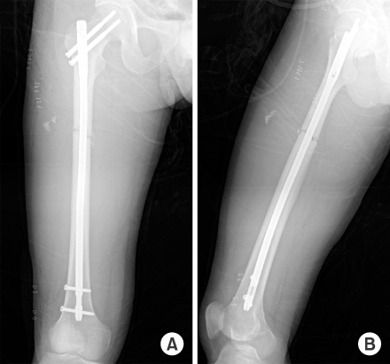
Postoperative anteroposterior (A) and lateral (B) image show a fixed state of fracture with antegrade intramedullary nail.
Fig. 3

Axial (A) and coronal (B) image of computed tomography demonstrate a small bony fragment on the medial aspect of the lateral femoral condyle. And coronal (C), sagittal (D), axial (E), and oblique coronal (F) images show avulsion fracture at the femoral attachment of the anterior cruciate ligament (white arrows).
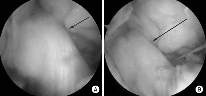

Figure & Data
REFERENCES
Citations
Citations to this article as recorded by 
 Cite
CiteAvulsion of the Femoral Attachment of Anterior Cruciate Ligament Associated with Ipsilateral Femoral Shaft Fracture in Skeletally Mature Patient: A Case Report


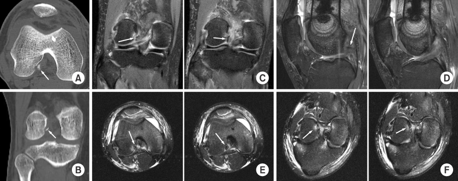


Fig. 1
Initial femur anteroposterior (A) and lateral (B) image show a right femoral shaft fracture. And initial knee anteroposterior (C) and lateral (D) image without a sign of fracture.
Fig. 2
Postoperative anteroposterior (A) and lateral (B) image show a fixed state of fracture with antegrade intramedullary nail.
Fig. 3
Axial (A) and coronal (B) image of computed tomography demonstrate a small bony fragment on the medial aspect of the lateral femoral condyle. And coronal (C), sagittal (D), axial (E), and oblique coronal (F) images show avulsion fracture at the femoral attachment of the anterior cruciate ligament (white arrows).
Fig. 4
Arthroscopic images show a grossly normal looking anterior cruciate ligament (A) and an intact anterior cruciate ligament with applied tension using probe (B) (black arrows).
Fig. 5
Postoperative 6 months femur anteroposterior (A) and lateral (B) image show healed femoral shaft fracture after the internal fixation. And postoperative 6 months knee anteroposterior (C) and lateral (D) image show no abnormal findings.
Fig. 1
Fig. 2
Fig. 3
Fig. 4
Fig. 5
Avulsion of the Femoral Attachment of Anterior Cruciate Ligament Associated with Ipsilateral Femoral Shaft Fracture in Skeletally Mature Patient: A Case Report

