 E-submission
 TOTA
TOTA TOTS
TOTS
Articles
- Page Path
- HOME > J Musculoskelet Trauma > Volume 21(4); 2008 > Article
-
Review Article
- Volar Plating of Distal Radius Fractures
- Kwang-Hyun Lee, M.D.
-
Journal of the Korean Fracture Society 2008;21(4):325-333.
DOI: https://doi.org/10.12671/jkfs.2008.21.4.325
Published online: October 31, 2008
Department of Orthopedic Surgery, Hanyang University Hospital, Seoul, Korea.
- Address reprint requests to: Kwang-Hyun Lee, M.D. Department of Orthopedic Surgery, Hanyang University Hospital, 17, Haengdang-dong, Seongdong-gu, Seoul 133-792, Korea. Tel: 82-2-2290-8485, Fax: 82-2-2299-3774, leegh@hanyang.ac.kr
Copyright © 2008 The Korean Fracture Society. All rights reserved.
This is an Open Access article distributed under the terms of the Creative Commons Attribution Non-Commercial License (http://creativecommons.org/licenses/by-nc/3.0/) which permits unrestricted non-commercial use, distribution, and reproduction in any medium, provided the original work is properly cited.
- 1,098 Views
- 1 Download
- 5 Crossref
Abstract
- Volar plating seems to indicate that many surgeons believe it leads to superior results, and is attractive because of the ease of the operative approach and the soft tissue sleeve to protect digital and wrist tendons. And also it have a locking mechanism to produce the fixed angle device with a low profile and may be thought to be a new era in the surgical treatment of dorsally displaced distal radius fractures even in the face of comminuted or osteoporotic bone. Locked volar plating allows direct fracture reduction, stable fixation and provides stability enough to allow early mobilization and function. The results with volar locking or fixed angle fixation for the general treatment of unstable distal radius fractures in elderly patients has been favorable. Volar plating has fewer complications than external fixation and dorsal plating and allow for earlier return to function. The current indications, technical aspects, clinical results, and complications of the volar plating are being reviewed.
- 1. Arora R, Lutz M, Hennerbichler A, Krappinger D, Espen D, Gabl M. Complications following internal fixation of unstable distal radius fracture with a palmar locking-plate. J Orthop Trauma, 2007;21:316-322.Article
- 2. Baratz ME, Des Jardins J, Anderson DD, Imbriglia JE. Displaced intra-articular fractures of the distal radius: the effect of fracture displacement on contact stresses in a cadaver model. J Hand Surg Am, 1996;21:183-188.
- 3. Cho CH, Bae KC, Kwon DH. Volar T-locking compression plate for treatment of unstable distal radius fractures. J Korean Fract Soc, 2008;21:220-224.Article
- 4. Choi CH, Jung JH, Lee KH, Sung IH, Son KH. Volar locking T-plate fixation of dorsally displaced unstable distal radius fractures. J Korean Soc Surg Hand, 2005;10:123-128.
- 5. Disegi JA, Wyss H. Implant materials for fracture fixation: a clinical perspective. Orthopedics, 1989;12:75-79.Article
- 6. Freeland AE, Luber KT. Biomechanics and biology of plate fixation of distal radius fractures. Hand Clin, 2005;21:329-339.Article
- 7. Gehrmann SV, Windolf J, Kaufmann RA. Distal radius fracture management in elderly patients: a literature review. J Hand Surg Am, 2008;33:421-429.Article
- 8. Gerald G, Karl G, Christian G, et al. Volar plate fixation of AO type C2 and C3 distal radius fractures, a single-center study of 55 patients. J Orthop Trauma, 2008;22:467-472.Article
- 9. Gesensway D, Putnam MD, Mente PL, Lewis JL. Design and biomechanics of a plate for the distal radius. J Hand Surg Am, 1995;20:1021-1027.Article
- 10. Haidukewych GJ. Innovations in locking plate technology. J Am Acad Orthop Surg, 2004;12:205-212.Article
- 11. Lee KH. Clinical biomechanics of the locking plate. J Korean Fract Soc, 2008;21:186-188.Article
- 12. McKay SD, MacDermid JC, Roth JH, Richards RS. Assessment of complications of distal radius fractures and development of a complication checklist. J Hand Surg Am, 2001;26:916-922.Article
- 13. Muller M, Allgower M, Schneider R, Willenegger H. Manual of internal fixation. 3rd ed. New York: Springer Verlag; 1991. p. 1-3.
- 14. Nam IH, Ahn GY, Jang JH, Lee JW, Yun HH. Surgical treatment of dorsally displaced unstable distal radius fracture with volar plate fixation. J Korean Orthop Assoc, 2005;40:814-820.ArticlePDF
- 15. Orbay J. Volar plate fixation of distal radius fractures. Hand Clin, 2005;21:347-354.Article
- 16. Orbay JL. The treatment of unstable distal radius fractures with volar fixation. Hand Surg, 2000;5:103-112.Article
- 17. Osada D, Kamei S, Masuzaki K, Takai M, Kameda M, Tamai K. Prospective study of distal radius fractures treated with a volar locking plate system. J Hand Surg Am, 2008;33:691-700.Article
- 18. Peterson ED, Dennison DG. Fixed-angle volar plate fixation for distal radius fractures in immunosuppressed patients. Hand (NY), 2008.ArticlePDF
- 19. Pichon H, Chergaoui A, Jager S, et al. Volar fixed angle plate LCP 3.5 for dorsally distal radius fracture. About 24 cases. Rev Chir Orthop Reparatrice Appar Mot, 2008;94:152-159.
- 20. Putnam MD, Seitz WH Jr. Advances in fracture management in the hand and distal radius. Hand Clin, 1989;5:455-470.Article
- 21. Rozental TD, Blazar PE. Functional outcome and complications after volar plating for dorsally displaced, unstable fractures of the distal radius. J Hand Surg Am, 2006;31:359-365.Article
- 22. Stein H, Perren SM, Cordey J, Kenwright J, Mosheiff R, Francis MJ. The muscle bed--a crucial factor for fracture healing: a physiological concept. Orthopedics, 2002;25:1379-1383.Article
- 23. Strauss EJ, Banerjee D, Kummer FJ, Tejwani NC. Evaluation of a novel, nonspanning external fixator for treatment of unstable extra-articular fractures of the distal radius: biomechanical comparison with a volar locking plate. J Trauma, 2008;64:975-981.Article
- 24. Yamazaki H, Hattori Y, Doi K. Delayed rupture of flexor tendons caused by protrusion of a screw head of a volar plate for distal radius fracture: a case report. Hand Surg, 2008;13:27-29.Article
REFERENCES
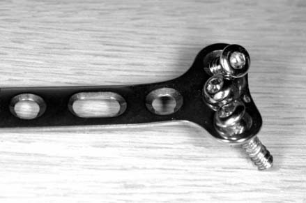
Fig. 2
(B) At 3 months follow-up, the union is advanced with collapsed dorsal cortex.

(A) The distal radius fracture was fixed with conventional plate, and the volar tilt was acceptable immediately after operation.
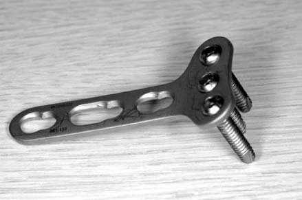
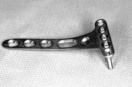
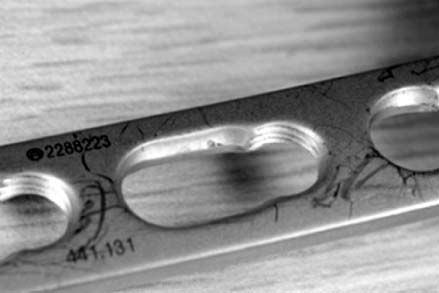

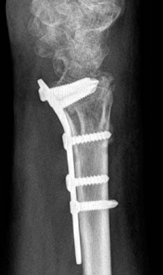


Fig. 10
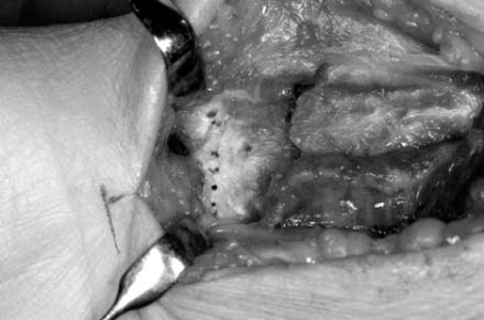
A dotted line indicates transverse ridge, and the fracture site is exposed after elevation of pronator quadratus.
Fig. 11
(B) But they don't violate the joint on tilt view.
(C) At lateral view, it is not sure that the plate and scres involve the joint line.
(D) It can be confirmed that they don't violate the joint at radial tilt lateral view.
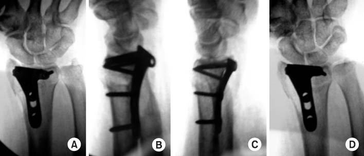
(A) On anteroposterior view, plate and screws look like to violate the joint line.

Figure & Data
REFERENCES
Citations
Citations to this article as recorded by 
- Ultrasonographic Assessment of the Pronator Quadratus Muscle after Surgical Treatment for Distal Radius Fractures
Dong Hyuk Choi, Hyun Kyun Chung, Ji Won Lee, Cheol Hwan Kim, Yong Soo Choi
Journal of the Korean Fracture Society.2017; 30(2): 69. CrossRef - The Fate of Pronator Quadratus Muscle after Volar Locking Plating of Unstable Distal Radius Fractures
Chae-Hyun Lim, Heun-Guyn Jung, Ju-Yeong Heo, Young-Jae Jang, Yong-Soo Choi
Journal of the Korean Fracture Society.2014; 27(3): 191. CrossRef - Comparison of Operative Management in Distal Radius Fractures Using 3.5 mm Versus 2.4 mm Volar Locking Compression Plates
Sung-Sik Ha, Tae-Ho Kim, Ki-Do Hong, Jae-Chun Sim, Jong Hyun Kim
Journal of the Korean Fracture Society.2011; 24(2): 156. CrossRef - Treatment for Unstable Distal Radius Fracture with Osteoporosis -Internal Fixation versus External Fixation-
Jin Rok Oh, Tae Yean Cho, Sung Min Kwan
Journal of the Korean Fracture Society.2010; 23(1): 76. CrossRef - Short Term Results of Operative Management with 2.4 mm Volar Locking Compression Plates in Distal Radius Fractures
Ki-Chul Park, Chang-Hun Lee
Journal of the Korean Fracture Society.2009; 22(4): 264. CrossRef
 Cite
CiteVolar Plating of Distal Radius Fractures

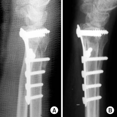


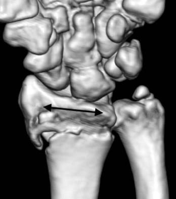

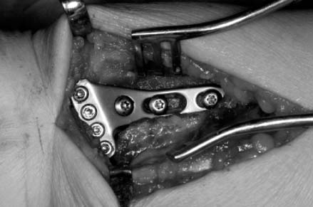
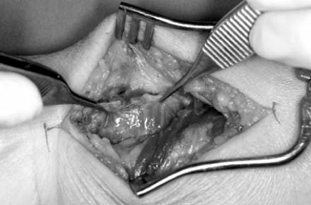


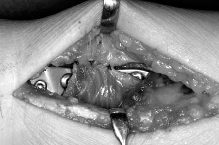
Fig. 1
Non-locking conventional volar plate for distal radius fractures.
Fig. 2
(A) The distal radius fracture was fixed with conventional plate, and the volar tilt was acceptable immediately after operation.
(B) At 3 months follow-up, the union is advanced with collapsed dorsal cortex.
Fig. 3
Volar locking plate.
Fig. 4
Volar juxtaarticular locking plate, Synthes®.
Fig. 5
Combihole, Synthes®.
Fig. 6
The arrow indicates transverse ridge line of the distal radius.
Fig. 7
The plate is fixed distally, and the flexor tendon can be ruptured by attrition.
Fig. 8
This juxtaarticular plate is fixed distal to the transverse ridge.
Fig. 9
The pronator quadrates is elevated from the lateral edge of radial orign.
Fig. 10
A dotted line indicates transverse ridge, and the fracture site is exposed after elevation of pronator quadratus.
Fig. 11
(A) On anteroposterior view, plate and screws look like to violate the joint line.
(B) But they don't violate the joint on tilt view.
(C) At lateral view, it is not sure that the plate and scres involve the joint line.
(D) It can be confirmed that they don't violate the joint at radial tilt lateral view.
Fig. 12
The pronator quadrates should be reattached its orign site.
Fig. 1
Fig. 2
Fig. 3
Fig. 4
Fig. 5
Fig. 6
Fig. 7
Fig. 8
Fig. 9
Fig. 10
Fig. 11
Fig. 12
Volar Plating of Distal Radius Fractures

