 E-submission
 TOTA
TOTA TOTS
TOTS
Articles
- Page Path
- HOME > J Musculoskelet Trauma > Volume 21(4); 2008 > Article
-
Original Article
- Treatment of Failed Intertrochanteric Fractures to Maintain the Reduction in Elderly Patients
- Soon-Yong Kwon, M.D., Hyun-Woo Park, M.D., Sang-Uk Lee, M.D., Soo-Hwan Kang, M.D., Jae-Young Kwon, M.D., Jung-Hoon Do, M.D., Seung-Koo Rhee, M.D.
-
Journal of the Korean Fracture Society 2008;21(4):267-273.
DOI: https://doi.org/10.12671/jkfs.2008.21.4.267
Published online: October 31, 2008
Department of Orthopedic Surgery, The Catholic University of Korea College of Medicine, Seoul, Korea.
*Department of Orthopedic Surgery, Dankook University College of Medicine, Seoul, Korea.
- Address reprint requests to: Seung-Koo Rhee, M.D. Department of Orthopedic Surgery, St. Mary's Hospital, The Catholic University of Korea, 62, Yeouido-dong, Yeongdeungpo-gu, Seoul 150-713, Korea. Tel: 82-2-3779-1192, Fax: 82-2-783-0252, skrhee@catholic.ac.kr
• Received: July 26, 2008 • Revised: August 19, 2008 • Accepted: September 22, 2008
Copyright © 2008 The Korean Fracture Society. All rights reserved.
This is an Open Access article distributed under the terms of the Creative Commons Attribution Non-Commercial License (http://creativecommons.org/licenses/by-nc/3.0/) which permits unrestricted non-commercial use, distribution, and reproduction in any medium, provided the original work is properly cited.
- 1,078 Views
- 1 Download
- 1 Crossref
Abstract
-
Purpose
- The aim of this study was to evaluate and report the new method with a cement augmented screw fixation again to treat the failed intertrochanteric fracture in elderly which were treated with ordinary compression hip screw initially.
-
Materials and Methods
- From Mar. 1988 to May 2007, 10 patients (mean age 69 years) with the failed intertrochanteric fracture which were treated with initial hip screw, were treated with a cement augmented compression hip screw again. The mean follow-up after surgery was over 18 months. The cause of failure, the period upto the reoperation, the neck-shaft angle after the reoperation, the position of lag screw in the femoral head, and the degree of union at last follow-up were analyzed. The change in the functional hip capacity were evaluated by the classification of Clawson.
-
Results
- Causes of failure were superior cutting-out in 6 cases, cortical anchorage failure in 3, and nonunion in one case. The period upto the reoperation was average 7.8 months. Valgus reduction of average 5.7° was achieved, and the positions of lag screw were postero-inferior in 6 cases, center in 3, infero-center in one case. We obtained complete union in 9 cases. The functional outcome showed moderate in 6 cases, good in 3 and poor degree in one case.
-
Conclusion
- Cement augmented compression hip screw treatment will possibly reduce cutting-out of screw and bring more stability in fixation for intertrochanteric fractures in old osteoporotic patients, as well, even in failed cases treated with initial compression hip screw, but proper selection of patients is important.
- 1. Bartucci EJ, Gonzalez MH, Cooperman DR, Freedberg HI, Barmada R, Laros GS. The effect of adjunctive methylmethacrylate on failures of fixation and function in patients with intertrochanteric fractures and osteoporosis. J Bone Joint Surg Am, 1985;67:1094-1107.Article
- 2. Boyd HB, Lipinski SW. Nonunion of trochanteric and subtrochanteric fractures. Surg Gynecol Obstet, 1957;104:463-470.
- 3. Choueka J, Koval KJ, Kummer FJ, Crawford G, Zuckerman JD. Biomechanical comparison of the sliding hip screw and the dome plunger. Effects of material and fixation design. J Bone Joint Surg Br, 1995;77:277-283.ArticlePDF
- 4. Clawson DK. Intertrochanteric fracture of the hip. Am J Surg, 1957;93:580-587.Article
- 5. Coran AG, Banks HH, Aliapoulios MA, Wilson RE. The mangement of pathologic fractures in patients with metastatic carcinoma of the breast. Surg Gynecol Obstet, 1968;127:1225-1230.
- 6. Davis TR, Sher JL, Horsman A, Simpson M, Porter BB, Checketts RG. Intertrochanteric femoral fractures. Mechanical failure after internal fixation. J Bone Joint Surg Br, 1990;72:26-31.ArticlePDF
- 7. Haidukewych GJ, Berry DJ. Salvage of failed internal fixation of intertrochanteric hip fractures. Clin Orthop Relat Res, 2003;412:184-188.Article
- 8. Harrington KD. The use of methylmethacrylate as an adjunct in the internal fixation of unstable communited intertrochanteric fractures in osteoporotic patients. J Bone Joint Surg Am, 1975;57:744-750.
- 9. Hwang DS, Kwak SG, Kim YM, Nam DC, Hong UP. Fixation failure of compression hip screw in unstable intertrochanteric fracture of femur. J Korean Soc Fract, 2003;16:600-604.Article
- 10. Jensen JS. Classification of trochanteric fractures. Acta Orthop Scand, 1980;51:803-810.Article
- 11. Kim BJ, Lee SJ, Kwon SY, Track GR. A biomechanical study on a new surgical procedure for the treatment of intertrochanteric fractures in relation to osteoporosis of carrying degrees. J Biomed Eng Res, 2003;24:401-410.
- 12. Kim SS, Sohn SK, Lee MJ, Kang MS, Kim SK. Analysis of fractures of union of the intertrochanteric femoral fractures. J Korean Soc Fract, 2003;16:456-464.Article
- 13. Kyle RF. Intertrochanteric fractures. In: Chapman MW, editor. Operative orthopaedics. Philadelphia, JB: Lippincott; 1988. p. 353-359.
- 14. Laros GS, Moore JF. Complications of fixation in intertrochanteric fractures. Clin Orthop Relat Res, 1974;101:110-119.
- 15. Laskin RS, Gruber MA, Zimmerman AJ. Intertrochanteric fractures of the hip in the elderly: a retrospective analysis of 236 cases. Clin Orthop Relat Res, 1979;141:188-195.
- 16. Madsen JE, Naess L, Aune AK, Alho A, Ekeland A, Strømøse K. Dynamic hip screw with trochanteric stabilizing plate in the treatment of unstable proximal femoral fractures: a comparative study with the Gamma nail and compression hip screw. J Orthop Trauma, 1998;12:241-248.Article
- 17. Mariani EM, Rand JA. Nonunion if Intertrochanteric fractures of the femur following open reduction and internal fixation. Results of second attempts to gain union. Clin Orthop Relat Res, 1987;218:81-89.
- 18. Mariani EM, Rand JA. Subcapital fractures after open reduction and internal fixation of Intertrochanteric fractures of the hip. Report of three cases. Clin Orthop Relat res, 1989;245:165-168.
- 19. Simpson AH, Varty K, Dodd CA. Sliding hip screw: modes of failure. Injury, 1989;20:227-231.Article
- 20. Wilson HJ Jr, Rubin BD, Helbig FE, Fielding JW, Unis GL. Treatment of intertrochanteric fractures with jewett nail: experience with 1,015 cases. Clin Orthop Relat Res, 1980;148:186-191.
- 21. Wu CC, Shih CH, Chen WJ, Tai CL. Treatment of cutout of a lag screw of a dynamic hip screw in an intertrochanteric fracture. Arch Orthop Trauma Surg, 1998;117:193-196.ArticlePDF
REFERENCES


Fig. 4
(B) Immediate postoperative radiograph shows a good compression hip screw fixation.
(C) Cortical anchorage failure and reduction loss in varus position after 11 months later.
(D) 2nd postoperative state with cement augmented fixation again.
(E) Complete union was achieved 2 years 7 months later.

(A) Initial radiograph of 55 years old man shows the unstable femur intertrochanteric comminuted fracture (Evans type II).
Fig. 5

AP & lateral radiographs of 63 year old male patient show (A) unstable intertrochanteric fracture, (B) postoperative state with compression hip screw, (C) nonunion state at 1 year, (D) 2nd postoperative state with cement augmented fixation, and (E) achievement of complete union at 2 years and 6 months later.
Fig. 6

AP & lateral radiographs of 74 year old male patient show (A) unstable intertrochanteric fracture, (B) postoperative X-ray with compression hip screw, (C) superior cut-out at 3 months, (D) 2nd postoperative X-rays with cement augmented fixation again, (E) screw penetration at 1 year 7 months later, and (F) 3rd postoperative X-ray with total hip replacement arthroplasty.
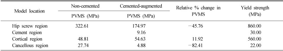
Figure & Data
REFERENCES
Citations
Citations to this article as recorded by 
- Safety and Effectiveness of the Anchor Augmentation with Bone Cement on Osteoporotic Femoral Fracture: A Systematic Reviews
So Young Kim
Journal of the Korean Fracture Society.2019; 32(2): 89. CrossRef
 Cite
CiteTreatment of Failed Intertrochanteric Fractures to Maintain the Reduction in Elderly Patients
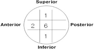
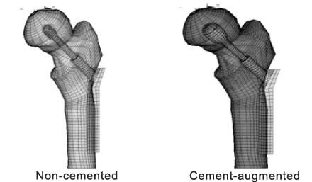
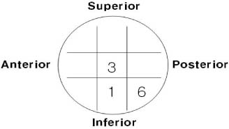
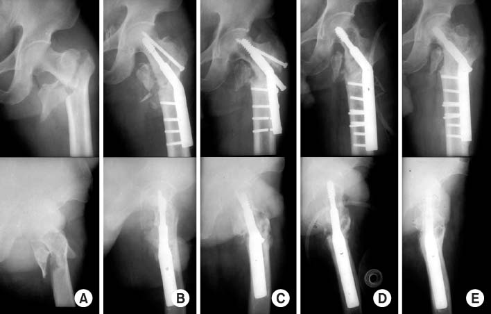
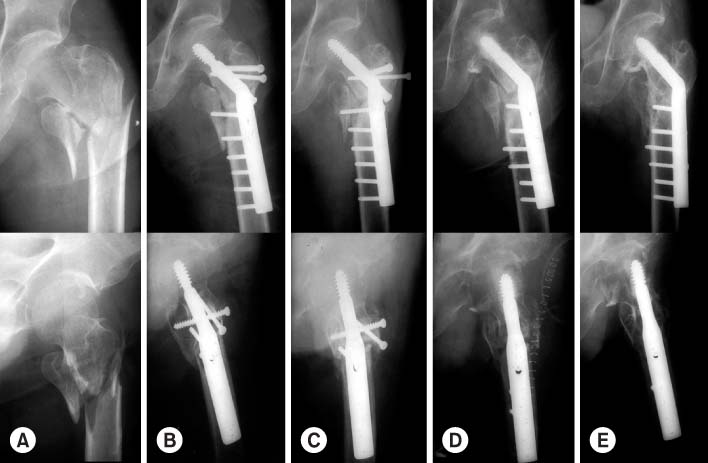
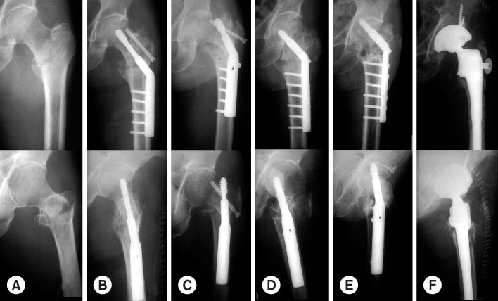
Fig. 1
The position of lag screw on femoral head at premetallic failure.
Fig. 2
Finite Element Method (FEM) model.
Fig. 3
The position of lag screw on femoral head after reoperation.
Fig. 4
(A) Initial radiograph of 55 years old man shows the unstable femur intertrochanteric comminuted fracture (Evans type II).
(B) Immediate postoperative radiograph shows a good compression hip screw fixation.
(C) Cortical anchorage failure and reduction loss in varus position after 11 months later.
(D) 2nd postoperative state with cement augmented fixation again.
(E) Complete union was achieved 2 years 7 months later.
Fig. 5
AP & lateral radiographs of 63 year old male patient show (A) unstable intertrochanteric fracture, (B) postoperative state with compression hip screw, (C) nonunion state at 1 year, (D) 2nd postoperative state with cement augmented fixation, and (E) achievement of complete union at 2 years and 6 months later.
Fig. 6
AP & lateral radiographs of 74 year old male patient show (A) unstable intertrochanteric fracture, (B) postoperative X-ray with compression hip screw, (C) superior cut-out at 3 months, (D) 2nd postoperative X-rays with cement augmented fixation again, (E) screw penetration at 1 year 7 months later, and (F) 3rd postoperative X-ray with total hip replacement arthroplasty.
Fig. 1
Fig. 2
Fig. 3
Fig. 4
Fig. 5
Fig. 6
Treatment of Failed Intertrochanteric Fractures to Maintain the Reduction in Elderly Patients
Comparison of PVMS* for non-cemented and cement-augmented cases
*PVMS (MPa): peak-Von Mises stress (Mega Pascal).
Table 1
Comparison of PVMS* for non-cemented and cement-augmented cases
*PVMS (MPa): peak-Von Mises stress (Mega Pascal).

