 E-submission
 TOTA
TOTA TOTS
TOTS
Articles
- Page Path
- HOME > J Musculoskelet Trauma > Volume 24(3); 2011 > Article
-
Original Article
- Treatment of the Intertrochanteric Femoral Fracture with Proximal Femoral Nail: Nailing Using the Provisional K-wire Fixation
- Gu-Hee Jung, M.D.
-
Journal of the Korean Fracture Society 2011;24(3):223-229.
DOI: https://doi.org/10.12671/jkfs.2011.24.3.223
Published online: July 15, 2011
Department of Orthopedic Surgery, Kosin University Gospel Hospital, Busan, Korea.
- Address reprint requests to: Gu-Hee Jung, M.D. Department of Orthopaedic Surgery, Kosin University Gospel Hospital, 34, Amnam-dong, Seo-gu, Busan 602-030, Korea. Tel: 82-51-990-6785, Fax: 82-51-243-0181, jyujin2001@kosin.ac.kr
• Received: September 3, 2010 • Revised: December 7, 2010 • Accepted: April 11, 2011
Copyright © 2011 The Korean Fracture Society
- 1,406 Views
- 6 Download
- 2 Crossref

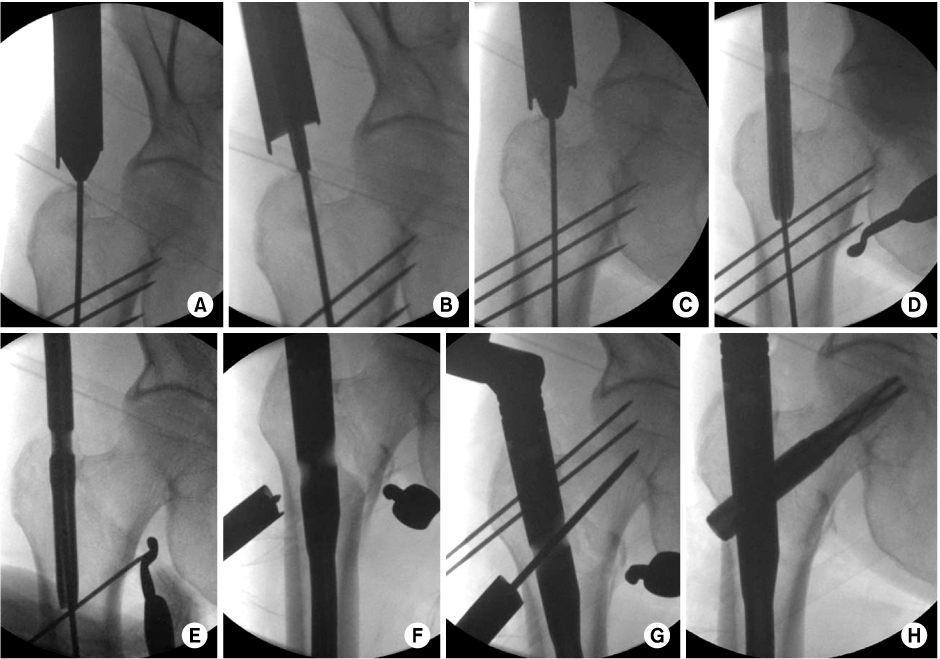


Figure & Data
REFERENCES
Citations
Citations to this article as recorded by 
- Analysis of Low-Energy Trochanter Fracture Using the Multiplanar Computed Tomography Image: Application for Intramedullary Nail Fixation
Gu-Hee Jung, Sung-Keun Heo, Hyun-Je Seo
Journal of the Korean Fracture Society.2015; 28(3): 155. CrossRef - Morbidity and Mortality of the Elderly after Early Operation for Trochanteric Fractures
Se-Ang Jang, Young-Ho Cho, Young-Soo Byun, Ki-Hong Park, Hyun-Seong Yoo, Chul Jung
Journal of the Korean Fracture Society.2013; 26(3): 199. CrossRef
 Cite
CiteTreatment of the Intertrochanteric Femoral Fracture with Proximal Femoral Nail: Nailing Using the Provisional K-wire Fixation
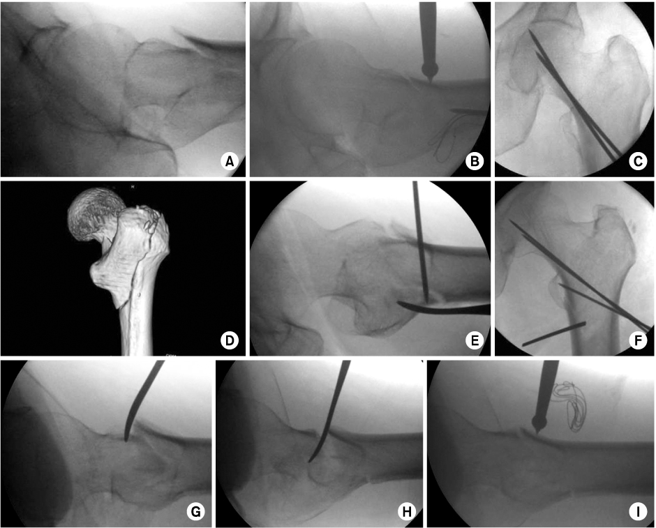

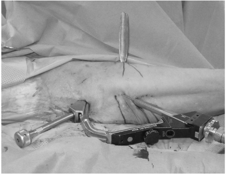
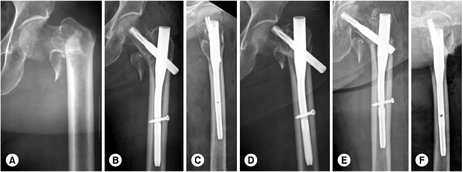
Fig. 1
Percutaneous reduction technique.
(A~C) The medial anatomy was reduced directly by pushing the proximal fragment using the ball-spike instrument.
(D, E) A fracture of greater trochanter was percutaneously reduced and fixed with K-wire.
(F) The intertrochatneric fracture was provisionally fixed with K-wires.
(G~I) The overlap of the head and neck fragment from the shaft was disimpacted using the Langenbeck elevator.
Fig. 2
Surgical Technique.
(A~C) After percutaneous reduction and provisional K-wires fixation, the reamer for lag screw was firstly inserted to make the entry portal and then, reamer for nail.
(D, E) the K-wire was removed during nail insertion.
(F, G) After nail insertion, the reduction was lost and provisionally fixed with K-wire for reduction.
(H) The lag screw was inserted in deep and central area of femoral head.
Fig. 3
Clinical photo revealed the nail insertion through minimal incision after provisional K-wire fixation and percutaneous reduction.
Fig. 4
(A) Initial radiographs showed the transverse intertrochanteric fracture.
(B, C) The postoperative radiographs showed a good reduction and accurate point of nail insertion but the lag screw was not located in deep area of the femoral head (TAD=37.2 mm).
(D) After 7 days, the lag screw was penetrated the femoral head and the fracture was collapsed.
(E, F) the lag screw was revised.
Fig. 1
Fig. 2
Fig. 3
Fig. 4
Treatment of the Intertrochanteric Femoral Fracture with Proximal Femoral Nail: Nailing Using the Provisional K-wire Fixation

