 E-submission
 TOTA
TOTA TOTS
TOTS
Search
- Page Path
- HOME > Search
Original Articles
- Computational simulation of coracoclavicular screw insertion through the superior distal clavicular plate for clinical applications in Korean cadavers
- Hyung-Lae Cho, Ji Han Choi, Se-Lin Jeong, Gu-Hee Jung
- J Musculoskelet Trauma 2025;38(3):143-151. Published online July 22, 2025
- DOI: https://doi.org/10.12671/jmt.2025.00122
-
 Abstract
 PDF
PDF - Background
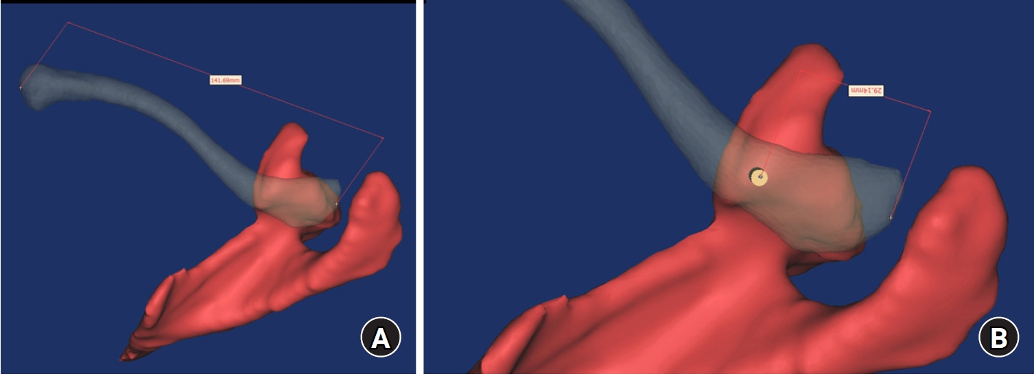
The study was conducted to determine the practical area for inserting the coracoclavicular (CC) screw through the plate by analyzing three-dimensional (3D) shoulder models featuring virtually implanted, actual-size plates and screws.
Methods
Ninety cadaveric shoulders (41 males and 49 females) underwent continuous 1.0-mm slice computed tomography scans. The data were imported into image-processing software to generate a 3D shoulder model, including the scapula and clavicle. The overlapping area between the clavicle and the horizontal portion of the coracoid process (horizontal portion_CP) was analyzed in the cranial view. A curved pelvic recon plate was virtually placed on the upper surface of the distal clavicle, and an actual-size (3.5 mm) CC screw was inserted through the plate.
Results
The distal clavicle directly overlapped with the horizontal portion_CP in the vertical direction. The overlapping area was sufficient to place the 3.5 mm and 4.5 mm-sized screws. In all shoulder models, the CC screw could be inserted through the plate into the vertical direction, with an average length of 35.5 mm (range, 26.2–62.5 mm; standard deviation, 1.2 mm). In 87 models, the CC screw was inserted through the third hole from the lateral end of the plate. Two models were inserted through the second hole, and one model through the fourth hole.
Conclusions
The upper surface of the clavicle has sufficient overlapping area to place CC screws through the plate in the vertical direction in the corresponding hole. Supplemental CC screw fixation through the plate can be performed without additional or special equipment. Level of evidence: IV
- 1,253 View
- 26 Download

- Three-Dimensional Analysis of the Morphological Features in the Femur of Atypical Fracture and Practical Implications of Intramedullary Nailing
- Yong Uk Kwon, Kyung-Jae Lee, Joo Young Choi, Gu-Hee Jung
- J Korean Fract Soc 2020;33(2):87-95. Published online April 30, 2020
- DOI: https://doi.org/10.12671/jkfs.2020.33.2.87
-
Abstract
PDF
- Purpose
This study analyzed the morphological features of the contralateral femur without an atypical fracture by constructing a three-dimensional model with an actual size medullary canal.
Materials and Methods
Lateral and anterior bowing of the shaft were measured for 21 models, and the shape of the medullary canal was analyzed. To eliminate the projection error, the anteroposterior (AP) femur was rotated internally to the extent that the centerline of the head and neck, which is the ideal position of cephalomedullary nail screw, was neutral, and the lateral femur matched the medial and lateral condyle exactly.
Results
The lateral bowing and anterior bowing was an average of 5.5° (range, 2.8°-10.7°; standard deviation [SD], 2.4°) and 13.1° (range, 6.2°-21.4°; SD, 3.2°), respectively. In the area where lateral bowing increased, the lateral cortex became thicker, and the medullary canal was straightened. On the lateral femur, the anterior angle was increased significantly, and the diameter of curvature averaged 1,370.2 mm (range, 896-1,996 mm; SD, 249.5 mm).
Conclusion
Even if the anterolateral bowing increases in the atypical femur, the medullary canal tends to be straightened in the AP direction. So, it might be considered as a reference to the modification of an intramedullary nail to increase the conformity.
- 840 View
- 6 Download
- Three-Dimensional Computed Tomography of Acetabular Fractures
- Poong Taek Kim, Joo Chul Ihn, Chang Wug Oh, Seung Hoon Oh
- J Korean Soc Fract 2000;13(1):46-51. Published online January 31, 2000
- DOI: https://doi.org/10.12671/jksf.2000.13.1.46
-
Abstract
PDF
- PURPOSE
In the evaluation of acetebular fractures, conventional radiography is limited by distortion, magnification, and overlap of fracture fragments. Computed tomography(CT) has already been shown to be superior in this field. The purpose of this paper was to use 3D reformations for classification of acetabular fractures and planning of operation.
MATERIALS AND METHODS
From July 1994 to December 1998, we reviewed 40 acetabular fractures. We evaluated fractures as plain X-ray(inlet & outlet view, AP view, obturator foramen & illiac wing view), axial CT with 3 mm slices, and 3D reformations. We classified fractures by classification of Letournel.
RESULTS
32 cases of 40 cases were displaced fractures, We recognized fracture easily in 3D reformations. 12 cases were posteior wall fracture. 9 cases were both column frctures. We interpretated both column fractures difficultly in plain X-ray, but we had many informations about rotation & displacement of fracture fragment by 3D reformations. Undisplaced fracture was 8 cases. We interpretated undisplaced fracture difficultly in 3D reformations and distinguished difficultly from normall 3D reformations.
CONCLUSION
3D reformations were useful for analysis of complex displaced fracture but not useful for analysis of undisplaced fracture. Acetabular internal oblique view was useful for analysis of quadrilateral space & posterior wall fractures. Acetabular external view was useful for decision of surgical approach.
- 747 View
- 0 Download
 First
First Prev
Prev

