
 E-submission
 KOTA
KOTA TOTA
TOTA TOTS
TOTS
Search
- Page Path
- HOME > Search
Review Article
- Locked plating in older patients with distal femur fractures: strategies to avoid complications
- Je-Hyun Yoo
- Received June 2, 2026 Accepted June 29, 2026 Published online August 24, 2026
- DOI: https://doi.org/10.12671/jmt.2026.00234 [Epub ahead of print]
-
 Abstract
 PDF
PDF - Distal femur fractures in older patients with osteoporosis are difficult to treat because poor bone quality reduces screw purchase and fixation stability. Minimally invasive locked plating with an angularly stable locking compression plate is widely used to obtain secure fixation in osteoporotic distal femur fractures and offers improved biomechanical performance. However, complications such as implant failure and peri-implant fracture continue to occur. This narrative review summarizes current principles of minimally invasive lateral locked plating for osteoporotic distal femur fractures in older patients and discusses practical strategies to reduce fixation-related complications based on the available literature and the author’s clinical experience.
- 392 View
- 3 Download

Original Articles
- Open reduction and internal fixation for distal humerus fractures in older adults: a retrospective comparative study by osteoporosis status
- Tong Joo Lee, Hee Beom Shin, Yongseok Lee
- J Musculoskelet Trauma 2026;39(3):227-235. Published online July 15, 2026
- DOI: https://doi.org/10.12671/jmt.2026.00101
-
Abstract
PDF
- Background
Distal humerus fractures in older patients, particularly those with osteoporosis, pose substantial treatment challenges because of increased fracture complexity and compromised bone stock. Open reduction and internal fixation (ORIF) is the preferred treatment but may be complicated by fixation failure. This study investigated the outcomes of ORIF in older osteoporotic and nonosteoporotic patients with complete articular distal humerus fractures.
Methods
This retrospective study included 19 patients with Arbeitsgemeinschaft für Osteosynthesefragen/ Orthopaedic Trauma Association (AO/OTA) 13C distal humerus fractures who underwent surgical treatment between 2012 and 2024. The mean patient age was 79.1 years. Patients were divided into osteoporotic (n=11) and nonosteoporotic (n=8) groups based on the lowest recorded dual-energy X-ray absorptiometry T-score at the femoral neck or lumbar spine. Osteoporosis was defined as a T-score of −2.5 or lower, and the nonosteoporotic group included patients with osteopenia. All fractures were treated with ORIF using bicolumnar plating. All included patients completed 12 months of clinical follow-up for visual analog scale (VAS) and Mayo Elbow Performance Score (MEPS) assessments. Radiographic follow-up was recorded separately and continued until union was confirmed; imaging follow-up extended to 12 months or longer in 14 patients and was limited to 6 months in five asymptomatic patients after confirmed union.
Results
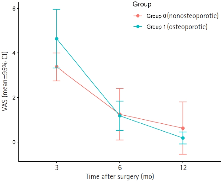
Both groups showed significant within-group improvement in pain, as assessed using the VAS, and function, as assessed using the MEPS, over time. Between-group comparisons at each follow-up time point showed no statistically significant differences in VAS or MEPS. No radiographic nonunion was observed during the available imaging follow- up, and complications were limited to one case of screw pullout/loosening and one case of postoperative stiffness.
Conclusions
ORIF provides reliable outcomes for older patients with complex distal humerus fractures, regardless of osteoporosis status, when stable reconstruction is achievable. These findings suggest that ORIF remains a viable treatment option, with satisfactory functional recovery and low complication rates in this population. Level of evidence: III.
- 645 View
- 18 Download
- Percutaneous Iliosacral Screw Fixation with Cement Augmentation in Osteoporotic Sacral Fracture
- Cheol hwan Kim, Young yool Chung, Seung woo Shim, Sung nyun Baek, Choong young Kim
- J Korean Fract Soc 2019;32(4):165-172. Published online October 31, 2019
- DOI: https://doi.org/10.12671/jkfs.2019.32.4.165
-
Abstract
PDF
- PURPOSE
The prevalence of osteoporotic sacral fractures is increasing. Traditionally, conservative treatment is the 1st option, but it can increase the risk of comorbidity in the elderly. To reduce the complications and allow early mobility, iliosacral screw fixation with cement augmentation will be one of the treatment options for patients with osteoporotic sacral fractures.
MATERIALS AND METHODS
This study reviewed 25 patients (30 cases) who had undergone percutaneous iliosacral screw fixation with cement augmentation for osteoporotic sacral fractures from July 2012 to December 2018 with a minimum follow up of six months. The clinical outcomes were assessed using the measures of pain (visual analogue scale [VAS] score), hospital stay and the date when weight-bearing started. All patients were evaluated radiologically for pull-out of screw, bone-union, and cement-leakage.
RESULTS
Bone union was achieved in 30 cases (100%). The mean duration of the hospital stay was 24 days (4–66 days); weight-bearing was performed on an average nine days after surgery. The VAS scores immediately (3.16) and three months after surgery (2.63) were lower than that of the preoperative VAS score (8.3) (p<0.05). No cases of cement-leakage or neurologic symptoms were encountered. Two patients (6.7%) experienced a pulling-out of the screw, but bone-union was accomplished without any additional procedures.
CONCLUSION
Percutaneous iliosacral fixation with cement augmentation will be an appropriate and safe surgical option for osteoporotic sacral fractures in the elderly in terms of early weight-bearing, pain reduction, and bone-union. -
Citations
Citations to this article as recorded by
- Role of Augmentation in the Fixation of Osteoporotic Fractures
Chinmoy Das, Partha Pratim Das
Indian Journal of Orthopaedics.2025; 59(3): 294. CrossRef
- Role of Augmentation in the Fixation of Osteoporotic Fractures
- 2,306 View
- 6 Download
- 1 Crossref
- Safety and Effectiveness of the Anchor Augmentation with Bone Cement on Osteoporotic Femoral Fracture: A Systematic Reviews
- So Young Kim
- J Korean Fract Soc 2019;32(2):89-96. Published online April 30, 2019
- DOI: https://doi.org/10.12671/jkfs.2019.32.2.89
-
Abstract
PDF
- PURPOSE
This paper reviewed the safety and effectiveness of anchor augmentation with bone cement in osteoporotic femoral fractures.
MATERIALS AND METHODS
A systematic review was conducted by searching multiple databases including five Korean databases, Ovid-MEDLINE, Ovid-EMBASE, and Cochrane Library. Safety was assessed through the incidence of complication. The effectiveness was assessed through the failure rate of anchor fixation, improvement of function and radiological assessment (sliding distance of lag screw and cutout). The safety and effectiveness of anchor augmentation with bone cement were assessed by reviewing all articles reporting on the treatment. Two researchers carried out independently each stage from the literature search to data extraction. The tools of Scottish Intercollegiate Guidelines Networks were used to assess the quality of studies.
RESULTS
Six studies were considered eligible. The safety results revealed a small amount of cement leakage (1 case), but no other severe complications were encountered. Regarding the effectiveness, the failure rate of anchor fixation was 16.7% and the Harris's hip score showed no significant improvement. The sliding distance of the anchor was similar in the cement augmentation group and non-cement group but there was no cutout.
CONCLUSION
The results of the assessment suggest that the safety is acceptable, but further research will be needed to verify the effectiveness of the treatment.
- 1,472 View
- 5 Download
- Vertebral Recompression after Vertebroplasty or Kyphoplasty
- Deuk Soo Jun, Do Hyun Moon, Young Kyu Ko, Jang Seok Choi, Byoung Keun An, Je Won Paik, Min Ho Park
- J Korean Fract Soc 2015;28(2):110-117. Published online April 30, 2015
- DOI: https://doi.org/10.12671/jkfs.2015.28.2.110
-
Abstract
PDF
- PURPOSE
The purpose of this study was to examine incidence of recompression and risk factors in the patients with osteoporotic vertebral compression fracture (OVCF) after vertebroplasty or kyphoplasty.
MATERIALS AND METHODS
This study was conducted on 179 vertebral bodies of 126 patients who underwent vertebroplasty or kyphoplasty on OVCF from January 2004 to August 2013.
RESULTS
When anterior vertebral height of fractured vertebrae declined by more than 3 mm from the height immediately after vertebroplasty or kyphoplasty, it was judged that recompression had occurred. Recompression was observed in a total of 58 vertebrae (32.4%). Recompression occurrences were found to be decreasing significantly when fractured vertebrae were the thoracic spine. In addition, osteonecrosis occurred in the preoperative vertebrae and restoration degree of anterior vertebral height immediately after vertebroplasty or kyphoplasty affected recompression occurrences significantly. The other factors (age, sex, bone mineral density, steroid medication history, follow-up duration, cement volume, vertebroplasty or kyphoplasty, and approach method) were compared, but no statistical significance was found.
CONCLUSION
The risk of vertebral recompression is more common, especially when osteonecrosis occurred in preoperative vertebrae or when vertebroplasty or kyphoplasty achieved remarkable restoration of anterior vertebra height. When performing vertebroplasty or kyphoplasty, such conditions should be considered carefully. -
Citations
Citations to this article as recorded by- Effect of Weekly Teriparatide Administration Followed by Percutaneous Balloon Kyphoplasty on Post-Menopausal Osteoporotic Compression Fracture Treatment
Sung-Ha Hong, Seung-Pyo Suh, Woo Jin Shin, Seung Gi Lee, Byung Jun Kang
Journal of the Korean Orthopaedic Association.2022; 57(1): 35. CrossRef - Analysis of the Cement Distribution Pattern and Other Risk Factors that Affect the Incidence of Recompression Fractures of Vertebral Bodies after Vertebroplasty or Kyphoplasty
Deuk Soo Jun, Jong Min Baik, Young Hyun Yoon
Journal of the Korean Orthopaedic Association.2022; 57(3): 204. CrossRef - Clinical Characteristics of Elderly People with Osteoporotic Vertebral Compression Fracture Based on a 12-Year Single-Center Experience in Korea
Seung-Kwan Lee, Deuk-Soo Jun, Dong-Keun Lee, Jong-Min Baik
Geriatrics.2022; 7(6): 123. CrossRef - Risk Factors for Recollapse of the Augmented Vertebrae After Percutaneous Vertebral Augmentation: A Systematic Review and Meta-Analysis
Weibo Yu, Weixing Xu, Xiaobing Jiang, De Liang, Wang Jian
World Neurosurgery.2018; 111: 119. CrossRef
- Effect of Weekly Teriparatide Administration Followed by Percutaneous Balloon Kyphoplasty on Post-Menopausal Osteoporotic Compression Fracture Treatment
- 1,533 View
- 2 Download
- 4 Crossref
- Surgical Fitness for Trochanteric Fracture in Elderly: Prospective Study
- Gu Hee Jung, Jong Seo Lee, Sung Gun Heo, Jae Do Kim, Hyun Ik Cho
- J Korean Fract Soc 2014;27(4):261-266. Published online October 31, 2014
- DOI: https://doi.org/10.12671/jkfs.2014.27.4.261
-
Abstract
PDF
- PURPOSE
The purpose of this study was to evaluate the risks of undergoing intramedullary nailing with minimum surgical optimization (fast-track) for geriatric trochanter fracture due to fall from a standing height.
MATERIALS AND METHODS
From May 2006 to August 2013, 48 fractures were enrolled in fast-track, and were an average age of patients was 77.6 years (range, 62-97 years). They underwent primary testing for anesthesia, including basic body fluid test, arterial blood, electrocardiography, and chest radiographs. The time from visit to surgery was 28.9 hours (range, 1-96 hours).
RESULTS
During hospitalization, there was one case of stress-induced cardiac arrest; however, other complications, infection, and 30-day mortality did not occur. According to preoperative classic test, the average albumin was 3.45 g/dl, blood sugar, 169 mg/dl, blood urea nitrogen, 20.5 mg/dl, Cr, 1.5 mg/dl, Na, 135.3 mEq/L, and K, 4.21 mEq/L. The average PaCO2 of arterial blood was 37.6 mmHg.
CONCLUSION
We found that the fast-track for trochanteric fracture due to slip-down was relatively safe, and could be considered as a therapeutic approach.
- 1,000 View
- 3 Download
 First
First Prev
Prev


