 E-submission
 TOTA
TOTA TOTS
TOTS
Search
- Page Path
- HOME > Search
Original Article
- Clinical and radiographic outcomes of elastic stable intramedullary nailing for pediatric humeral shaft fractures: a retrospective case series
- Kang-San Lee, Dongju Shin, Sang Hee Kim, Il Seo, Tae-Hoon Kim, Sung Jung Kim
- J Musculoskelet Trauma 2026;39(2):156-161. Published online March 10, 2026
- DOI: https://doi.org/10.12671/jmt.2025.00381
-
 Abstract
 PDF
PDF - Background
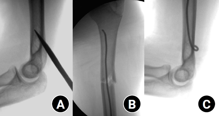
Pediatric humeral shaft fractures are uncommon and are generally treated conservatively, with satisfactory clinical outcomes reported in most cases. However, conservative management often necessitates prolonged immobilization and frequent outpatient follow-up visits, and it carries an inherent risk of residual angular or translational deformity. Elastic stable intramedullary nailing (ESIN) provides a simple and minimally invasive method of fracture fixation that offers adequate stability without disrupting the periosteal blood supply, thereby permitting early mobilization and promoting rapid bone union. The purpose of this study was to evaluate the clinical and radiological outcomes of ESIN fixation in pediatric patients with humeral shaft fractures.
Methods
The medical records of pediatric patients with humeral shaft fractures who underwent ESIN fixation between January 2015 and November 2025 were retrospectively reviewed. Data collected included patient demographics, mechanism of injury, fracture location, number of elastic nails used, time to union, degree of residual angulation, range of motion (ROM), and postoperative complications.
Results
The mean age of the patients was 10.0 years (range, 7 to 15 years). The mean time to radiographic union was 5.4 weeks (range, 2.4 to 10.4 weeks). The mean coronal angulation was 0.2° (range, −9.1° to 5.8°), while the mean sagittal angulation was −1.3° (range, −6.9° to 5.3°). No cases of infection, nerve injury, or nail migration were observed during the follow-up period. At the final follow-up assessment, all patients demonstrated full shoulder and elbow ROM, with no residual deformity or pain reported.
Conclusions
In this small retrospective case series, ESIN fixation resulted in favorable union rates and excellent functional outcomes in pediatric humeral shaft fractures. Level of evidence: IV.
- 480 View
- 19 Download

Case Report
- Snapping Metacarpo-Phalangeal Joint after Depressed Fracture of Metacarpal Neck: Case Report
- Hyun Seok Song, Nam Yong Choi, Sung Jin Park, Suk Ku Han, Ki Ho Nah, Sang Il Seo, Do Sung Lee
- J Korean Fract Soc 2004;17(4):359-361. Published online October 31, 2004
- DOI: https://doi.org/10.12671/jkfs.2004.17.4.359
-
Abstract
PDF
- We report one case of snapping metacarpo-phalangeal joint after depressed fracture of metacarpal neck which could be diagnosed by exploration for the snapping during extension in spite of conservative treatments.
- 539 View
- 1 Download
Original Articles
- Treatment of the Intertrochanteric Fractures of the Femur in Elderly Patients: Comparision of Wayne-County Reduction and Anatomical Reduction
- Nam Yong Choi, Kee Ho Nah, Hyun Seok Song, Sang Il Seo, Jung Keun Choi, Suk Ku Han
- J Korean Fract Soc 2004;17(4):301-307. Published online October 31, 2004
- DOI: https://doi.org/10.12671/jkfs.2004.17.4.301
-
Abstract
PDF
- PURPOSE
To compare the radiological and clinical results of Wayne-County reduction with anatomical reduction in treatment of the intertrochanteric fractures of the femur in elderly patients.
MATERIALS AND METHODS
Among one hundred-three of intertrochanteric fractures treated with 135- degree angled compression hip scresw, eighty three cases treated by Wayne-County reduction (Group 1, 42 cases) and anatomical reduction (Group 2, 41 cases) with at least 1 year follow-up were reviewed. The average pateint ages were 72.4 (65~92) in group 1, 71.6 (65~89) in group 2, respectively. 33 cases (75.2%) in group 1 and 31 cases (77.5%) displayed unstable fractures by Jensen classification. The radiological observation was included neck-shaft angle, penetrating length of lag screw into head, sliding length of lag screw and time of bony union. The clinical results were evaluated by Koval criteria, Kyle's functional evaluation, leg length inequality and complications.
RESULTS
There were no significant changes between group 1 and group 2 in stable fractures in the radiological and clinical results. In unstable fractures, the neck-shaft angle averaged 132.2 degree in group 1 and 129.4 degree in group 2 in the final follow-up films. The penetrating length of lag screw into head were 2.2 mm in group 1 and 3.1 mm in group 2 (p<005). But there were little differences in the sliding length of lag screw, the time of bony union and complication rates between groups. In post- operative evaluation of walking abilility by Koval, 31 patients (73.8%) in group 1 and 28 (68.3%) recovered the activity level before injury by the postoperative 1 year follow-up. Leg length discrepancy at final follow-up was 4.1+/-6 mm shortening in group 1 and 6.5+/-8 mm in group 2, respectively.
CONCLUSION
Both Wayne-County reduction and anatomical reduction had a favorable results after treatment of stable intertrochanteric fractures of the femur, but Wayne-County reduction may be a better method in treatment of unstable fractures, especially in elderly patients, in which it is difficult to obtain anatomical reduction. -
Citations
Citations to this article as recorded by
- High Fixation Failure Rate of Cephalomedullary Nail Fixation in Patients with Low-Energy Basicervical Femoral Fractures: Do We Need Extramedullary Reduction?
Chang-Jin Yon, Ki-Cheor Bae, Young-Hun Kim, Kyung-Jae Lee
Medicina.2025; 61(1): 112. CrossRef - New Approach in the Treatment of Intertrochanteric Fracture Using a Cephalomedullary Nail
Junyoung Kim, Kihong Choi, Kyu Hyun Yang
Journal of the Korean Orthopaedic Association.2020; 55(3): 193. CrossRef - The Effect of Valgus Reduction on the Position of the Blade of the Proximal Femoral Nail Antirotation in Intertrochanteric Hip Fractures
Eui Yub Jung, In Taek Oh, Sang Yeup Shim, Byung Ho Yoon, Yerl Bo Sung
Clinics in Orthopedic Surgery.2019; 11(1): 36. CrossRef - Effectiveness of the Valgus Reduction Technique in Treatment of Intertrochanteric Fractures Using Proximal Femoral Nail Antirotation
Ji-Kang Park, Hyun-Chul Shon, Yong-Min Kim, Eui-Sung Choi, Dong-Soo Kim, Kyoung-Jin Park, Byung-Ki Cho, Jung-Kwon Cha, Sang-Woo Kang
Journal of the Korean Orthopaedic Association.2013; 48(6): 441. CrossRef
- High Fixation Failure Rate of Cephalomedullary Nail Fixation in Patients with Low-Energy Basicervical Femoral Fractures: Do We Need Extramedullary Reduction?
- 918 View
- 7 Download
- 4 Crossref
- The Effect of Platelet-Rich Plasma (PRP) on the Healing of Allograft for the Treatment of Segmental Bone Defect of the Ulna in Rabbits
- Joo Hyun Song, Jinyoung Jeong, Yong Koo Kang, Han Yong Lee, Mun Ik Son, Sang Il Seo
- J Korean Soc Fract 2003;16(3):416-422. Published online July 31, 2003
- DOI: https://doi.org/10.12671/jksf.2003.16.3.416
-
Abstract
PDF
- PURPOSE
The purpose of this study was to evaluate the effect of Platelet-Rich Plasma (PRP) on the healing of the allograft for the treatment of the segmental bone defect of the ulna in Rabbits.
MATERIALS AND METHODS
About 2 cm-sized segmental bone defects were created on both ulna of twenty rabbits. The rabbits were divided into two groups, even and odd number group after numbering them from 1 to 20. The segmental bone from the odd numbered animal was transplanted to the even numbered animal, and the even numbered to the odd numbered. The left side of the ulna of the animal is grafted with a segmental allograft only. The right side of the ulna was grafted with a segmental allograft and 0.7 cc of PRP. Radiographs obtained at 0, 4, 8, and 12 weeks postoperatively were graded for radiologic union.
RESULTS
The use of the combination of PRP and segmental allograft demonstrated improved healing on radiolographic study compared with that demonstrated after use of allogrft alone.
CONCLUSION
The results of the study suggests that the use of the combination of PRP and segmental allograft can be considered as an alternative method to manage the segmental defect of the long bone. -
Citations
Citations to this article as recorded by- Effect of Different Bone Substitutes on the Concentration of Growth Factors in Platelet-rich Plasma
Hee Soon Cho, So-Young Park, Sukyoung Kim, Sang Keun Bae, Duk Seop Shin, Myun-Whan Ahn
Journal of Biomaterials Applications.2008; 22(6): 545. CrossRef
- Effect of Different Bone Substitutes on the Concentration of Growth Factors in Platelet-rich Plasma
- 900 View
- 0 Download
- 1 Crossref
- Avulsion Fracture of The Medial Meniscus: A Case Report
- Hyoung Soo Kim, Seung Rim Park, Joon Soon Kang, Woo Hyeong Lee, Kil Seok Ko
- J Korean Soc Fract 2000;13(1):109-112. Published online January 31, 2000
- DOI: https://doi.org/10.12671/jksf.2000.13.1.109
-
Abstract
PDF
- Post-traumatic meniscal ossicle due to avulsion fracture of medial meniscus was very rare. They were often associated with meniscal tear, but caused symptoms without a tear, by mass effect from protruding meniscal contour. so it had to be differential diagnosised with free loose body in the knee joint. We experienced a symptomatic meniscal ossicle due to post-traumatic avulsion fracture of the posterior horn of medial meniscus, and managed with open reduction, internal fixation with screw and washer after arthroscopic examination. We report a rare case of meniscal ossicle in detail with literature
- 496 View
- 0 Download
- External Fixation and Limited Internal Fixation in AO Type C Pilon Fracture: Report of Five Cases
- Eun Woo Lee, Soo Yong Kang, Il Seok Kim
- J Korean Soc Fract 1995;8(1):243-247. Published online January 31, 1995
- DOI: https://doi.org/10.12671/jksf.1995.8.1.243
-
Abstract
PDF
- Techniaue of biologic fixation and external fifation are playing an crucial role in the management of the severe comminuted fracture with soft tissue injuries. To evaluate the treatment of severe pilon fracture by a conbination internal and external fixation, five high Pilon fractures with open or severs soft tissue injries were treated by a medial external fixator with an articulated ankle hinge(EBI) and limited internal flxation. Two AO C2 fractures and three AO C3 fractures were followed for a minimum of 1 year. All fractures united and had good functional results without any serious complication. We believe that external fixation and limited internal fixation using biologic principle is an excellent alternative method in high energy, complex fracture with diaphyseal comminution.
- 517 View
- 1 Download
 First
First Prev
Prev

