 E-submission
 TOTA
TOTA TOTS
TOTS
Search
- Page Path
- HOME > Search
Original Article
- Association between decreased bone mineral density and Pauwels angle in femoral neck fractures: a cross-sectional study
- Soo-Hwan Jung, Yong-Uk Kwon, Ji-Hun Park
- J Musculoskelet Trauma 2026;39(1):20-29. Published online January 25, 2026
- DOI: https://doi.org/10.12671/jmt.2025.00269
-
 Abstract
 PDF
PDF Supplementary Material
 - Background
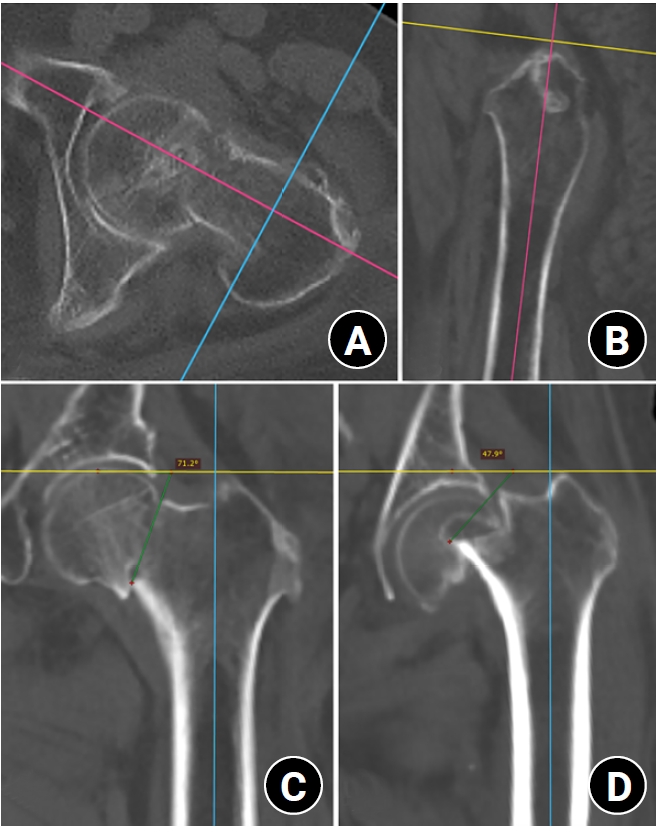
Progressive osteoporosis reduces the trabecular structures of the proximal femur, whereas the primary compression trabeculae (PCTs) are relatively preserved. We hypothesize that the loss of the vertically oriented PCTs in osteoporosis, which act as a mechanical barrier, affects fracture line propagation and influences the Pauwels angle. This study investigated the association between bone mineral density (BMD) and Pauwels angles in low-energy femoral neck fractures (FNFs).
Methods
This cross-sectional study included 150 patients (mean age, 75.3 years; range, 50–94 years) diagnosed with intracapsular FNFs between May 2019 and May 2023. BMD was measured within 1 month of the injury date using dual-energy X-ray absorptiometry, and modified Pauwels angles were assessed using a computed tomography-based multiplanar reconstruction program. Multiple linear regression analysis was performed to evaluate the factors influencing the Pauwels angles. The dependent variable was the Pauwels angle, while the independent variables included sex, age, height, body weight, body mass index, American Society of Anesthesiologists score, Charlson comorbidity index score, smoking status, alcohol use, preinjury walking ability, and femoral neck BMD T-scores.
Results
Higher femoral neck BMD T-scores were significantly associated with increased Pauwels angles (β=3.449, P<0.001). Greater body weight was independently associated with increased Pauwels angles (β=0.213, P=0.007).
Conclusions
The Pauwels angle demonstrated a significant association with BMD, with lower BMD associated with less steep Pauwels angles. In the absence of BMD measurement, the Pauwels angle may indicate osteoporosis severity in patients with low-energy FNFs. Level of evidence: III.
- 909 View
- 23 Download

Review Article
- Current Concepts in Management of Phalangeal Fractures
- Yohan Lee, Sunghun Park, Jun-Ku Lee
- J Korean Fract Soc 2022;35(4):169-181. Published online October 31, 2022
- DOI: https://doi.org/10.12671/jkfs.2022.35.4.169
-
Abstract
PDF
- This review focused on the research published to date on the treatment of phalangeal fractures according to the anatomical location of the finger bones, excluding the thumb. In many finger fracture cases, conservative treatment should be prioritized over surgical treatment. The three determinants of surgical treatment are the presence of an intra-articular fracture, the stability of the fracture itself, and the degree of damage to the surrounding soft tissues. Surgical treatment is recommended when bone fragments of 3 mm or more and distal phalanx subluxation are present in the bony mallet finger, and the main surgical treatment is closed reduction and extension block pin fixation. It is essential to pay attention to rotational deformation asf ractures occur proximally. Since intra-articular fractures can cause stiffness and arthritis in the future, a computed tomography scan is recommended to confirm the fracture pattern. These fractures require anatomical reduction of the bone fragments within the joint, and the instability of the joint itself must be corrected. There are no superior surgical treatment methods. It is therefore advantageous for the surgeon to select a surgical method that he is familiar with and confident of performing, considering the fracture itself and various patient-related clinical factors. Nonunion is rare as a complication of a finger fracture, and finger stiffness is the most common complication. Ensuring rapid joint movement as soon as possible can reduce finger stiffness.
- 1,347 View
- 26 Download
Original Articles
- Treatment of the Femoral Fracture Using Sirus(R) Nail: A Comparison of Complication according to the Entry Potal
- Young Yool Chung, Dong Hyuk Choi, Dae Hyun Yoon, Jung Ho Lee, Ji Hun Park
- J Korean Fract Soc 2015;28(2):103-109. Published online April 30, 2015
- DOI: https://doi.org/10.12671/jkfs.2015.28.2.103
-
Abstract
PDF
- PURPOSE
The purpose of this study is to analyze the clinical results of fixation using Sirus(R) nail in patients with femoral subtrochanteric and shaft fracture and the difference in the frequency of complications according to the entry portal.
MATERIALS AND METHODS
From July 2006 to August 2013, at least 1-year clinical follow-up, we retrospectively analyzed 36 cases with femoral subtrochanteric (15 cases) and shaft fracture (21 cases) who underwent surgery using Sirus(R) nail. We reviewed the records of operation time, intra-operative amounts of bleeding and complications. At last follow-up, we reviewed clinical results by Ray-Sanders criteria and analyzed the periods of bone union on the radiograph. We also measured changing of the femoral neck-shaft angle in the subtrochanteric fractures and angulation in the shaft fractures, respectively. Considering anatomical variation of the trochanter and fracture position of subtrochanteric and femoral shaft, entry points were divided into subgroups, and the clinical results were compared.
RESULTS
The mean Ray-Sanders score was 27.4, 27 cases (75.0%) were good or excellent. The mean periods of bone union was 21.1 weeks in 31 cases. The mean neck-shaft angles were 135.7o preoperatively, 130.2o postoperatively. The mean angulation of the femur was 24.4o preoperatively, 2.4o postoperatively in patients of femoral shaft fractures. Despite no statistical significance, greater trochanter tip entry point and lateral entry point had a higher rate of frequency than medial entry point, with respect to the occurrence of iatrogenic fracture and malalignment.
CONCLUSION
Using Sirus(R) nail for femoral subtrochanteric and shaft fractures showed good clinical and radiographic results and a high rate of union. Medial entry point yielded slightly better results in the occurrence of iatrogenic fracture and malalignment, compared to greater trochanter tip entry point and lateral entry point.
- 982 View
- 8 Download
- Injury Severity and Patterns of Accompanying Injury in Spinal Fracture
- Hun Park, Kyung Jin Song, Kwang Bok Lee, Joo Hyun Sim
- J Korean Fract Soc 2012;25(3):203-207. Published online July 31, 2012
- DOI: https://doi.org/10.12671/jkfs.2012.25.3.203
-
Abstract
PDF
- PURPOSE
To examine the relationship between injury severity and patterns of associated injury in spinal fracture.
MATERIALS AND METHODS
From March 2004 to March 2010, a retrospective study was conducted on 291 patients who had undergone surgeries due to spinal fractures. Spinal fractures were categorized as upper cervical, lower cervical, thoracic, thoracolumbar, and lumbar region, and the severity of fracture was measured using the Abbreviated Injury Scale and Injury Severity Score (ISS). We evaluated the correlation between the fracture site and the incidence and injury severity of the associated injury, and compared the neurologic damage according to the presence/absence of the associated injury.
RESULTS
Spinal fracture occurred in the thoracic (43.5%) and lower cervical (30.0%) levels, and associated injury developed in 134 patients (47%). The area of associated injury was in the extremity (41.2%), thorax (25.5%), head, neck, and face (21.9%). Lower cervical fracture (34.5%) had a lower prevalence than thoracic (81%) and lumbar fracture (61%). The average ISS of the associated injury was 17.14 for the thoracic fracture, 12.30 for the lower cervical fracture, 8.7 for the thoracolumbar fracture and 5.69 for the lumbar fracture. Neurologic damage was highly frequent in the lower cervical fracture and included 54 patients (62.1%) and was less frequent in the upper cervical fracture, which included 7 patients (17.9%) (p=0.032).
CONCLUSION
Although the associated injury was less frequent in the lower cervical spine among the spinal fractures that underwent surgical treatment, there was a high risk of neurologic damage in the case of associated injury; therefore, there is a need to pay special attention to patients that suffer damage in this area. In addition, since the degree of the associated injury in the thoracic and lower cervical fracture is significant, an appropriate management strategy for the associated injury must be considered. -
Citations
Citations to this article as recorded by
- The Clinical Effects of Complex Korean Medicine Treatment in Patients with Cervical Spine Fracture Caused by Traffic Accident: A Report of 2 Cases
Si-Hoon Han, Gi-Eon Lee, Kyeong-Sang Jo, Da-Young Byun, Min-Seok Oh
Journal of Korean Medicine Rehabilitation.2018; 28(2): 113. CrossRef - Clinical results of early stabilization of spine fractures in polytrauma patients
Ki-Chul Park, Ye-Soo Park, Wan-Sik Seo, Jun-Ki Moon, Bo-Hyun Kim
Journal of Critical Care.2014; 29(4): 694.e7. CrossRef
- The Clinical Effects of Complex Korean Medicine Treatment in Patients with Cervical Spine Fracture Caused by Traffic Accident: A Report of 2 Cases
- 1,003 View
- 2 Download
- 2 Crossref
Case Report
- Bilateral Open Transcalcaneal Fracture with Talonavicular Dislocation: A Case Report
- Hun Park, Sung Jin Shin, Sang Rim Kim, Kwang Woo Nam, Sung Wook Choi, Kyu Bum Seo, Jun Young Seo
- J Korean Fract Soc 2011;24(1):87-91. Published online January 31, 2011
- DOI: https://doi.org/10.12671/jkfs.2011.24.1.87
-
Abstract
PDF
- Although calcaneal fracture is relatively common in ankle injury, open intraarticular calcaneal fracture with dorsal dislocation of the navicular from talus is extremely rare and severe injury. There are few data which are available concerning the injury mechanism and treatment options. The purpose of this report is to describe a case with bilateral open transcalcaneal fracture with talonavicular dislocation and to discuss the prevalence, mechanism of this injury, and treatment options.
-
Citations
Citations to this article as recorded by- Results in Operative Treatment of Open Calcaneal Fracture
Ba Rom Kim, Jun Young Lee, Donghyuk Cha
Journal of Korean Foot and Ankle Society.2021; 25(3): 133. CrossRef
- Results in Operative Treatment of Open Calcaneal Fracture
- 904 View
- 2 Download
- 1 Crossref
Original Article
- Bilateral traumatic didlocation of the tibialis posterior tendons a case report
- Jin Young Kim, Chan Hee Park, Jong Who Kang, Jong Hun Park
- J Korean Soc Fract 1992;5(1):157-160. Published online May 31, 1992
- DOI: https://doi.org/10.12671/jksf.1992.5.1.157
- 590 View
- 0 Download
 First
First Prev
Prev

