 E-submission
 TOTA
TOTA TOTS
TOTS
Articles
- Page Path
- HOME > J Musculoskelet Trauma > Volume 20(4); 2007 > Article
-
Original Article
- Mid-term Results of Distal Tibial Fractures Treated with Ilizarov External Fixator
- Suk Kyu Choo, M.D., Kyung Wook Nha, M.D., Hyoung Keun Oh, M.D., Dong Bong Lee, M.D.
-
Journal of the Korean Fracture Society 2007;20(4):323-329.
DOI: https://doi.org/10.12671/jkfs.2007.20.4.323
Published online: June 14, 2016
Department of Orthopedic Surgery, Inje University, Ilsan Paik Hospital, Goyang, Korea.
- Address reprint requests to: Kyung Wook Nha, M.D. Department of Orthopedic Surgery, Ilsan Paik Hospital, Inje University, 2240, Daehwa-dong, Ilsanseo-gu, Goyang-si 411-706, Korea. Tel: 82-31-910-7968, Fax: 82-31-910-7967, kwnha@ilsanpaik.ac.kr
Copyright © The Korean Fracture Society. All rights reserved
- 1,117 Views
- 3 Download
Abstract
-
Purpose
- We analyed the mid-term results of distal tibial fractures treated with ilizarov external fixator and functional results according to delayed metaphyseal healing and fracture pattern.
-
Materials and Methods
- We reviewed 23 distal tibial fractures treated with ilizarov external fixator followed for minimum two year (mean 53 months). There were 10 A fractures, 2 B fractures, and 11 C fractures according to the AO classification. Radiographically, we analyzed bony union time according to translation of diaphyseal-metaphyseal fracture line and assessed arthritic score. Functional results was assessed with AOFAS score and analyzed according to delayed healing and fracture pattern.
-
Results
- Average union time was 21 weeks. Delayed healing of metaphyseal fracture line was associated translational displacement >3 mm (p=0.01). AOFAS scrore was averaged to 68 and there was no stastical significance between delayed metaphyseal healing and functional results (p=0.31). But, low AOFAS score and arthritis score was related to fracture type (p=0.02). In 11 C fractures, radiographic arthritic change were developed in 6 cases (55%).
-
Conclusion
- The main prognosis of distal tibial fractures depends on articular involvement and to shorten the external fixation time, metaphyseal fracture should be reduced within 3mm.
- 1. Bonar SK, Marsh JL. Tibia plafond fractures, changing principles of treatment. J Am Acad Orthop Surg, 1994;2:297-305.
- 2. Bone LB. Fractures of the tibial plafond. Orthop Clin North Am, 1987;18:95-104.Article
- 3. Borg T, Larsson S, Lindsjo U. Percutaneous plating of distal tibial fractures. Preliminary results in 21 patients. Injury, 2004;35:608-614.Article
- 4. Bradley GW, Mckenna GB, Dunn HK, Daniels AU, Statton WO. Effects of flexural rigidity of plates on bone healing. J Bone Joint Surg Am, 1979;61:866-872.Article
- 5. Chrisovitsinos JP, Xenakis T, Papakostides KG, Skaltsoyannis N, Grestas A, Soucacos PN. Bridge plating osteosynthesis of 20 comminuted fractures of the femur. Acta Orthop Scand Suppl, 1997;275:72-76.Article
- 6. Claes L., Krischak G, Braun H, Hierholzer G. Fixation technique influences osteogenesis of comminuted fractures. Clin Orthop Relat Res, 1999;365:221-229.Article
- 7. Crenshaw AH. Campbell's operative orthopaedics. Vol. 2:8th ed. St. Louse: Mosby-Year Book, Inc; 1992. p. 794-797.
- 8. Farouk O, Krettek C, Miclau T, Schandelmaier P, Tscherne H. The topography of the perforating vessels of the deep femoral artery. Clin Orthop Relat Res, 1999;368:255-259.Article
- 9. Green SA, Gibbs P. The relationship of angulation to translation in fracture deformities. J Bone Joint Surg Am, 1994;76:390-397.Article
- 10. Helfet DL, Suk M. Minimally invasive percutaneous plate osteosynthesis of fractures of the distal tibia. Instr Course Lect, 2004;53:471-475.
- 11. Ibrahim T, Beiri A, Azzabi M, Best AJ, Taylor GJ, Menon DK. Reliability and validity of the subjective component of the American orthopaedic foot and ankle society clinical rating scales. J Foot Ankle Surg, 2007;46:65-74.Article
- 12. Johner R, Wruhs O. Classification of tibial shaft fractures and correlation with results after rigid internal fixation. Clin Orthop Relat Res, 1983;178:7-25.Article
- 13. Karas EH, Weiner LS. Displaced pilon fracures. An update. Orthop Clin North Am, 1994;25:651-663.
- 14. Krackhardt T, Dilger J, Flesch I, Hontzsch D, Eingartner C, Weise K. Fractures of the distal tibia treated with closed reduction and minimally invasive plating. Arch Orthop Trauma Surg, 2005;125:87-94.ArticlePDF
- 15. Lee SC, Yoo MJ, Seo HS. Treatment of the distal metaphyseal fractures of tibia; comparison between internal fixation with a plate and screws and external fixation with Ilizarov device. J Korean Soc Fract, 2002;15:371-378.Article
- 16. Mast JW, Teipner WA. A reproducible approach to the internal fixation of adult ankle fractures: rational, technique and early results. Orthop Clin North Am, 1980;11:661-679.
- 17. McFerran MA, Smith SW, Boulas HJ, Schwartz HS. Complications encountered in the treatment of pilon fractures. J Orthop Trauma, 1992;6:195-200.Article
- 18. Mckibbin B. The biology of fracture healing in long bones. J Bone Joint Surg Br, 1978;60:150-162.ArticlePDF
- 19. Moon MS, Ha KY, Kim HG. The use of ender nails in distal tibial fractures. J Korean Orthop Assoc, 1990;25:61-68.ArticlePDF
- 20. Nicoll EA. Fractures of the tibial shaft. A surgery of 705 cases. J Bone Joint Surg Br, 1964;46:373-387.
- 21. Oh CW, Park BC, Ihn JC, Kim SJ, Kim HS, Lee SG. Ilizarov/Hybrid external fixation in the management of tibial plafond fractures. J Korean Soc Fract, 2000;13:244-251.Article
- 22. Ovadia DN, Beals RK. Fractures of the tibial plafond. J Bone Joint Surg Am, 1986;68:543-551.Article
- 23. Patterson MJ, Cole JD. Two-staged delayed open reduction and internal fixation of severe pilon fractures. J Orthop Trauma, 1999;13:85-91.Article
- 24. Pierce RO, Heinrich JH. Comminuted intraarticular fractures of the distal tibia. J Trauma, 1979;19:828-832.Article
- 25. Redfern DJ, Syed SU, Davies SJ. Fractures of the distal tibia: minimally invasive plate osteosynthesis. Injury, 2004;35:615-620.Article
- 26. Ristiniemi J, Flinkkilä T, Hyvönen P, et al. Two-ring hybrid external fixation of distal tibial fractures: a review of 47 cases. J Trauma, 2007;62:174-183.Article
- 27. Rockwood CA, Green DP. Fractures in adults. Vol. 2. Philadelphia: J.B. Lippincott Co; 1975.
- 28. Rozbruch SR, Muller U, Gautier E, Ganz R. The evolution of femoral shaft plating technique. Clin Orthop Relat Res, 1998;354:195-208.Article
- 29. Ruedi T. Fractures of the lower end of the tibia into the ankle-joint: results 9 years after open reduction and internal fixation. Injury, 1973;5:130-134.Article
- 30. Ruedi TP, Allogower M. The operative treatment of intraarticular fractures of the lower end of the tibia. Clin Orthop Relat Res, 1979;138:105-110.
- 31. Sarmiento A. Functional belew knee cast for tibial fracture. J Bone Joint Surg Am, 2004;86:2777.
- 32. Shtarker H, DAvid R, Stolero J, Grimberg B, Soudry M. Treatment of open tibial fractures with primary suture and Ilizarov fixation. Clin Orthop Relat Res, 1997;335:268-274.Article
- 33. Sirkin M, Sanders R, DiPasquale T, Herscovici D Jr. A staged protocol for soft tissue management in the treatment of complex pilon fractures. J Orthop Trauma, 1999;13:78-84.Article
- 34. Teeny SM, Wiss DA. Open reduction and internal fixation of tibial plafond fractures: variables contributing to poor results and complications. Clin Orthop Relat Res, 1993;292:108-117.
- 35. Toms AD, McMurtie A, Maffulli N. Percutaneous plating of the distal tibia. J Foot Ankle Surg, 2004;43:199-203.Article
- 36. Tornetta P, Weiner L, Bergman M, et al. Pilon fracturs: treatment with combined internal and external fixation. J Orthop Trauma, 1993;7:489-496.
- 37. Watson-Jones R. Fractures and Joint injuries. 6th ed. New York: Churchill Livingstone Co; 1982. p. 1130-1133.
- 38. Williams TM, Nepola JV, DeCoster TA, Hurwitz SR, Dirschl DR, Marsh JL. Factors affecting outcome in tibial plafond fractures. Clin Orthop Relat Res, 2004;423:93-98.Article
REFERENCES
Fig. 1Displacement of the diaphyseal-metaphyseal fracture line was measured by the method described by Green and Gibbs.

Displacement of the diaphyseal-metaphyseal fracture line was measured by the method described by Green and Gibbs. Translation m m = APtrans m m 2 + LATtrans m m 2 
Fig. 2
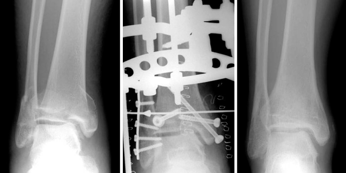
A 17-year-old male was suffered from right distal tibial fracture. He was treated with Ilizarov external fixator and minimal invasive screw fixation for intra-articular fracture. The last follow-up x-ray show complete union of fracture.
Fig. 3

This graph shows that residual translational displacement is closely related to delayed metaphyseal healing (p=0.01).
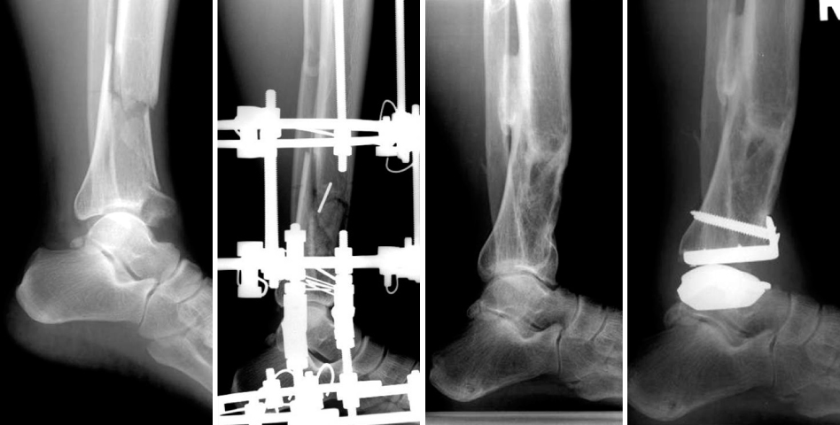
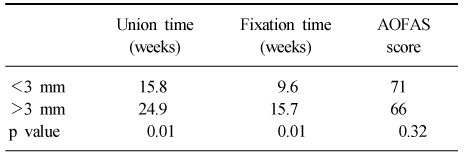
Figure & Data
REFERENCES
Citations
Citations to this article as recorded by 
 Cite
CiteMid-term Results of Distal Tibial Fractures Treated with Ilizarov External Fixator
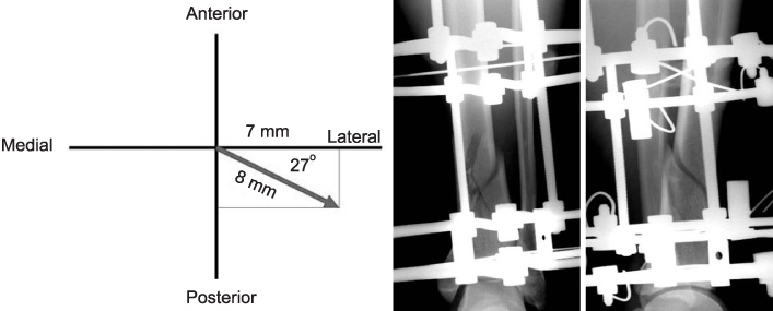

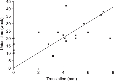

Fig. 1
Displacement of the diaphyseal-metaphyseal fracture line was measured by the method described by Green and Gibbs. Translationmm=APtransmm2+LATtransmm2
Fig. 2
A 17-year-old male was suffered from right distal tibial fracture. He was treated with Ilizarov external fixator and minimal invasive screw fixation for intra-articular fracture. The last follow-up x-ray show complete union of fracture.
Fig. 3
This graph shows that residual translational displacement is closely related to delayed metaphyseal healing (p=0.01).
Fig. 4
A 47-year-old female was suffered from right distal tibial fracture. She was treated with Ilizarov external fixator. But, after removal of external fixator, anterior angulation and posttraumatic arthrosis was developed. Then, total ankle arthroplasty was performed.
Fig. 1
Fig. 2
Fig. 3
Fig. 4
Mid-term Results of Distal Tibial Fractures Treated with Ilizarov External Fixator
Results according to 3 mm translation
Table 1
Results according to 3 mm translation

