
 E-submission
 KOTA
KOTA TOTA
TOTA TOTS
TOTS
Articles
- Page Path
- HOME > > Ahead-of print articles > Article
-
Review Article
- Locked plating in older patients with distal femur fractures: strategies to avoid complications
- Je-Hyun Yoo
-
DOI: https://doi.org/10.12671/jmt.2026.00234
Published online: August 24, 2026
Department of Orthopedic Surgery, Hallym University Sacred Heart Hospital, Anyang, Korea
- Correspondence to: Je-Hyun Yoo Department of Orthopedic Surgery, Hallym University Sacred Heart Hospital, Hallym University College of Medicine, 22 Gwanpyeong-ro 170beon-gil, Dongan-gu, Anyang 14068, Korea Tel: +82-31-380-1885 E-mail: oships@hallym.ac.kr
• Received: June 2, 2026 • Revised: June 23, 2026 • Accepted: June 29, 2026
© 2026 The Korean Orthopaedic Trauma Association
This is an Open Access article distributed under the terms of the Creative Commons Attribution Non-Commercial License (http://creativecommons.org/licenses/by-nc/4.0/) which permits unrestricted noncommercial use, distribution, and reproduction in any medium, provided the original work is properly cited.
- 352 Views
- 3 Download


Abstract
- Distal femur fractures in older patients with osteoporosis are difficult to treat because poor bone quality reduces screw purchase and fixation stability. Minimally invasive locked plating with an angularly stable locking compression plate is widely used to obtain secure fixation in osteoporotic distal femur fractures and offers improved biomechanical performance. However, complications such as implant failure and peri-implant fracture continue to occur. This narrative review summarizes current principles of minimally invasive lateral locked plating for osteoporotic distal femur fractures in older patients and discusses practical strategies to reduce fixation-related complications based on the available literature and the author’s clinical experience.
Introduction
Distal femur fractures in elderly patients are prone to fixation failure due to decreased bone quality and fixation strength associated with osteoporosis [1-3]. Surgical treatment options commonly include intramedullary (IM) nailing and locked plating. Although IM nailing offers certain biomechanical advantages over plate fixation, it has several limitations. These include difficulty in achieving stable fixation and preventing malalignment due to the wide metaphyseal canal of the distal femur, often necessitating additional procedures such as Poller screw placement. Furthermore, its use is restricted in cases with comminuted metaphyseal fractures, intra-articular extension, or in patients with prior hip or knee arthroplasty.
Consequently, plate fixation remains the most common osteosynthesis method for distal femur fractures in the elderly. Recently, with the introduction of advanced techniques such as bridging plating and minimally invasive surgery, as well as anatomically contoured plates designed to match the distal femur morphology, locked plating has become widely adopted.
Recent studies have emphasized the importance of individualized fixation strategies based on fracture pattern, bone quality, and patient frailty. In particular, augmentation techniques such as cement augmentation, dual plating, and nail-plate combination constructs have gained attention as viable options to enhance fixation stability in osteoporotic distal femur fractures [4-6]. However, except for far distal femur fractures, lateral locked plating alone is generally sufficient to achieve favorable clinical outcomes, as it provides adequate fixation stability by ensuring sufficient screw purchase in the distal fragment and maintaining an appropriate working length in most fracture patterns [7,8].
When performing lateral locked plating in elderly patients with distal femur fractures, it is crucial to prevent malalignment through indirect reduction and to achieve proper stabilization based on a precise understanding of locking plate mechanics. Therefore, this narrative review aims to summarize the therapeutic principles of lateral locked plating in elderly patients with distal femur fractures and to provide practical recommendations for avoiding surgical complications based on published evidence and the author’s clinical experience.
This review is an expanded and updated version of the author’s previous review published in the Journal of the Korean Fracture Society in 2019 [9]. Compared with the earlier article, the present review incorporates recent evidence regarding augmentation techniques such as nail-plate combination constructs, far cortical locking, and controlled dynamization strategies, and provides an updated discussion of fixation biomechanics and complication-prevention strategies in elderly patients with osteoporotic distal femur fractures.
Written informed consent was obtained from the patients for publication of their images in this review.
Characteristics of locking plates
After Mast et al. [10] described the advantages of indirect reduction without exposing the fracture site and introduced the concept of biological plate fixation, several studies [11-13] have reported that bridging plating can reduce the time to bone union, decrease the need for bone grafting, lower the incidence of complications such as infection, and yield better functional outcomes. Locking plates provide stability through the locking mechanism between the screw and the plate, which minimizes compression of the plate against the cortical bone. This helps preserve periosteal blood supply while maintaining mechanical stability, making them particularly useful for minimally invasive percutaneous plate osteosynthesis (MIPO). In addition, with the development of anatomical pre-shaped plates, precise intraoperative anatomical contouring of the plate is no longer necessary. This reduces operative time and prevents primary reduction loss caused by inaccurate plate contouring during surgery [14].
Furthermore, the locking mechanism prevents micromotion between fracture fragments, resulting in relatively higher pull-out strength [15]. Consequently, secondary loss of reduction during postoperative follow-up is less frequent. Because the structural stability of a locking plate construct does not rely solely on the screw’s purchase in the bone, it is exceptionally effective for treating osteoporotic distal femur fractures in elderly patients [16-19].
Despite these advantages, locking plates may create excessively rigid constructs, potentially suppressing interfragmentary micromotion necessary for secondary bone healing. Recent biomechanical and clinical evidence suggests that controlled dynamization strategies, including far cortical locking or selective screw omission near the fracture site, may improve healing outcomes by optimizing construct flexibility [20,21].
Indirect reduction
In indirect reduction of distal femur fractures, the key considerations include preservation of femoral length, rotation, and functional alignment. Femoral length and rotation can be restored by comparison with the contralateral limb, while the use of an anatomical plate can aid in maintaining functional alignment in both coronal and sagittal planes.
For reduction in the sagittal plane, a bump placed beneath the distal thigh can facilitate reduction. If this method is insufficient to maintain reduction, a Schanz screw may be additionally used to hold the reduction (Fig. 1). Femoral length should be restored and maintained through manual or skeletal traction.
For reduction in the coronal plane, instruments such as a long hemostatic clamp or pusher can be used. Temporary maintenance of reduction can be achieved with a collinear reduction clamp or percutaneous cerclage wiring. In addition, indirect reduction can be facilitated by compressing the proximal fragment to the plate using a conventional cortical screw (Fig. 2).
Internal fixation using locking plates
In conventional plating, surgeons tended to select shorter plates to minimize soft tissue damage around the fracture. However, when performing MIPO using locking plates for distal femur fractures, it is, conversely, important to use the longest possible plate [22]. The minimum length of a locking plate is influenced by two factors: plate span width and plate-screw density (Fig. 3) [23]. Plate span width is defined as the ratio of plate length to the overall fracture length. For comminuted fractures, a plate length of 2‒3 times the fracture length is recommended, whereas for simple fractures, a plate length of 8‒10 times the fracture length is advised.
Elderly patients with distal femur fractures are at risk of subsequent hip fractures, in which case space for hip nailing or screw fixation is required. Therefore, the plate should be extended as far as possible to just below the lower margin of the lesser trochanter. The use of a short locking plate in simple fractures results in a reduced working length, leading to high strain at the fracture site on the plate and an increased risk of fixation failure (Fig. 4). To prevent this, the use of a sufficiently long plate or dual plating is recommended.
Plate-screw density is defined as the ratio of the number of inserted screws to the total number of screw holes in the plate. A density of 0.4‒0.5, meaning that fewer than half of the available screw holes are filled, is recommended [24]. In plates with fewer than 12 screw holes, maintaining a plate-screw density below 0.5 is difficult; therefore, the use of locking plates with at least 12 screw holes is recommended.
In a normal distal femur, the lateral cortex is inclined approximately 10° medially relative to the sagittal plane in the axial plane, and an anatomical locking plate is positioned on the anterolateral aspect of the distal femur. Therefore, during lateral locked plating, the plate should be positioned such that the screw holes are slightly visible on the anteroposterior view, while on the lateral view, the anterior surface of the plate appears parallel to or slightly anterior to the anterior cortex of the femur. This positioning allows the plate to be placed parallel and in close contact with the anterolateral cortical surface of the femur (Fig. 5).
In locking plates, four types of screws can be used. Conventional cortical and cancellous screws allow insertion at various angles, whereas locking screws require precise insertion because the stability is significantly reduced if the insertion angle deviates by more than 5°. Therefore, the use of a locking drill guide is essential for accurate screw placement [25]. In cases where locking screws achieve unicortical fixation, the working length of the screw becomes shorter and the construct is more vulnerable to torsional forces; thus, bicortical fixation is generally recommended in elderly patients with distal femur fractures [24].
Regarding screw positioning, Stoffel et al. [22] reported that the working length of the plate—defined as the distance between the two nearest screws on either side of the fracture—affects axial stiffness and torsional rigidity. In osteoporotic distal femur fractures, it is important to secure a sufficient working length by leaving at least one empty screw hole on each side of the fracture site. This strategy reduces local stress concentration on the plate, improves fatigue strength, and allows controlled micromotion at the fracture site, thereby promoting callus formation and secondary bone healing.
With regard to the number of screws, increasing the screw number beyond a certain point does not significantly improve stability and may even hinder secondary bone healing. In general, the use of at least 3‒4 screws per main fragment is recommended. Stoffel et al. [22] demonstrated that while the fourth screw in each fragment contributes little to bending stiffness, it does enhance torsional stability. In elderly patients with distal femur fractures, it is recommended to distribute four screws (at least three) in the proximal fragment, avoid screw insertion in the intermediate plate segment spanning the fracture to maintain sufficient working length, and insert as many screws as possible in the distal segment to secure the femoral condyle. This approach ensures stable fixation while adhering to the recommended plate-screw density.
In general, structural stability decreases as the distance between the cortical bone and the plate increases [16,22]. However, some studies have reported that when contact between the plate and cortical bone is reduced (when the distance between the bone and plate increases) and greater strain is applied, the cross-sectional area of the construct increases, which can promote secondary bone healing and ultimately result in greater in vivo strength after surgery [26]. A recent study comparing locking plates with dynamic compression plates recommended maintaining the distance between the cortical bone and the locking plate at less than 2 mm [27].
To improve fixation strength in osteoporotic distal femur fractures, several augmentation strategies have been introduced. Cement augmentation around screws has been shown to increase pull-out strength and improve fixation in severely osteoporotic bone [28]. Dual plating, particularly the addition of a medial plate, provides enhanced resistance to varus collapse and is especially useful in comminuted fractures or those with medial cortical deficiency [29]. In addition, nail-plate combination constructs have recently emerged as an effective strategy, combining the biomechanical advantages of IM nailing and lateral locked plating. These constructs improve load sharing and reduce the risk of implant failure, particularly in highly unstable fracture patterns [30]. However, these techniques require careful surgical planning and may increase operative time and soft tissue dissection.
Precautions for lateral locked plating
Lateral locked plating, when used in a minimally invasive manner for complex distal femur fractures, may be associated with several complications. Reported rates of nonunion or delayed union range from 0% to 32%, while fixation failure has been reported to occur in up to 75% of the failures after 3 months and approximately 50% after 6 months [31]. To address these issues, it is essential to establish a thorough preoperative plan and to strictly adhere to the principles of fixation using a locking plate [19].
When performing indirect reduction, achieving reduction of the fracture site is a prerequisite. Attempting to obtain reduction using the plate itself increases the risk of malalignment; therefore, the use of various traction devices, a Schanz pin, or conventional screws is recommended to facilitate reduction. However, with indirect reduction techniques, there remains a risk of angular or rotational deformities due to imperfect overall alignment. In particular, osteoporotic femurs in elderly patients often exhibit significant bowing, which can predispose to malalignment. Thus, intraoperative assessment of length and rotational and angular alignments is critical. To prevent this malalignment, comparison with the contralateral limb and obtaining an accurate lateral view is necessary both preoperatively and intraoperatively. In some cases, a temporary external fixator may be used to maintain reduction and preserve overall alignment and length.
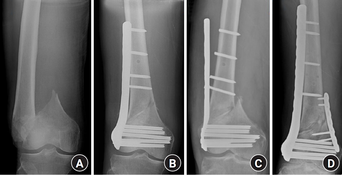
Regarding screw insertion, when a locking screw is used for the most proximal screw fixation in the proximal fragment, its larger core diameter provides greater strength compared to a cortical screw; however, it also leads to increased stress concentration, which may result in a stress-riser fracture at that site (Fig. 6). Therefore, in elderly patients with distal femur fractures treated with a locking plate, it is recommended to use a conventional cortical screw rather than a locking screw in the most proximal screw hole. Bottlang et al. [32] reported that using a cortical screw instead of a locking screw in the most proximal screw hole reduces stress concentration at the proximal end of the plate and increases resistance to bending forces. To achieve optimal fracture reduction or to ensure close apposition of the plate to the proximal fragment, cortical screws must be inserted prior to locking screws [24,33]. If a cortical screw is tightened after a locking screw has already been engaged, especially when a gap exists between the plate and bone, it can impose excessive stress on the locked screw-hole interface or the bone-screw construct. This may lead to compromised fixation strength or potential screw failure. In cases where a stress-riser fracture occurs due to locking screw fixation at the most proximal screw hole, stable fixation can be readily achieved using an overlapping IM nailing (Fig. 6) [34].
When performing locked plating, additional screw insertion at the fracture site to achieve precise reduction may increase structural stiffness at the fracture site, thereby inhibiting callus formation and leading to delayed union or nonunion. Therefore, screw insertion at the fracture site should be avoided whenever possible, and a sufficient working length should be maintained (Fig. 7) [35,36]. In locked plating, screw heads lock into the plate holes, preventing screw pull-out or loosening typically seen with conventional compression plating. However, in osteoporotic bone with reduced cortical strength, failure may occur in the bone surrounding the plate and screws, and in the distal femur, screws may even penetrate into the joint. In addition, early weight-bearing before sufficient bone healing may result in screw breakage, and the use of a short plate with insufficient working length may lead to plate breakage [36].
If locking screws inserted into the distal fragment protrude beyond the medial cortex, they may cause soft tissue irritation and pain during joint motion. Although intraoperative fluoroscopy is used to confirm appropriate screw length, screws are often inserted longer than intended, resulting in medial protrusion. Because the medial cortical surface of the distal femur is inclined approximately 25° relative to the sagittal plane, it is important to internally rotate the femur by about 25° and obtain an anteroposterior radiograph intraoperatively to confirm whether distal locking screws have penetrated the medial cortex (Fig. 8).
Excessive distance between the plate and cortical bone may also lead to soft tissue irritation and decreased structural stability, requiring careful attention. Even in simple fractures, failure to adhere to the principles of locking plate fixation may result in delayed union, nonunion, malunion, and fixation failure [15,37]. It should also be recognized that the use of a locking plate alone does not necessarily guarantee superior mechanical strength in osteoporotic fractures; rather, a more stable construct can be achieved biomechanically by appropriately combining conventional cortical screws with locking screws [32,38].
In the case of anatomically precontoured plates, additional intraoperative contouring is not required, and indirect fracture reduction can be achieved using the plate. However, if the entry point is improperly selected and the locking plate is not accurately positioned on the anterolateral aspect of the femur, the plate may assume an eccentric position. This can result in locking screws, which have predetermined insertion angles, failing to properly engage the cortical bone. Consequently, the plate may be positioned farther from the femur, reducing structural stability and causing soft tissue irritation. Therefore, careful attention to proper plate positioning is essential [24].
Finally, patient-related factors such as frailty, comorbidities, nutritional status, and bone metabolism should also be considered. Optimization of systemic conditions, including correction of vitamin D deficiency and appropriate osteoporosis management, may contribute to an improved biological environment for fracture healing and potentially reduce the risk of fixation failure [39].
Conclusions
With the development of anatomically precontoured locking plates and the adoption of MIPO techniques, satisfactory outcomes using lateral locked plating have been reported in the treatment of distal femur fractures in elderly patients. However, meticulous caution is required, as fixation failure and impaired bone healing frequently occur due to poor bone quality and inadequate fixation. Therefore, more effective treatment of distal femur fractures in elderly patients can be achieved by selecting an appropriately sized plate according to the fracture pattern and bone quality and adhering to the fundamental principles of locked plating.
-
Author contribution
All the work was done by Je-Hyun Yoo.
-
Conflict of interests
No potential conflict of interest relevant to this article was reported.
-
Funding
None.
-
Data availability
Not applicable.
-
Acknowledgments
None.
-
Supplementary materials
None.
Article Information
Fig. 1.
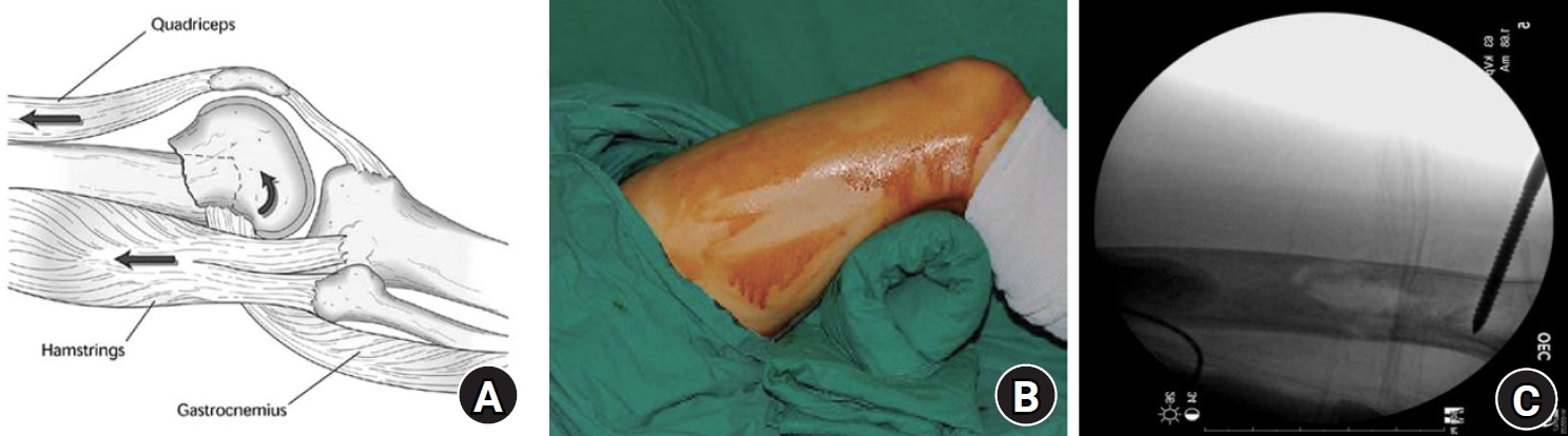
Indirect sagittal-plane reduction of a distal femur fracture. (A) Posterior angulation caused by deforming forces of the surrounding muscles. (B, C) Reduction using a bump beneath the distal thigh or a Schanz screw.
Fig. 2.
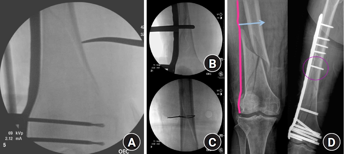
Coronal-plane reduction methods for distal femur fractures using (A) a long hemostatic clamp, (B) a collinear reduction clamp, (C) percutaneous cerclage wiring, or (D) a conventional screw. In (D), the arrow indicates the insertion site of the conventional cortical screw, and the circle indicates the screw inserted to reduce the fracture site.
Fig. 3.
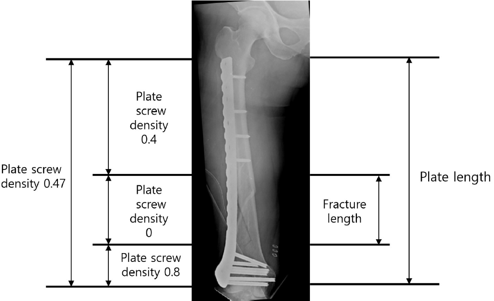
Appropriate plate length and plate-screw density after minimally invasive plate osteosynthesis using a locking plate for an osteoporotic distal femur fracture.
Fig. 4.
(A) Distal femur fracture in a 70-year-old male patient. (B) Postoperative radiographs after minimally invasive locked plating. (C) Fixation failure with broken locking screws 2 months after surgery. (D) Anteroposterior and lateral radiographs obtained 7 months after double plating.
Fig. 5.
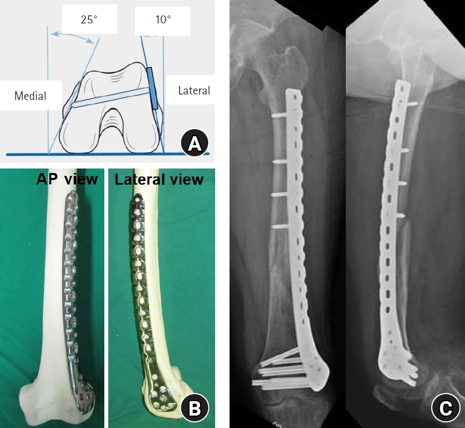
(A) Schematic drawing showing the axial morphology of the distal femur and appropriate distal locking screw length. (B) Anteroposterior (AP) and lateral photographs showing the appropriate position of a lateral anatomical locking plate in a distal femur model. (C) Anteroposterior and lateral radiographs showing appropriate locking plate position after fixation of a distal femur fracture in a 72-year-old female patient.
Fig. 6.
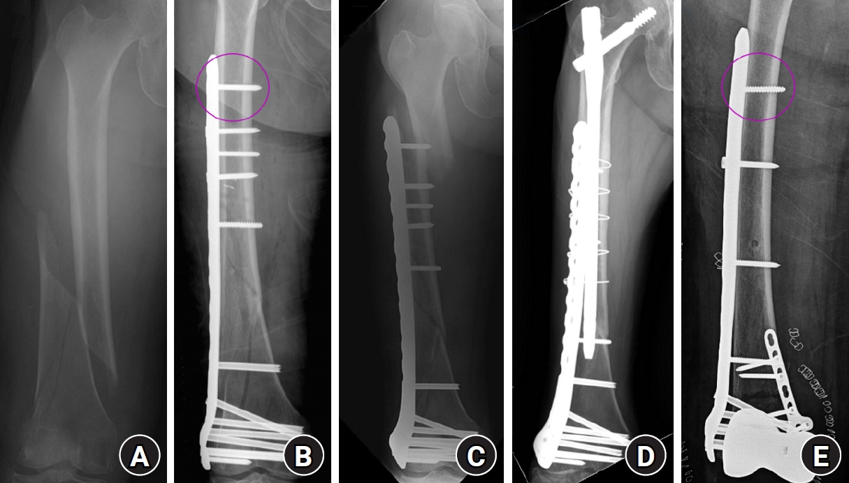
(A) Radiograph of a distal femur fracture in a 74-year-old female patient after a ground-level fall. (B) Postoperative radiograph after closed reduction and minimally invasive locked plating, showing a locking screw in the proximal outermost screw hole (circle). (C) Six-week postoperative radiograph showing a stress-riser fracture around the outermost locking screw after a slip-and-fall injury. (D) Radiograph showing bony union 2 years after overlapping intramedullary nailing. (E) Postoperative radiograph showing locked plating with a conventional screw (circle), rather than a locking screw, in the proximal outermost screw hole to minimize the stress riser.
Fig. 7.
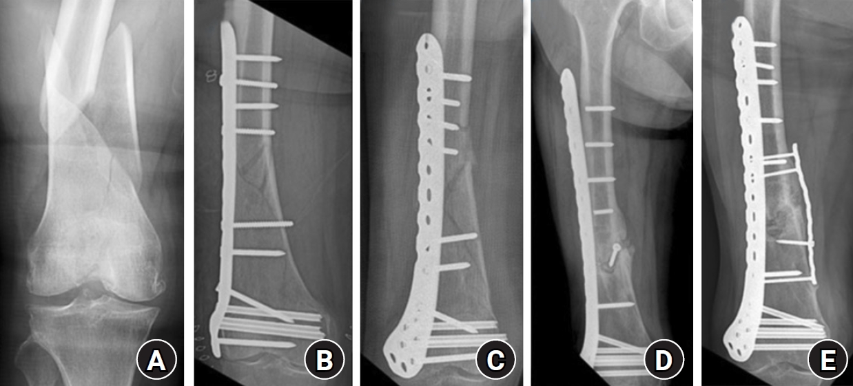
(A) Radiograph of a 75-year-old female patient showing a spiral fracture with a butterfly fragment of the distal femur. (B) Lateral locked plating performed with two appositional screws at the fracture site. (C) Collapse of the fracture site with pull-out of the plate and proximal screws 2 months after surgery. (D) Additional fixation failure with breakage of the proximal screws after revision surgery using a longer locking plate. (E) Bony union achieved 9 months after the third operation, which included medial support plating and autologous iliac bone grafting.
Fig. 8.
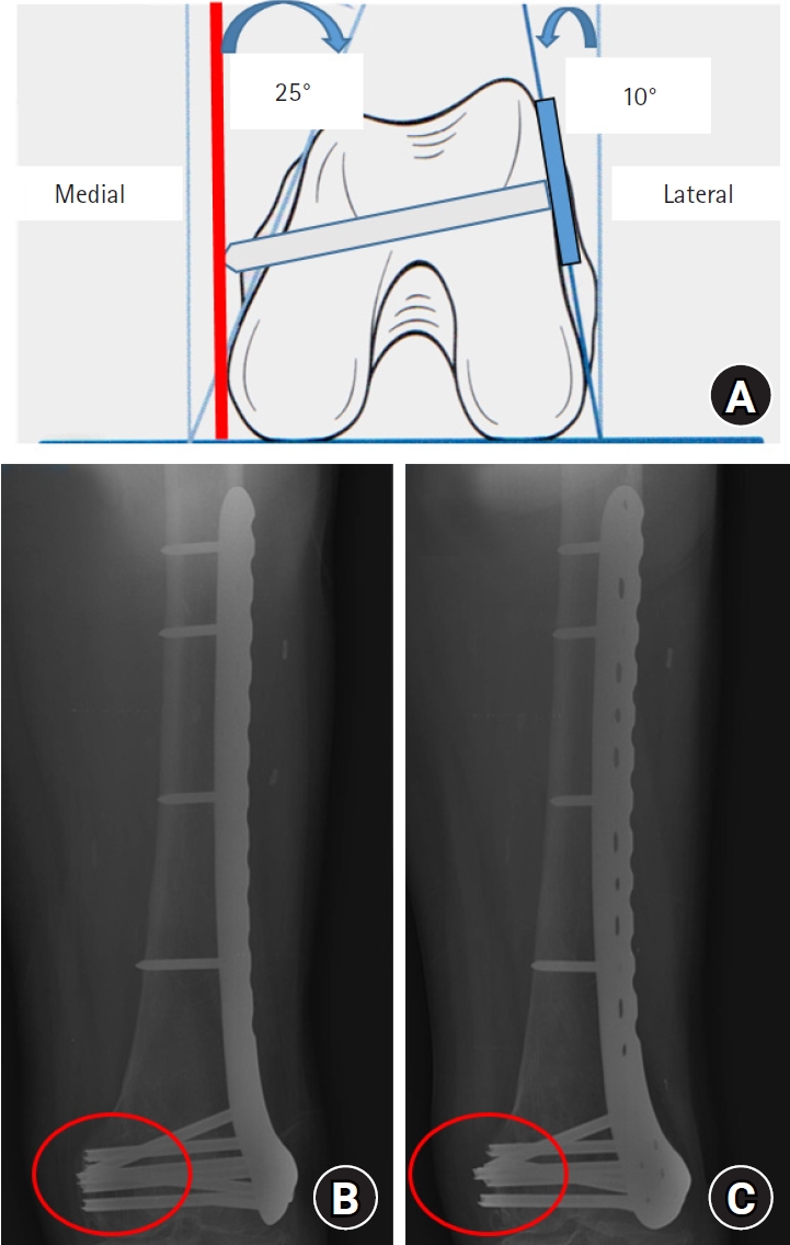
(A) Schematic drawing showing the axial morphology of the distal femur and protrusion of a distal locking screw. (B) True anteroposterior radiograph showing appropriate distal locking screw length (circle). (C) Radiograph obtained after approximately 25° of internal thigh rotation, showing protrusion of the distal screws across the medial cortex (circle).
- 1. Krettek C, Müller M, Miclau T. Evolution of minimally invasive plate osteosynthesis (MIPO) in the femur. Injury 2001;32 Suppl 3:SC14-23.ArticlePubMed
- 2. Stover M. Distal femoral fractures: current treatment, results and problems. Injury 2001;32 Suppl 3:SC3-13.ArticlePubMed
- 3. Wong MK, Leung F, Chow SP. Treatment of distal femoral fractures in the elderly using a less-invasive plating technique. Int Orthop 2005;29:117-20.ArticlePubMedPMCPDF
- 4. Kammerlander C, Riedmuller P, Gosch M, et al. Functional outcome and mortality in geriatric distal femoral fractures. Injury 2012;43:1096-101.ArticlePubMed
- 5. Adams JD, Tanner SL, Jeray KJ. Far cortical locking screws in distal femur fractures. Orthopedics 2015;38:e153-6.ArticlePubMed
- 6. Liporace FA, Yoon RS. Nail plate combination technique for native and periprosthetic distal femur fractures. J Orthop Trauma 2019;33:e64-8.ArticlePubMed
- 7. Dunbar RP, Egol KA, Jones CB, et al. Locked lateral plating versus retrograde nailing for distal femur fractures: a multicenter randomized trial. J Orthop Trauma 2023;37:70-6.ArticlePubMed
- 8. Ehlinger M, Ducrot G, Adam P, Bonnomet F. Distal femur fractures: surgical techniques and a review of the literature. Orthop Traumatol Surg Res 2013;99:353-60.ArticlePubMed
- 9. Jang CY, Yoo JH. Locked plating in elderly patients with distal femur fracture: how to avoid complications? J Korean Fract Soc 2019;32:112-9.ArticlePDF
- 10. Mast J, Jakob R, Ganz R. Planning and reduction technique in fracture surgery. Springer-Verlag; 1989.
- 11. Helfet DL, Shonnard PY, Levine D, Borrelli J. Minimally invasive plate osteosynthesis of distal fractures of the tibia. Injury 1997;28 Suppl 1:A42-8.ArticlePubMed
- 12. Kinast C, Bolhofner BR, Mast JW, Ganz R. Subtrochanteric fractures of the femur: results of treatment with the 95 degrees condylar blade-plate. Clin Orthop Relat Res 1989;238:122-30.PubMed
- 13. Krettek C, Schandelmaier P, Miclau T, Tscherne H. Minimally invasive percutaneous plate osteosynthesis (MIPPO) using the DCS in proximal and distal femoral fractures. Injury 1997;28 Suppl 1:A20-30.ArticlePubMed
- 14. Wagner M. General principles for the clinical use of the LCP. Injury 2003;34 Suppl 2:B31-42.ArticlePubMed
- 15. Goyal T, Nag HL, Tripathy SK. Dynamization of locked plating on distal femur fracture. Arch Orthop Trauma Surg 2011;131:1331-2.ArticlePubMedPDF
- 16. Fulkerson E, Egol KA, Kubiak EN, Liporace F, Kummer FJ, Koval KJ. Fixation of diaphyseal fractures with a segmental defect: a biomechanical comparison of locked and conventional plating techniques. J Trauma 2006;60:830-5.ArticlePubMed
- 17. Gardner MJ, Griffith MH, Demetrakopoulos D, et al. Hybrid locked plating of osteoporotic fractures of the humerus. J Bone Joint Surg Am 2006;88:1962-7.ArticlePubMed
- 18. Greiwe RM, Archdeacon MT. Locking plate technology: current concepts. J Knee Surg 2007;20:50-5.ArticlePubMed
- 19. Lill H, Hepp P, Korner J, et al. Proximal humeral fractures: how stiff should an implant be?: a comparative mechanical study with new implants in human specimens. Arch Orthop Trauma Surg 2003;123:74-81.ArticlePubMedPDF
- 20. Bottlang M, Lesser M, Koerber J, et al. Far cortical locking can improve healing of fractures stabilized with locking plates. J Bone Joint Surg Am 2010;92:1652-60.ArticlePubMedPMC
- 21. Doornink J, Fitzpatrick DC, Madey SM, Bottlang M. Far cortical locking enables flexible fixation with periarticular locking plates. J Orthop Trauma 2011;25 Suppl 1:S29-34.ArticlePubMedPMC
- 22. Stoffel K, Dieter U, Stachowiak G, Gächter A, Kuster MS. Biomechanical testing of the LCP--how can stability in locked internal fixators be controlled? Injury 2003;34 Suppl 2:B11-9.ArticlePubMed
- 23. Rozbruch SR, Müller U, Gautier E, Ganz R. The evolution of femoral shaft plating technique. Clin Orthop Relat Res 1998;354:195-208.ArticlePubMed
- 24. Gautier E, Sommer C. Guidelines for the clinical application of the LCP. Injury 2003;34 Suppl 2:B63-76.ArticlePubMed
- 25. Kaab MJ, Frenk A, Schmeling A, Schaser K, Schutz M, Haas NP. Locked internal fixator: sensitivity of screw/plate stability to the correct insertion angle of the screw. J Orthop Trauma 2004;18:483-7.PubMed
- 26. Stoffel K, Klaue K, Perren SM. Functional load of plates in fracture fixation in vivo and its correlate in bone healing. Injury 2000;31 Suppl 2:S-B37-50.ArticlePubMed
- 27. Ahmad M, Nanda R, Bajwa AS, Candal-Couto J, Green S, Hui AC. Biomechanical testing of the locking compression plate: when does the distance between bone and implant significantly reduce construct stability? Injury 2007;38:358-64.ArticlePubMed
- 28. Bliemel C, Oberkircher L, Bockmann B, et al. Impact of cement-augmented condylar screws in locking plate osteosynthesis for distal femoral fractures: a biomechanical analysis. Injury 2016;47:2688-93.ArticlePubMed
- 29. Steinberg EL, Elis J, Steinberg Y, Salai M, Ben-Tov T. A double-plating approach to distal femur fracture: a clinical study. Injury 2017;48:2260-5.ArticlePubMed
- 30. Garala K, Ramoutar D, Li J, et al. Distal femoral fractures: a comparison between single lateral plate fixation and a combined femoral nail and plate fixation. Injury 2022;53:634-9.ArticlePubMed
- 31. Henderson CE, Kuhl LL, Fitzpatrick DC, Marsh JL. Locking plates for distal femur fractures: is there a problem with fracture healing? J Orthop Trauma 2011;25 Suppl 1:S8-14.ArticlePubMed
- 32. Bottlang M, Doornink J, Byrd GD, Fitzpatrick DC, Madey SM. A nonlocking end screw can decrease fracture risk caused by locked plating in the osteoporotic diaphysis. J Bone Joint Surg Am 2009;91:620-7.ArticlePubMed
- 33. Egol KA, Kubiak EN, Fulkerson E, Kummer FJ, Koval KJ. Biomechanics of locked plates and screws. J Orthop Trauma 2004;18:488-93.ArticlePubMed
- 34. Yoo JH, Kim SW, Kwak YH, Kim HJ. Overlapping intramedullary nailing after failed minimally invasive locked plating for osteoporotic distal femur fractures: report of 2 cases. Injury 2015;46:1174-7.ArticlePubMed
- 35. Kim SM, Yeom JW, Song HK, Hwang KT, Hwang JH, Yoo JH. Lateral locked plating for distal femur fractures by low-energy trauma: what makes a difference in healing? Int Orthop 2018;42:2907-14.ArticlePubMedPDF
- 36. Park JY, Yoo JH. Selection of plate in internal fixation of fractures: locking plate and compression plate. J Korean Fract Soc 2013;26:92-102.Article
- 37. Oh JK, Hwang JH, Lee SJ, Kim JI. Dynamization of locked plating on distal femur fracture. Arch Orthop Trauma Surg 2011;131:535-9.ArticlePubMedPDF
- 38. Doornink J, Fitzpatrick DC, Boldhaus S, Madey SM, Bottlang M. Effects of hybrid plating with locked and nonlocked screws on the strength of locked plating constructs in the osteoporotic diaphysis. J Trauma 2010;69:411-7.ArticlePubMed
- 39. Calori GM, Albisetti W, Agus A, Iori S, Tagliabue L. Risk factors contributing to fracture non-unions. Injury 2007;38 Suppl 2:S11-8.Article
References
Figure & Data
REFERENCES
Citations
Citations to this article as recorded by 
 ePub Link
 Cite
Cite XML Download
Locked plating in older patients with distal femur fractures: strategies to avoid complications
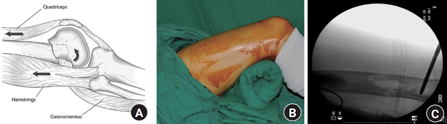
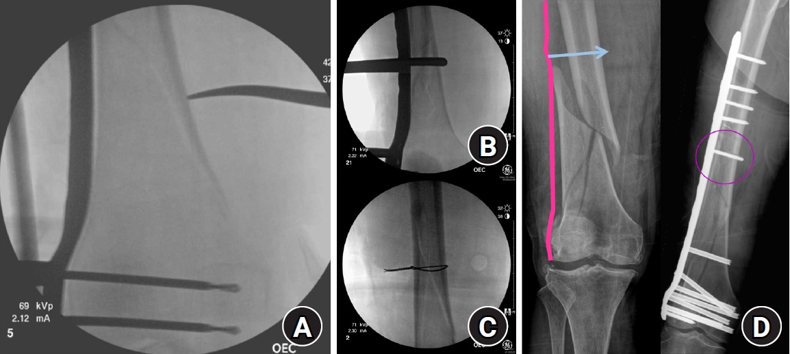
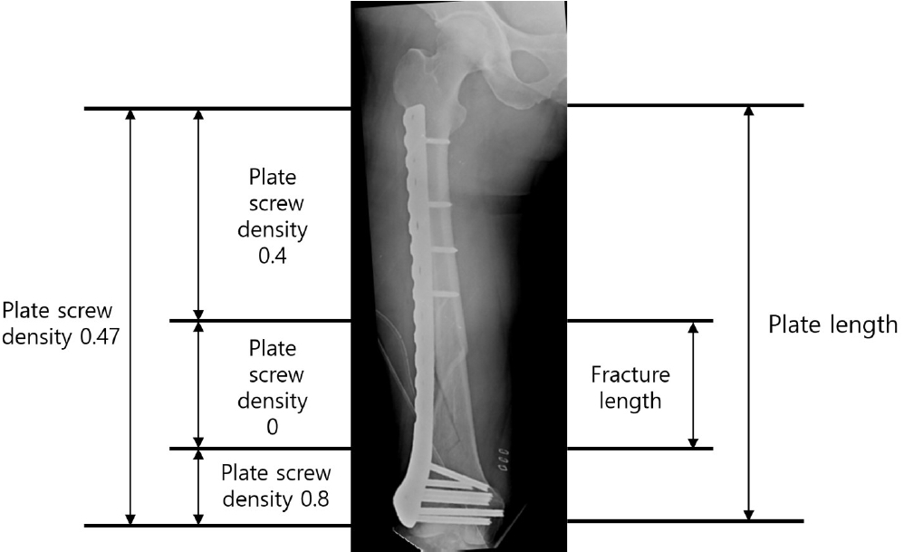
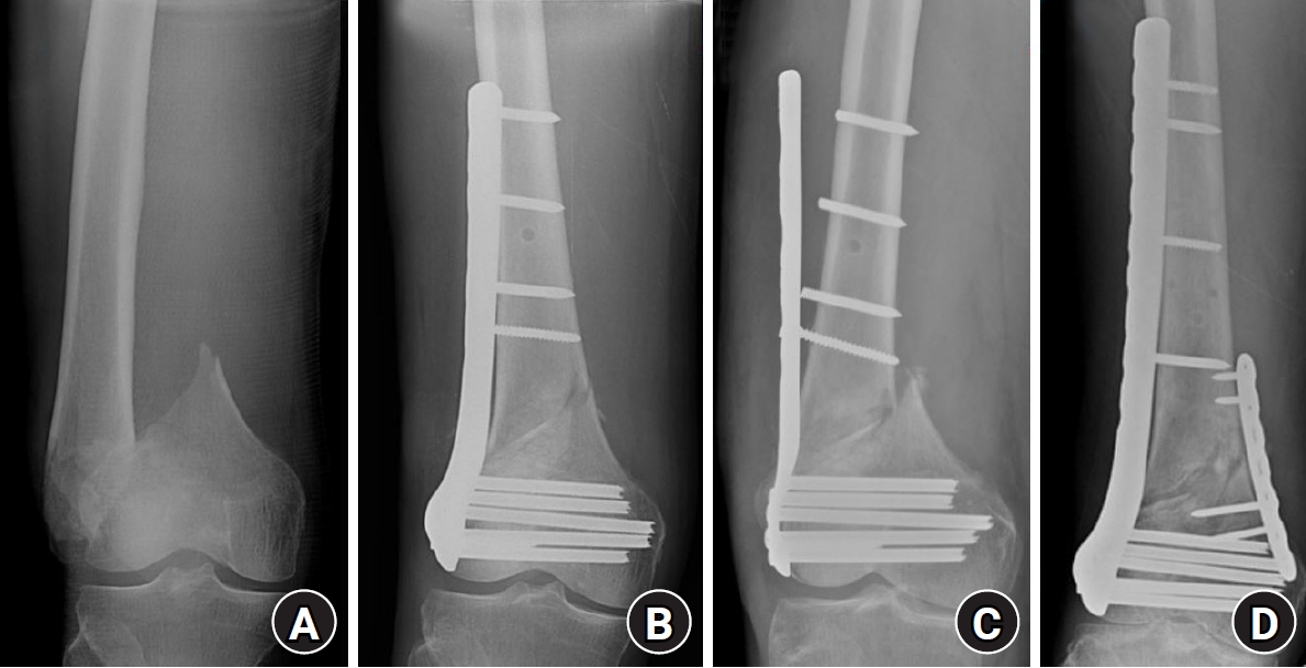
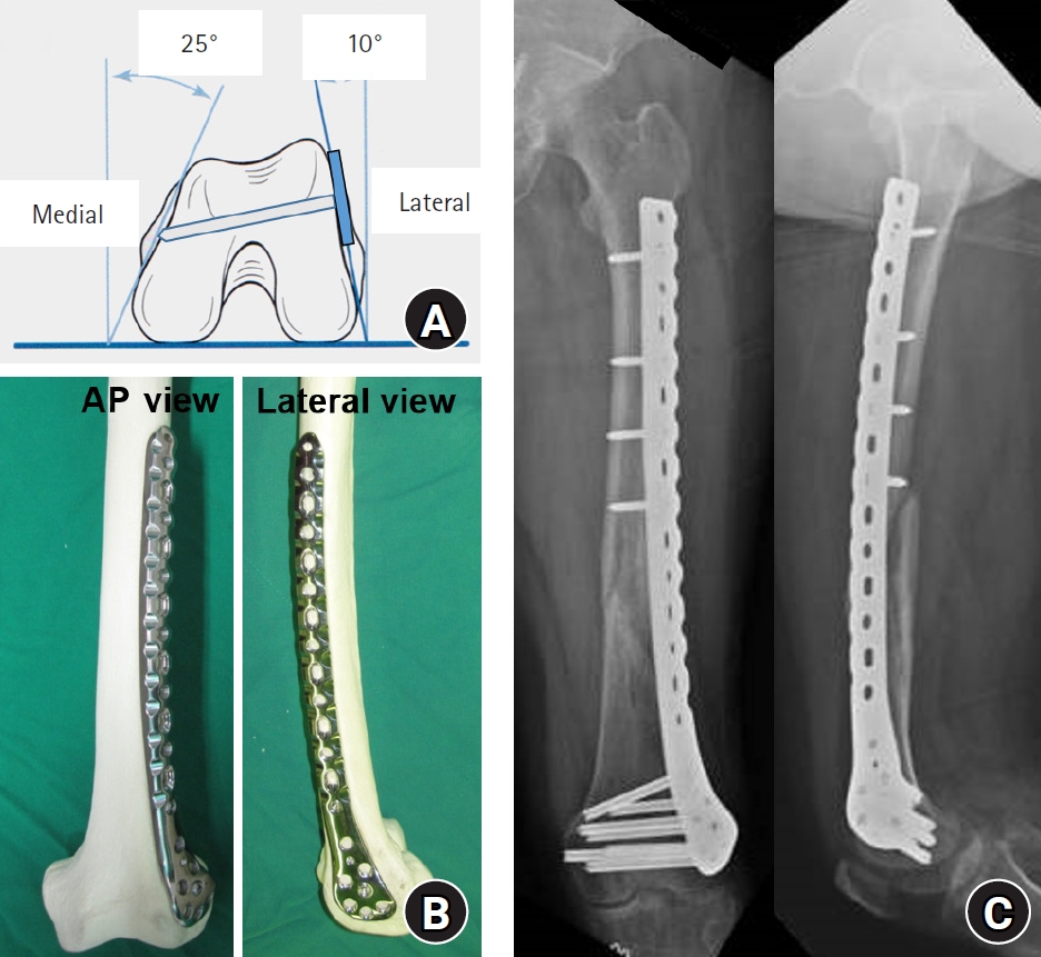
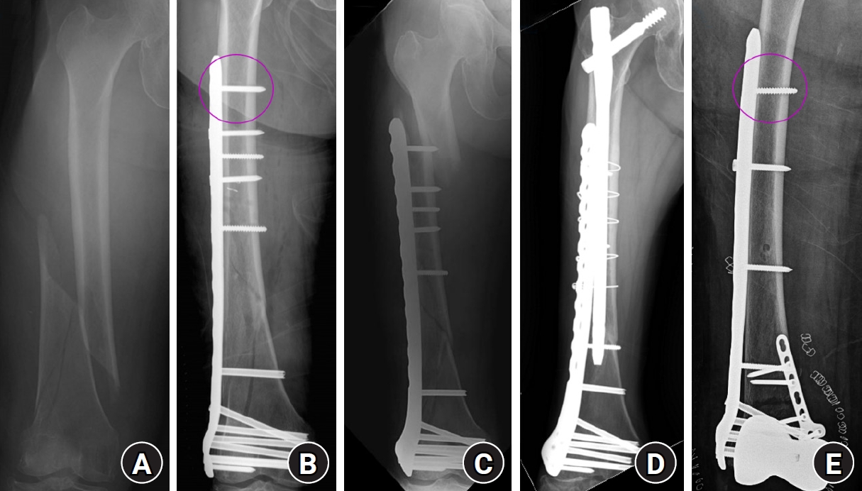
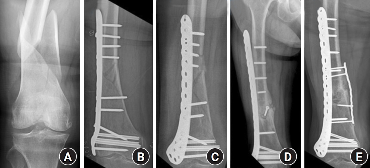
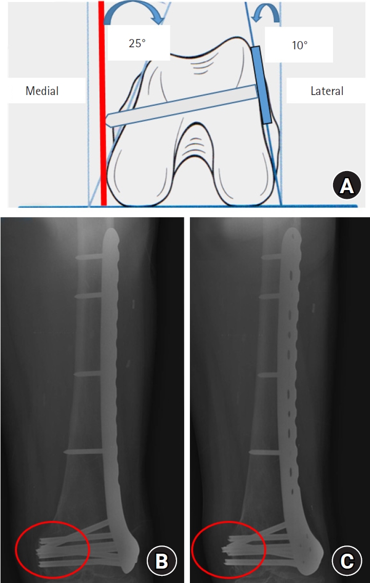
Fig. 1. Indirect sagittal-plane reduction of a distal femur fracture. (A) Posterior angulation caused by deforming forces of the surrounding muscles. (B, C) Reduction using a bump beneath the distal thigh or a Schanz screw.
Fig. 2. Coronal-plane reduction methods for distal femur fractures using (A) a long hemostatic clamp, (B) a collinear reduction clamp, (C) percutaneous cerclage wiring, or (D) a conventional screw. In (D), the arrow indicates the insertion site of the conventional cortical screw, and the circle indicates the screw inserted to reduce the fracture site.
Fig. 3. Appropriate plate length and plate-screw density after minimally invasive plate osteosynthesis using a locking plate for an osteoporotic distal femur fracture.
Fig. 4. (A) Distal femur fracture in a 70-year-old male patient. (B) Postoperative radiographs after minimally invasive locked plating. (C) Fixation failure with broken locking screws 2 months after surgery. (D) Anteroposterior and lateral radiographs obtained 7 months after double plating.
Fig. 5. (A) Schematic drawing showing the axial morphology of the distal femur and appropriate distal locking screw length. (B) Anteroposterior (AP) and lateral photographs showing the appropriate position of a lateral anatomical locking plate in a distal femur model. (C) Anteroposterior and lateral radiographs showing appropriate locking plate position after fixation of a distal femur fracture in a 72-year-old female patient.
Fig. 6. (A) Radiograph of a distal femur fracture in a 74-year-old female patient after a ground-level fall. (B) Postoperative radiograph after closed reduction and minimally invasive locked plating, showing a locking screw in the proximal outermost screw hole (circle). (C) Six-week postoperative radiograph showing a stress-riser fracture around the outermost locking screw after a slip-and-fall injury. (D) Radiograph showing bony union 2 years after overlapping intramedullary nailing. (E) Postoperative radiograph showing locked plating with a conventional screw (circle), rather than a locking screw, in the proximal outermost screw hole to minimize the stress riser.
Fig. 7. (A) Radiograph of a 75-year-old female patient showing a spiral fracture with a butterfly fragment of the distal femur. (B) Lateral locked plating performed with two appositional screws at the fracture site. (C) Collapse of the fracture site with pull-out of the plate and proximal screws 2 months after surgery. (D) Additional fixation failure with breakage of the proximal screws after revision surgery using a longer locking plate. (E) Bony union achieved 9 months after the third operation, which included medial support plating and autologous iliac bone grafting.
Fig. 8. (A) Schematic drawing showing the axial morphology of the distal femur and protrusion of a distal locking screw. (B) True anteroposterior radiograph showing appropriate distal locking screw length (circle). (C) Radiograph obtained after approximately 25° of internal thigh rotation, showing protrusion of the distal screws across the medial cortex (circle).
Fig. 1.
Fig. 2.
Fig. 3.
Fig. 4.
Fig. 5.
Fig. 6.
Fig. 7.
Fig. 8.
Locked plating in older patients with distal femur fractures: strategies to avoid complications


