 E-submission
 TOTA
TOTA TOTS
TOTS
Articles
- Page Path
- HOME > J Musculoskelet Trauma > Volume 23(1); 2010 > Article
-
Original Article
- Surgical Treatment of AO Type C Distal Femoral Fractures Using Locking Compression Plate (LCP-DF, Synthes(R))
- Kap-Jung Kim, M.D., Sang Ki Lee, M.D., Won-Sik Choy, M.D., Won-Cho Kwon, M.D., Do Hyun Lee, M.D.
-
Journal of the Korean Fracture Society 2010;23(1):20-25.
DOI: https://doi.org/10.12671/jkfs.2010.23.1.20
Published online: January 31, 2010
Department of Orthopedic Surgery, Eulji University College of Medicine, Daejeon, Korea.
- Address reprint requests to: Kap-Jung Kim, M.D. Department of Orthopedic Surgery, Eulji University College of Medicine, 1306, Dunsan-dong, Seo-gu, Daejeon 302-799, Korea. Tel: 82-42-611-3279, Fax: 82-42-259-1289, oskkj@eulji.ac.kr
• Received: September 15, 2009 • Revised: November 23, 2009 • Accepted: December 4, 2009
Copyright © 2010 The Korean Fracture Society
- 1,337 Views
- 4 Download
- 2 Crossref
Abstract
-
Purpose
- To analyze the surgical results of AO type C distal femoral fractures using locking compression plate.
-
Materials and Methods
- From February 2006 to June 2008, 14 patients 15 cases were included. Injury mechanisms, combined injuries, radiologic and clinical results and postoperative complications were analyzed.
-
Results
- The mean age was 59.6 (30~77) years. The mean follow up period was 25 (12~40) months. AO types were 3 of C1, 5 of C2 and 7 of C3. Injury mechanisms were 9 of traffic accident, 5 of slip down and 1 of fall from a height. Four cases were combined with other extremity injuries or fractures. The mean radiologic union was obtained at postoperative 15 (13~20) weeks. The mean Neer's functional score was 74.2 (58~97); 3 of excellent, 5 of satisfactory and 7 of unsatisfactory. Postoperative complications were 2 of infection and 1 of nonunion. There were no mechanical failures or fixation loss with locking compression plate at the final follow up.
-
Conclusion
- Internal fixation using locking compression plate for AO type C distal femoral fractures provided excellent fixations. At the final follow up, the clinical results were variable. The affecting factors on the final results seemed to be joint congruencies after anatomical reduction and active rehabilitation.
- 1. Bolhofner BR, Carmen B, Clifford P. The results of open reduction and internal fixation of distal femur fractures using a biologic (indirect) reduction technique. J Orthop Trauma, 1996;10:372-377.Article
- 2. Brown A, D'Arcy JC. Internal fixation for supracondylar fractures of the femur in the elderly patient. J Bone Joint Surg Br, 1971;53:420-424.ArticlePDF
- 3. Choi HR, Song JM, Kwon H, Ko YG, Lee JG, Yoon CH. Operative treatment for AO type C supracondylar fracture of the distal femur. J Korean Soc Fract, 2002;15:166-172.Article
- 4. Fracture and dislocation compendium. Orthopaedic Trauma association committee for coding and classification. J Orthop Trauma, 1996;10:Suppl 1. v-ix. 1-154.
- 5. Haidukewych GJ. Innovations in locking plate technology. J Am Acad Orthop Surg, 2004;12:205-212.Article
- 6. Kim SJ, Oh CW, Jeon IH, et al. Minimally invasive plate osteosynthesis for distal femoral fractures. J Korean Soc Fract, 2003;16:474-481.Article
- 7. Krettek C, Schandelmaier P, Miclau T, Tscherne H. Minimally invasive percutaneous plate osteosynthesis (MIPPO) using the DCS in proximal and distal femoral fractures. Injury, 1997;28:Suppl 1. A20-A30.Article
- 8. Mize RD, Bucholz RW, Grogan DP. Surgical treatment of displaced, comminuted fractures of the distal end of the femur. J Bone Joint Surg Am, 1982;64:871-879.Article
- 9. Müller ME, Allgower M, Schneider R, Willenegger H. Manual of internal fixation. Technique recommended by the AO group, 1979;2nd ed. New York, Springer-Verlag. 118-120.
- 10. Neer CS 2nd, Grantham SA, Shelton ML. Supracondylar fracture of the adult femur. A study of one hundred and ten cases. J Bone Joint Surg Am, 1967;49:591-613.
- 11. Niemeyer P, Südkamp NP. Principles and clinical application of the locking compression plate (LCP). Acta Chir Orthop Traumatol Cech, 2006;73:221-228.Article
- 12. Olerud S. Operative treatment of supracondylar--condylar fractures of the femur. Technique and results in fifteen cases. J Bone Joint Surg Am, 1972;54:1015-1032.
- 13. Russell GV Jr, Smith DG. Minimally invasive treatment of distal femur fractures: report of a technique. J Trauma, 1999;47:799-801.Article
- 14. Schatzker J, Tile M. The rationale of operative fracture care, 1996;3rd ed. New York, Springer-Verlag. 415-416.
- 15. Starr AJ, Jones AL, Reinert CM. The "Swashbuckler": a modified anterior approach for fractures of the distal femur. J Orthop Trauma, 1999;13:138-140.Article
- 16. Stewart MJ, Sisk TD, Wallace SL. Fractures of the distal third of the femur. A comparison methods of treatment. J Bone Joint Surg Am, 1966;48:784-807.
- 17. Walling AK, Seradge H, Speigel PG. Injuries to the knee ligaments with fractures of the femur. J Bone Joint Surg Am, 1982;64:1324-1327.Article
REFERENCES
Fig. 1
(A) Initial radiographs show the distal femoral fracture of AO type C3.
(B) Initial CT scans show the intra-articular fracture fragments. Sagittal image shows Hoffa fracture fragments.
(C) Postoperative radiographs show very congruent articular surface with good alignment. Fracture fragments are reduced and stabilized with locking compression plate and additional Acutrak and cable.
(D) Radiographs 16 months after surgery show the healed fracture with callus in good alignment.
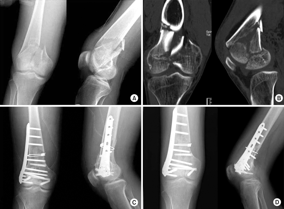

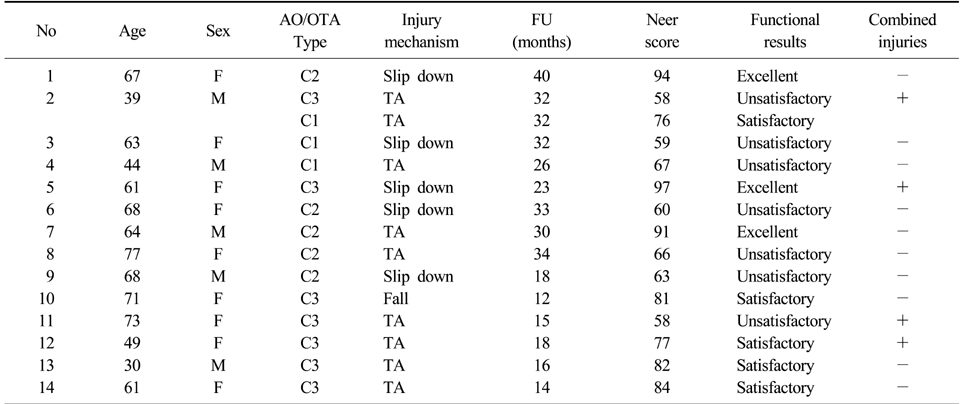
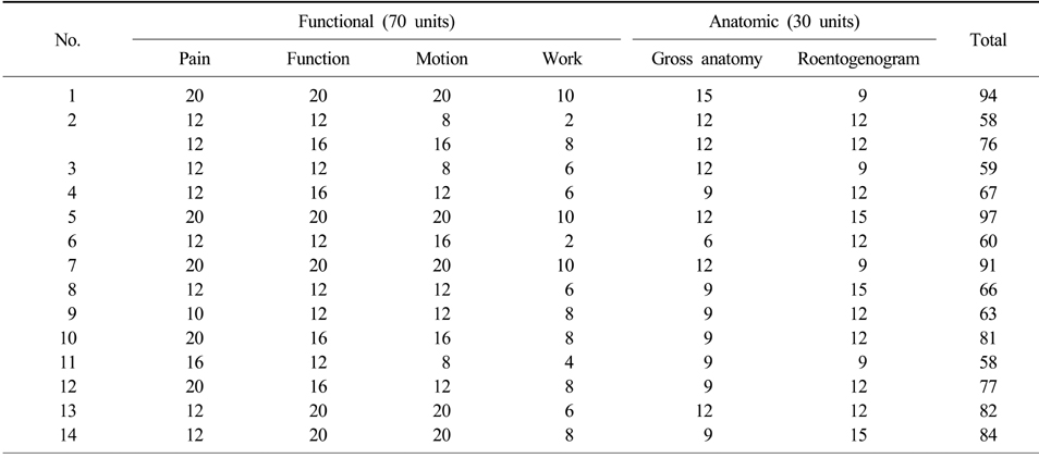
Figure & Data
REFERENCES
Citations
Citations to this article as recorded by 
- Functional outcome of distal femoral fractures treated with distal femoral locking compression plate: a cross-sectional study
Sandeep Kumar Kumar Deep, Varun Phogat, Sankar Debroy
International Journal of Research in Orthopaedics.2025; 11(5): 1089. CrossRef - A STUDY OF SURGICAL MANAGEMENT OF DISTAL FEMORAL FRACTURES BY DISTAL FEMORAL LOCKING COMPRESSION PLATE OSTEOSYNTHESIS
Dema Rajaiah, Yerukala Ramana, Kuppa Srinivas, Venkateswar Reddy S
Journal of Evidence Based Medicine and Healthcare.2016; 3(66): 3584. CrossRef
 Cite
CiteSurgical Treatment of AO Type C Distal Femoral Fractures Using Locking Compression Plate (LCP-DF, Synthes(R))

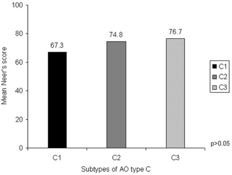
Fig. 1
(A) Initial radiographs show the distal femoral fracture of AO type C3.
(B) Initial CT scans show the intra-articular fracture fragments. Sagittal image shows Hoffa fracture fragments.
(C) Postoperative radiographs show very congruent articular surface with good alignment. Fracture fragments are reduced and stabilized with locking compression plate and additional Acutrak and cable.
(D) Radiographs 16 months after surgery show the healed fracture with callus in good alignment.
Fig. 2
Mean Neer's functional scores of each subtype of AO type C. It doesn't have statistical significance between subtypes of AO type C and mean Neer's scores.
Fig. 1
Fig. 2
Surgical Treatment of AO Type C Distal Femoral Fractures Using Locking Compression Plate (LCP-DF, Synthes(R))
Patients' data
No: Number, FU: Follow up, M: Male, F: Female, TA: Traffic accident.
Patients' Neer score
Table 1
Patients' data
No: Number, FU: Follow up, M: Male, F: Female, TA: Traffic accident.
Table 2
Patients' Neer score

