 E-submission
 TOTA
TOTA TOTS
TOTS
Articles
- Page Path
- HOME > J Musculoskelet Trauma > Volume 23(3); 2010 > Article
-
Case Report
- Operative Treatment of Bilateral Tibial Tuberosity Fractures in Adolescent: A Case Report
- Hong Kyun Kim, M.D., Jung Han Yoo, M.D., Yong Wook Park, M.D., Jin Soo Park, M.D., Kyu Cheol Noh, M.D., Kook Jin Chung, M.D, Keun Jong Jang, M.D., Ji Hyo Hwang, M.D.
-
Journal of the Korean Fracture Society 2010;23(3):317-320.
DOI: https://doi.org/10.12671/jkfs.2010.23.3.317
Published online: July 31, 2010
Department of Orthopedic Surgery, Kangnam Sacred Heart Hospital, College of Medicine, Hallym University, Seoul, Korea.
- Address reprint requests to: Ji Hyo Hwang, M.D. Department of Orthopedic Surgery, Kangnam Sacred Heart Hospital, Hallym University College of Medicine, 948-1, Daerim-1-dong, Yeongdeungpo-gu, Seoul 150-950, Korea. Tel: 82-2-829-5165, Fax: 82-2-834-1728, dr73@dreamwiz.com
• Received: February 18, 2010 • Revised: February 23, 2010 • Accepted: May 3, 2010
Copyright © 2010 The Korean Fracture Society
- 702 Views
- 1 Download
Abstract
- Bilateral avulsion fractures of the tibial tubercles are extremely rare. There is no case report about this in Korean literature. We present simultaneous bilateral tibial tuberosity fractures in 14-year-old adolescent male fell on the ground during running. These fractures were managed by open reduction and screw fixation. We gained complete union and removed metal after 6 months. Functional results were excellent 6 month after surgical treatment.
- 1. Ahmed D, Rangan R, Hagen J. Simultaneous bilateral tibial tuberosity avulsion-over 6-year follow-up. Eur J Trauma, 2006;32:78-79.ArticlePDF
- 2. Anja N, Oliver JM, Ulf B, Holger T. Bilateral avulsion fractures of the tibial tuberosity: A double case for open reduction and fixation. Eur J Trauma, 2008;34:83-87.ArticlePDF
- 3. Cyrus KK, Robert M, Micheal R, Norbert H. Simultaneous bilateral tibial tubercle avulsion fractures in a boy with osteogenesis imperfect. A case report and literature review. Eur J Trauma, 2003;29:164-167.ArticlePDF
- 4. Ergün M, Taşkiran E, Ozgürbüz C. Simultaneous bilateral tibial tubercle avulsion fracture in a basketball player. Knee Surg Sports Traumatol Arthrosc, 2003;11:163-166.ArticlePDF
- 5. Georgiou G, Dimitrakopoulou A, Siapkara A, Kazakos K, Provelengios S, Dounis E. Simultaneous bilateral tibial tubercle avulsion fracture in an adolescent: a case report and review of the literature. Knee Surg Sports Traumatol Arthrosc, 2007;15:147-149.ArticlePDF
- 6. Hamilton SW, Gibson PH. Simultaneous bilateral avulsion fractures of the tibial tuberosity in adolescence: a case report and review of over 50 years of literature. Knee, 2006;13:404-407.Article
- 7. Hanley C, Roche SJ, Chhabra J. Acute simultaneous bilateral avulsion fractures of the tibial tubercles in a 15-year-old male hurler: case report and literature review. Ir J Med Sci, 2008;12 04 [Epub ahead of print].ArticlePDF
- 8. Slobogean GP, Mulpuri K, Alvarez CM, Reilly CW. Comminuted simultaneous bilateral tibial tubercle avulsion fractures: a case report. J Orthop Surg (Hong Kong), 2006;14:319-321.ArticlePDF
- 9. Tulic G, Sopta J, Bumbasirevic M, Todorovic A, Vucetic C. Simultaneous bilateral avulsion fracture of the tibial tubercle in adolescent: a case report. J Pediatr Orthop B, 2010;19:118-121.Article
- 10. Yoon JR, Kim TS, Kim HJ, Noh HK, Oh JK, Yoo JC. Simultaneous patellar tendon avulsion fracture from both patella and tibial tuberosity: a case report. Knee Surg Sports Traumatol Arthrosc, 2007;15:225-227.ArticlePDF
REFERENCES
Figure 1
(A) (Rt): Fracture across the proximal tibial apophysis is completely displaced upward and rotated.
(B) (Lt): Fracture line propagates upward across the primary proximal tibial epiphysis into the knee joint.
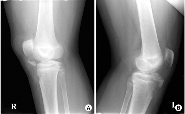
Figure 2
(A) (Rt) Avulsion fracture of the tibial tuberosity with infrapatellar tendon tear is definite in T1WI.
(B) (Lt) Tibial tuberosity shows comminuted intraarticular fracture with avulsion. Fracture related bone and soft tissue edema is also seen, especially anteromedial asepct. Patella tendon reveals diffuse swelling with normal continuity.
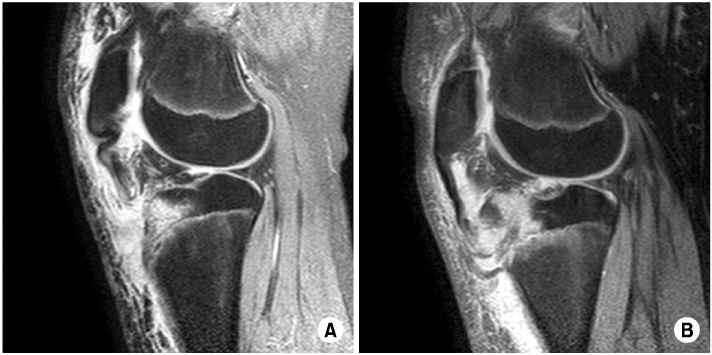
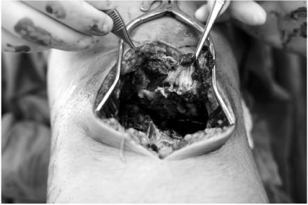

Figure & Data
REFERENCES
Citations
Citations to this article as recorded by 
 Cite
CiteOperative Treatment of Bilateral Tibial Tuberosity Fractures in Adolescent: A Case Report


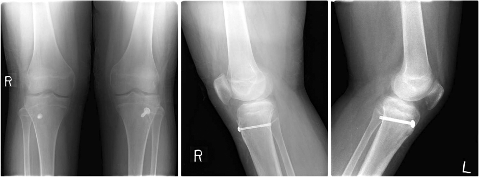
Figure 1
(A) (Rt): Fracture across the proximal tibial apophysis is completely displaced upward and rotated.
(B) (Lt): Fracture line propagates upward across the primary proximal tibial epiphysis into the knee joint.
Figure 2
(A) (Rt) Avulsion fracture of the tibial tuberosity with infrapatellar tendon tear is definite in T1WI.
(B) (Lt) Tibial tuberosity shows comminuted intraarticular fracture with avulsion. Fracture related bone and soft tissue edema is also seen, especially anteromedial asepct. Patella tendon reveals diffuse swelling with normal continuity.
Figure 3
Intraoperative photograph shows large communited avulsed fragment and ruptured retinaculum and thick periosteum in the left knee.
Figure 4
Completely union of previous avulsed fragments at 3 months after operations are seen in the plain AP and lateral view of both knees.
Figure 1
Figure 2
Figure 3
Figure 4
Operative Treatment of Bilateral Tibial Tuberosity Fractures in Adolescent: A Case Report

