 E-submission
 TOTA
TOTA TOTS
TOTS
Articles
- Page Path
- HOME > J Musculoskelet Trauma > Volume 25(2); 2012 > Article
-
Original Article
- Surgical Treatment Using a Transolecranon Approach with a Dual Locking Plate for Unstable Intercondylar Fractures of the Humerus
- Ji-Kang Park, M.D., Yong-Min Kim, M.D., Dong-Soo Kim, M.D., Eui-Sung Choi, M.D., Hyun-Chul Shon, M.D., Kyoung-Jin Park, M.D., Byung-Ki Cho, M.D.
-
Journal of the Korean Fracture Society 2012;25(2):129-135.
DOI: https://doi.org/10.12671/jkfs.2012.25.2.129
Published online: April 17, 2012
Department of Orthopedic Surgery, Chungbuk National University Hospital, Chungbuk National University College of Medicine, Cheongju, Korea.
- Address reprint requests to: Byung-Ki Cho, M.D. Department of Orthopedic Surgery, Chungbuk National University Hospital, 410, Seongbong-ro, Heungdeok-gu, Cheongju 361-711, Korea. Tel: 82-43-269-6077, Fax: 82-43-274-8719, titanick25@yahoo.co.kr
• Received: August 15, 2011 • Revised: September 25, 2011 • Accepted: December 16, 2011
Copyright © 2012 The Korean Fracture Society
- 1,051 Views
- 11 Download
Abstract
-
Purpose
- To evaluate the clinical outcomes of operative treatment using a transolecranon approach with a dual locking plate for unstable intercondylar fractures of the distal humerus.
-
Materials and Methods
- Eighteen patients were followed for more than 1 year after surgical treatment for unstable intercondylar fractures of the humerus. Anterior transpositioning of the ulnar nerve and an early rehabilitation program to allow range of motion (ROM) exercise from postoperative week 1 were used for all cases. The clinical and functional evaluation was performed according to the Mayo Elbow Performance Index and Cassebaum's classification of ROM.
-
Results
- The range of elbow joint motion was a flexion contracture mean of 12.8 degrees to a further flexion mean of 119.3 degrees at the final follow-up. The Mayo Elbow Performance Index was an average of 88.5 points. Among the results, 6 were excellent, 9 good, 2 fair, and 1 poor. Therefore, 15 cases (83.3%) achieved satisfactory results. Fourteen cases (77.7%) achieved a satisfactory ROM according to Cassebaum's classification. All cases achieved bone union, and the interval to union was an average of 14.2 weeks.
-
Conclusion
- Dual locking plate fixation through the transolecranon approach seems to be one of the effective treatment methods for unstable intercondylar fractures of the humerus because it enables the anatomical reduction and rigid fixation of articulation, and early rehabilitation exercise.
- 1. Arnander MW, Reeves A, MacLeod IA, Pinto TM, Khaleel A. A biomechanical comparison of plate configuration in distal humerus fractures. J Orthop Trauma, 2008;22:332-336.Article
- 2. Cassebaum WH. Open reduction of T & Y fractures of the lower end of the humerus. J Trauma, 1969;9:915-925.Article
- 3. Greiner S, Haas NP, Bail HJ. Outcome after open reduction and angular stable internal fixation for supra-intercondylar fractures of the distal humerus: preliminary results with the LCP distal humerus system. Arch Orthop Trauma Surg, 2008;128:723-729.ArticlePDF
- 4. Gupta R, Khanchandani P. Intercondylar fractures of the distal humerus in adults: a critical analysis of 55 cases. Injury, 2002;33:511-515.Article
- 5. Holdsworth BJ, Mossad MM. Fractures of the adult distal humerus. Elbow function after internal fixation. J Bone Joint Surg Br, 1990;72:362-365.ArticlePDF
- 6. Jung WS, Lee KH, Jung JH, Kim SM. Congruent plate fixation for intercondylar fractures of the humerus. J Korean Soc Surg Hand, 2006;11:87-91.
- 7. Jupiter JB. Internal fixation for fractures about the elbow. Techniques Orthop, 1994;4:31-48.Article
- 8. Kimball JP, Glowczewskie F, Wright TW. Intraosseous blood supply to the distal humerus. J Hand Surg Am, 2007;32:642-646.Article
- 9. Kom YK, Eom GS. Opreative treatment of intra-articula T or Y. J Korean Soc Fract, 2000;13:303-310.Article
- 10. Korner J, Diederichs G, Arzdorf M, et al. A biomechanical evaluation of methods of distal humerus fracture fixation using locking compression plates versus conventional reconstruction plates. J Orthop Trauma, 2004;18:286-293.Article
- 11. Lee MJ, Kim HJ, Sohn SK, et al. Clinical outcome of surgical treatment for intra-articular distal humerus fracture. J Korean Fract Soc, 2010;23:201-205.Article
- 12. O'Driscoll SW. Optimizing stability in distal humeral fracture fixation. J Shoulder Elbow Surg, 2005;14:186S-194S.
- 13. Park JY, Seo JB, Chun JY, Kim MH, Min SH, Lee JH. Treatment of intercondylar fractures of humerus with Y-plate. J Korean Fract Soc, 2006;19:443-448.Article
- 14. Ring D, Jupiter JB, Gulotta L. Articular fractures of the distal part of the humerus. J Bone Joint Surg Am, 2003;85-A:232-238.Article
- 15. Sanchez-Sotelo J, Torchia ME, O'Driscoll SW. Complex distal humeral fractures: internal fixation with a principle-based parallel-plate technique. J Bone Joint Surg Am, 2007;89:961-969.
- 16. Sanders RA, Raney EM, Pipkin S. Operative treatment of bicondylar intraarticular fractures of the distal humerus. Orthopedics, 1992;15:159-163.Article
- 17. Schemitsch EH, Tencer AF, Henley MB. Biomechanical evaluation of methods of internal fixation of the distal humerus. J Orthop Trauma, 1994;8:468-475.Article
- 18. Schuster I, Korner J, Arzdorf M, Schwieger K, Diederichs G, Linke B. Mechanical comparison in cadaver specimens of three different 90-degree double-plate osteosyntheses for simulated C2-type distal humerus fractures with varying bone densities. J Orthop Trauma, 2008;22:113-120.Article
- 19. Shin R, Ring D. The ulnar nerve in elbow trauma. J Bone Joint Surg Am, 2007;89:1108-1116.Article
- 20. Södergård J, Sandelin J, Böstman O. Mechanical failures of internal fixation in T and Y fractures of the distal humerus. J Trauma, 1992;33:687-690.Article
- 21. Wang KC, Shih HN, Hsu KY, Shih CH. Intercondylar fractures of the distal humerus: routine anterior subcutaneous transposition of the ulnar nerve in a posterior operative approach. J Trauma, 1994;36:770-773.
- 22. Wong AS, Baratz ME. Elbow fractures: distal humerus. J Hand Surg Am, 2009;34:176-190.Article
- 23. Zalavras CG, Vercillo MT, Jun BJ, Otarodifard K, Itamura JM, Lee TQ. Biomechanical evaluation of parallel versus orthogonal plate fixation of intra-articular distal humerus fractures. J Shoulder Elbow Surg, 2011;20:12-20.Article
REFERENCES
Fig. 1
(A) Operative photographs show olecranon osteotomy and extensive metaphyseal comminution. The ulnar nerve (tagged with a vessel loop) is protected and transposed anteriorly.
(B) Temporary K-wire fixation following the restoration of articulation.
(C) Internal fixation with dual locking-compression plates.

Fig. 2
(A~C) A 20 year-old man sustained a distal humeral intercondylar fracture classified as AO type C2.
(D~E) Three-month follow-up radiographs show complete bony union with dual locking-compression plates.
(F~G) Photographs show excellent functional results.

Fig. 3
(A~B) Three-month follow-up radiographs show complete bony union with heterotopic ossification (arrow).
(C~D) Postoperative radiographs after implant removal and bony spur excision.


Figure & Data
REFERENCES
Citations
Citations to this article as recorded by 
 Cite
CiteSurgical Treatment Using a Transolecranon Approach with a Dual Locking Plate for Unstable Intercondylar Fractures of the Humerus
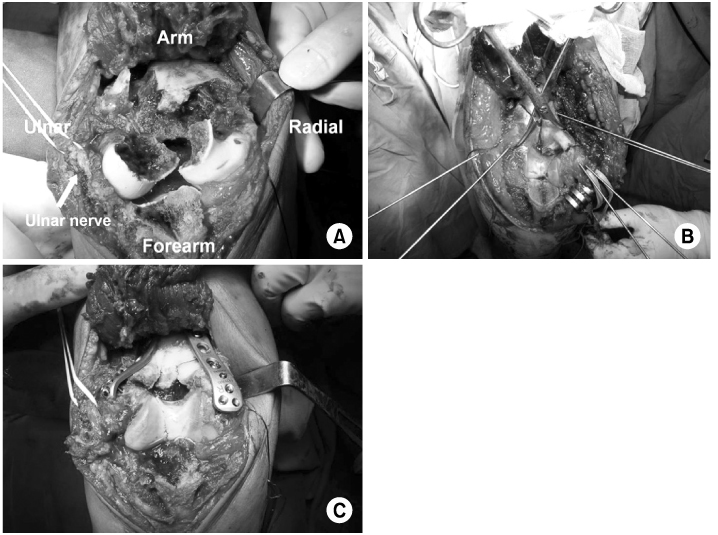
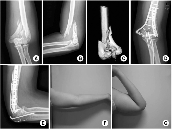
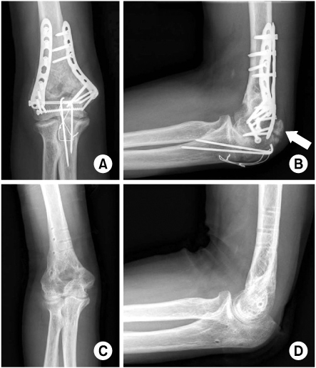
Fig. 1
(A) Operative photographs show olecranon osteotomy and extensive metaphyseal comminution. The ulnar nerve (tagged with a vessel loop) is protected and transposed anteriorly.
(B) Temporary K-wire fixation following the restoration of articulation.
(C) Internal fixation with dual locking-compression plates.
Fig. 2
(A~C) A 20 year-old man sustained a distal humeral intercondylar fracture classified as AO type C2.
(D~E) Three-month follow-up radiographs show complete bony union with dual locking-compression plates.
(F~G) Photographs show excellent functional results.
Fig. 3
(A~B) Three-month follow-up radiographs show complete bony union with heterotopic ossification (arrow).
(C~D) Postoperative radiographs after implant removal and bony spur excision.
Fig. 1
Fig. 2
Fig. 3
Surgical Treatment Using a Transolecranon Approach with a Dual Locking Plate for Unstable Intercondylar Fractures of the Humerus
Evaluation of clinical results with the Mayo Elbow Performance Index at final follow-up
SD: Standard deviation.
Evaluation of elbow range of motion with Cassebaum's classification system
Table 1
Evaluation of clinical results with the Mayo Elbow Performance Index at final follow-up
SD: Standard deviation.
Table 2
Evaluation of elbow range of motion with Cassebaum's classification system

