 E-submission
 TOTA
TOTA TOTS
TOTS
Articles
- Page Path
- HOME > J Musculoskelet Trauma > Volume 20(4); 2007 > Article
-
Case Report
- Stiff Knee by Entrapment of Quadriceps Femoris Tendon at Fracture Site in Paediatric Distal Femur Shaft Fracture
- Suk Kang, M.D., Jong Pil Kim, M.D., Chung Soo Hwang, M.D., Phil Hyun Chung, M.D., Young Sung Kim, M.D., Sang Ho Lee, M.D., Jin Wook Chung, M.D.
-
Journal of the Korean Fracture Society 2007;20(4):339-344.
DOI: https://doi.org/10.12671/jkfs.2007.20.4.339
Published online: June 14, 2016
Department of Orthopaedic Surgery, College of Medicine, Dongguk University, Gyeongju, Korea.
- Address reprint requests to: Jong Pil Kim, M.D. Department of Orthopaedic Surgery, College of Medicine, Dongguk University, 1090-1, Seokjang-dong, Gyeongju 780-350, Korea. Tel: 82-54-770-8225, Fax: 82-54-770-8378, kjpil@dongguk.ac.kr
Copyright © The Korean Fracture Society. All rights reserved
- 641 Views
- 1 Download
Abstract
- The complications following paediatric femur fracture are leg length discrepancy, angulation deformity, rotational deformity, ischemic limb. But, stiff knee is rarely expressed after trauma like paediatric femur fracture. We report a case of stiff knee due to entrapment of quadriceps femoris tendon at displaced fracture site after conservative treatment by Russel traction and hip spica cast in paediatric femur fracture. We treated successfully by resection of distal end of proximal segment of femur and release of quadriceps femoris tendon for flexion contracture of the knee.
- 1. Chung ES, Lee JB, Lee BH. Spontaneous correction of angular deformity after femoral shaft fracture in children. J Korean Soc Fract, 1998;11:644-649.Article
- 2. Chung KH, Shim JS, Sung KS, Park SJ. Treatment of the children's femur shaft fracture by early spica cast. J Korean Soc Fract, 2003;16:270-277.Article
- 3. Chung YK, Yoo JH, Song BY, Park YW, Roh GC. Spontaneous correction of the angular deformity after femoral shaft fracture in children - preliminery report -. J Korean Orthop Assoc, 1995;30:1382-1388.ArticlePDF
- 4. Lee HY, Rhyu KW, Chung JY, Sohn MI, Kim CK, Kang YK. Immediate hip spica cast application for femoral shaft fractures in children. J Korean Soc Fract, 2003;16:91-97.Article
- 5. Moon ES, Rowe SM, Kim OH. Treatment femoral fracture in children. J Korean Orthop Assoc, 1993;28:1084-1092.ArticlePDF
- 6. Nicoll EA. Quadricepsplasty. J Bone Joint Surg Br, 1963;45:483-490.ArticlePDF
- 7. Park ND, Ihnn JC, Lee SY, Kim ID. Overgrowth following fracture of the shaft of femur in childhood. J Korean Orthop Assoc, 1973;8:107-112.ArticlePDF
- 8. Suh JT, Choi SJ, Kim YG. Treatment of stiff knee. J Korean Knee Soc, 2004;16:59-64.
REFERENCES
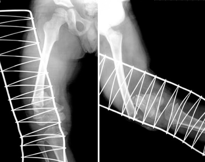
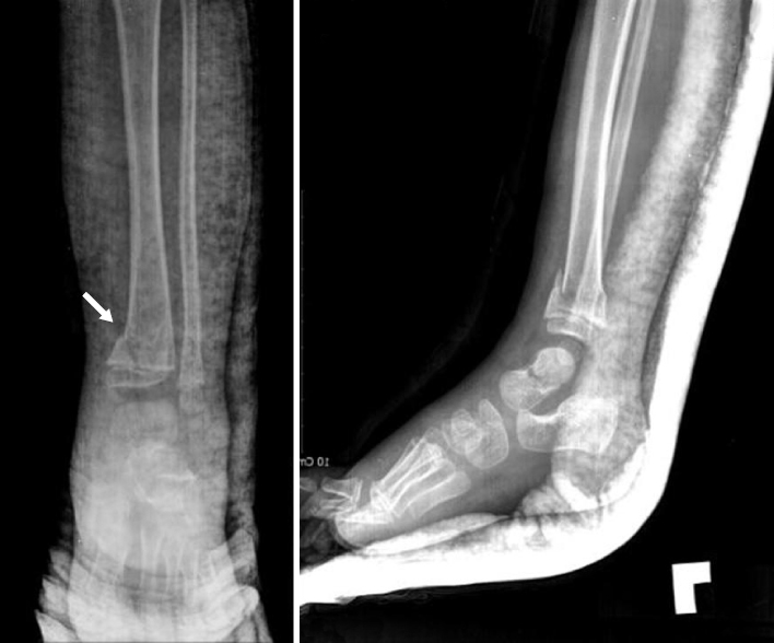
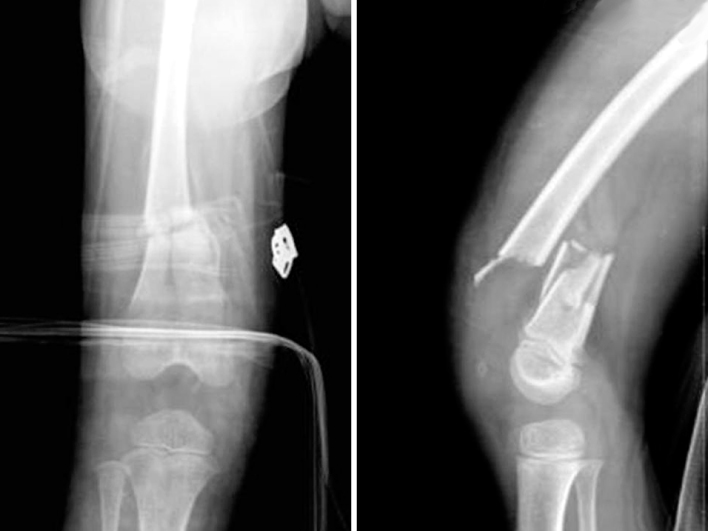
Fig. 4

The radiographs show bridging callus formation from distal fragment to side of proximal fragment at 4 months later.
Fig. 5

The MRI shows entrapment of quadriceps femoris tendon between proximal fragment and anterior cortex of distal fragment.


Figure & Data
REFERENCES
Citations
Citations to this article as recorded by 
 Cite
CiteStiff Knee by Entrapment of Quadriceps Femoris Tendon at Fracture Site in Paediatric Distal Femur Shaft Fracture


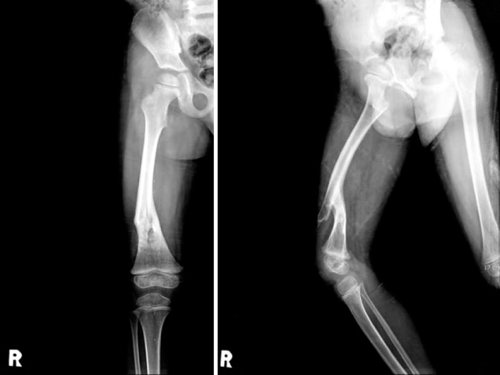
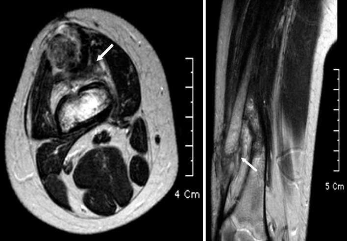
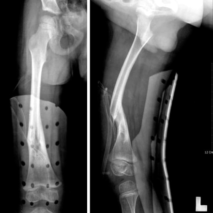
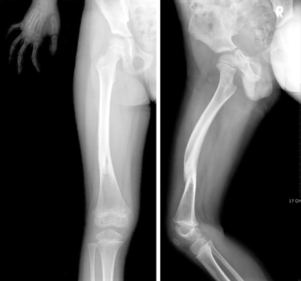
Fig. 1
Initial radiographs show comminuted fracture on distal one fourth of right femoral shaft.
Fig. 2
Initial radiographs show Salter-Harris type II fracture of left distal tibia.
Fig. 3
The radiographs show some callus along the periosteum at 2nd weeks after initial trauma.
Fig. 4
The radiographs show bridging callus formation from distal fragment to side of proximal fragment at 4 months later.
Fig. 5
The MRI shows entrapment of quadriceps femoris tendon between proximal fragment and anterior cortex of distal fragment.
Fig. 6
The radiographs are taken after resection of proximal fragment and release of quadriceps femoris tendon.
Fig. 7
The radiographs show solid union and remodeling on 1 year after operation.
Fig. 1
Fig. 2
Fig. 3
Fig. 4
Fig. 5
Fig. 6
Fig. 7
Stiff Knee by Entrapment of Quadriceps Femoris Tendon at Fracture Site in Paediatric Distal Femur Shaft Fracture

