 E-submission
 TOTA
TOTA TOTS
TOTS
Articles
- Page Path
- HOME > J Musculoskelet Trauma > Volume 27(3); 2014 > Article
-
Original Article
- The Fate of Pronator Quadratus Muscle after Volar Locking Plating of Unstable Distal Radius Fractures
- Chae-Hyun Lim, M.D., Heun-Guyn Jung, M.D., Ju-Yeong Heo, M.D., Young-Jae Jang, M.D., Yong-Soo Choi, M.D.
-
Journal of the Korean Fracture Society 2014;27(3):191-197.
DOI: https://doi.org/10.12671/jkfs.2014.27.3.191
Published online: July 16, 2014
Department of Orthopedic Surgery, Kwangju Christian Hospital, Gwangju, Korea.
- Address reprint requests to: Heun-Guyn Jung, M.D. Department of Orthopedic Surgery, Kwangju Christian Hospital, 37 Yangrim-ro, Nam-gu, Gwangju 503-715, Korea. Tel: 82-62-650-5064, Fax: 82-62-650-5066, handmicro@naver.com
• Received: November 18, 2013 • Revised: March 24, 2014 • Accepted: March 28, 2014
Copyright © 2014 The Korean Fracture Society. All rights reserved.
This is an Open Access article distributed under the terms of the Creative Commons Attribution Non-Commercial License (http://creativecommons.org/licenses/by-nc/3.0/) which permits unrestricted non-commercial use, distribution, and reproduction in any medium, provided the original work is properly cited.
- 788 Views
- 1 Download
Abstract
-
Purpose
- The purpose of this study is to evaluate the pronator quadrates muscle in patients who underwent internal fixation with a volar locking plate for unstable distal radius fractures.
-
Materials and Methods
- Forty patients who underwent internal fixation with a volar locking plate for unstable distal radius fracture were enrolled. We evaluated the clinical results according to the Mayo wrist score, the wrist range of motion, and the grip strength at the last follow-up. Using ultrasonography, muscle thickness of the pronator quadrates was compared between injured and uninjured arm.
-
Results
- Bone union was achieved in all cases. The mean Mayo wrist score was 82.79 points. The grip strength of the injured arm was decreased to 89.1% of the uninjured side. The decrease of pronation range of the injured wrist motions was significant (82.3°, p=0.004). There was significant atrophy of the pronator quadrates muscle on the injured side (injured side: 3.19 mm, uninjured side: 4.72 mm, p=0.001); and the decrement of muscle thickness in pronator quadrates showed an association with the Mayo wrist score (r=-0.35, p=0.042).
-
Conclusion
- These results suggest that continuity of the muscle is maintained after use of the volar locking plating for unstable distal radius fractures with repair of pronator quadrates; however, there is atrophy of pronator quadrates muscle and limitation of pronation in the injured wrist.
- 1. Cooney WP 3rd, Linscheid RL, Dobyns JH. External pin fixation for unstable Colles' fractures. J Bone Joint Surg Am, 1979;61:840-845.Article
- 2. Fernandez DL, Wolfe SW. Distal radius fractures. In: Geen DP, Hotchkiss RN, Pederson WC, Wolfe SW, editors. Green's operative hand surgery. 5th ed. Philadephia: Elsevier Churchill livingstone; 2005. p. 645-710.
- 3. Leung KS, Shen WY, Tsang HK, Chiu KH, Leung PC, Hung LK. An effective treatment of comminuted fractures of the distal radius. J Hand Surg Am, 1990;15:11-17.Article
- 4. Lee KH. Volar plating of distal radius fractures. J Korean Fract Soc, 2008;21:325-333.Article
- 5. Knox J, Ambrose H, McCallister W, Trumble T. Percutaneous pins versus volar plates for unstable distal radius fractures: a biomechanic study using a cadaver model. J Hand Surg Am, 2007;32:813-817.Article
- 6. Cho CH, Bae KC, Kwon DH. Volar T-locking compression plate for treatment of unstable distal radius fractures. J Korean Fract Soc, 2008;21:220-224.Article
- 7. Drobetz H, Kutscha-Lissberg E. Osteosynthesis of distal radial fractures with a volar locking screw plate system. Int Orthop, 2003;27:1-6.ArticlePDF
- 8. Jupiter JB, Ring D, Weitzel PP. Surgical treatment of redisplaced fractures of the distal radius in patients older than 60 years. J Hand Surg Am, 2002;27:714-723.Article
- 9. Orbay JL, Fernandez DL. Volar fixation for dorsally displaced fractures of the distal radius: a preliminary report. J Hand Surg Am, 2002;27:205-215.Article
- 10. Trumble TE, Culp RW, Hanel DP, Geissler WB, Berger RA. Intra-articular fractures of the distal aspect of the radius. Instr Course Lect, 1999;48:465-480.
- 11. Protopsaltis TS, Ruch DS. Volar approach to distal radius fractures. J Hand Surg Am, 2008;33:958-965.Article
- 12. Ahsan ZS, Yao J. The importance of pronator quadratus repair in the treatment of distal radius fractures with volar plating. Hand (N Y), 2012;7:276-280.Article
- 13. Hershman SH, Immerman I, Bechtel C, Lekic N, Paksima N, Egol KA. The effects of pronator quadratus repair on outcomes after volar plating of distal radius fractures. J Orthop Trauma, 2013;27:130-133.Article
- 14. McConkey MO, Schwab TD, Travlos A, Oxland TR, Goetz T. Quantification of pronator quadratus contribution to isometric pronation torque of the forearm. J Hand Surg Am, 2009;34:1612-1617.Article
- 15. Amadio PC, Berquist TH, Smith DK, Ilstrup DM, Cooney WP 3rd, Linscheid RL. Scaphoid malunion. J Hand Surg Am, 1989;14:679-687.Article
- 16. Chung KC, Squitieri L, Kim HM. Comparative outcomes study using the volar locking plating system for distal radius fractures in both young adults and adults older than 60 years. J Hand Surg Am, 2008;33:809-819.Article
- 17. Orbay JL. The treatment of unstable distal radius fractures with volar fixation. Hand Surg, 2000;5:103-112.Article
- 18. Leung F, Zhu L, Ho H, Lu WW, Chow SP. Palmar plate fixation of AO type C2 fracture of distal radius using a locking compression plate--a biomechanical study in a cadaveric model. J Hand Surg Br, 2003;28:263-266.ArticlePDF
- 19. Orbay JL, Fernandez DL. Volar fixed-angle plate fixation for unstable distal radius fractures in the elderly patient. J Hand Surg Am, 2004;29:96-102.Article
- 20. Stuart PR. Pronator quadratus revisited. J Hand Surg Br, 1996;21:714-722.ArticlePDF
- 21. Gordon KD, Dunning CE, Johnson JA, King GJ. Influence of the pronator quadratus and supinator muscle load on DRUJ stability. J Hand Surg Am, 2003;28:943-950.Article
- 22. Orbay JL, Badia A, Indriago IR, et al. The extended flexor carpi radialis approach: a new perspective for the distal radius fracture. Tech Hand Up Extrem Surg, 2001;5:204-211.Article
- 23. Armangil M, Bezirgan U, Başarır K, Bilen G, Demirtaş M, Bilgin SS. The pronator quadratus muscle after plating of distal radius fractures: is the muscle still working? Eur J Orthop Surg Traumatol, 2014;24:335-339.ArticlePDF
REFERENCES
Fig. 1During surgical procedures for pronator quadratus muscle, detached muscle was repaired with 4 or 5 interrupted matrix sutures with 3-0 Vicryl after the fracture fixation.
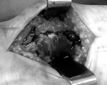
Fig. 2Ultrasound examinations were performed bilaterally at pronator quadrates muscle (asterisk). (A) Transverse view and longitudinal view of the uninjured side. (B) Transverse view and longitudinal view of the injured side.

Fig. 3In a 65-year-old man with an AO type C3 distal radius fracture, gross photo at plate removal showed atrophy of pronator quadratus muscle.
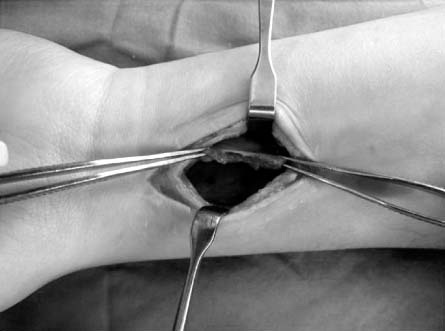

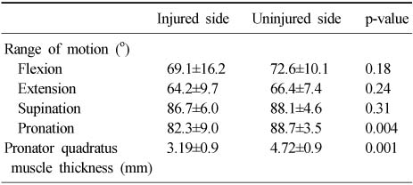
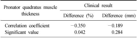
Figure & Data
REFERENCES
Citations
Citations to this article as recorded by 
 Cite
CiteThe Fate of Pronator Quadratus Muscle after Volar Locking Plating of Unstable Distal Radius Fractures

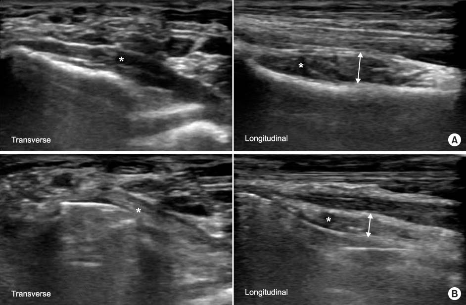

Fig. 1
During surgical procedures for pronator quadratus muscle, detached muscle was repaired with 4 or 5 interrupted matrix sutures with 3-0 Vicryl after the fracture fixation.
Fig. 2
Ultrasound examinations were performed bilaterally at pronator quadrates muscle (asterisk). (A) Transverse view and longitudinal view of the uninjured side. (B) Transverse view and longitudinal view of the injured side.
Fig. 3
In a 65-year-old man with an AO type C3 distal radius fracture, gross photo at plate removal showed atrophy of pronator quadratus muscle.
Fig. 1
Fig. 2
Fig. 3
The Fate of Pronator Quadratus Muscle after Volar Locking Plating of Unstable Distal Radius Fractures
Demographic Characteristics
Values are presented as number, number (%), or median (range).
Comparison of Range of Motion of the Wrist and Muscle Thickness of Pronator Quadratus between Injured Side and Uninjured Side at Last Follow Up
Values are presented as mean±standard deviation.
Correlation Analysis of Clinical Results and Difference of Pronator Quadratus Muscle Thickness
Table 1
Demographic Characteristics
Values are presented as number, number (%), or median (range).
Table 2
Comparison of Range of Motion of the Wrist and Muscle Thickness of Pronator Quadratus between Injured Side and Uninjured Side at Last Follow Up
Values are presented as mean±standard deviation.
Table 3
Correlation Analysis of Clinical Results and Difference of Pronator Quadratus Muscle Thickness

