 E-submission
 TOTA
TOTA TOTS
TOTS
Articles
- Page Path
- HOME > J Musculoskelet Trauma > Volume 24(1); 2011 > Article
-
Original Article
- Operative Treatment with Gamma 3 Nail in Femur Intertrochanteric Fracture
- Ki-Do Hong, M.D., Jae-Chun Sim, M.D., Sung-Sik Ha, M.D., Tae-Ho Kim, M.D., Yoon-Ho Choi, M.D., Jong-Hyun Kim, M.D.
-
Journal of the Korean Fracture Society 2011;24(1):7-15.
DOI: https://doi.org/10.12671/jkfs.2011.24.1.7
Published online: January 19, 2011
Department of Orthopedic Surgery, Sahmyook Medical Center, Seoul, Korea.
- Address reprint requests to: Jae-Chun Sim, M.D. Department of Orthopedic Surgery, Sahmyook Medical Center, 29-1, Hwigyoung 2-dong, Dongdaemun-gu, Seoul 130-711, Korea. Tel: 82-2-2210-3581·Fax: 82-2-2217-1897, kjh2789@hanmail.net
• Received: July 26, 2010 • Revised: October 20, 2010 • Accepted: November 10, 2010
Copyright © 2011 The Korean Fracture Society
- 2,660 Views
- 19 Download
- 4 Crossref
Abstract
-
Purpose
- To evaluate clinical and radiological results of surgical treatment of femur intertrochantenric fracture using Gamma 3 nail.
-
Materials and Methods
- With clinical study, 22 patients who were treated surgically by Gamma 3 nail were retrospectively evaluated. By postoperative radiograph and last follow up radiograph we measured Tip-apex distance, Cleveland index, Neck-shaft angle change Lag screw slippage and Union time. And By medical record review, the clinical results were evaluated with the operation time, intraperative estimated blood loss, amount of transfusion, change of mobility and complication.
-
Results
- The mean change of femur neck shaft angle was 5.18 degrees. The mean lag screw sliding was 5.43 mm. The mean bone union time was 11.8 weeks. From all of these examples shows bone union. The mean operative time was 41 min, blood loss was 161 ml and the transfusion amount was 0.3 pint. In Ceder et al mobility score, it showed 0.2 point decreased and in Jensen social function score, it showed 0.6 point increased. Comparing the results before and after operation, the results were satisfactory.
-
Conclusion
- Using the Gamma 3 nail, the treatment of fermur intertrochanteric fractures showed good results both radiologically and clinically.
- 1. Anglen JO, Baumgaertner MR, Smith WR, Tornetta Iii P, Ziran BH. Technical tips in fracture care: fractures of the hip. Instr Course Lect, 2008;57:17-24.
- 2. Aune AK, Ekeland A, Odegaard B, Grøgaard B, Alho A. Gamma nail vs compression screw for trochanteric femoral fractures. 15 reoperations in a prospective, randomized study of 378 patients. Acta Orthop Scand, 1994;65:127-130.Article
- 3. Baumgaertner MR, Curtin SL, Lindskog DM. Intramedullary versus extramedullary fixation for the treatment of intertrochanteric hip fractures. Clin Orthop Relat Res, 1998;348:87-94.Article
- 4. Bridle SH, Patel AD, Bircher M, Calvert PT. Fixation of intertrochanteric fractures of the femur. A randomised prospective comparison of the gamma nail and the dynamic hip screw. J Bone Joint Surg Br, 1991;73:330-334.ArticlePDF
- 5. Ceder L, Lindberg L, Odberg E. Differentiated care of hip fracture in the elderly Mean hospital days and results of rehabilitation. Acta Orthop Scand, 1980;51:157-162.Article
- 6. Chevalley F, Gamba D. Gamma nailing of pertrochanteric and subtrochanteric fractures: clinical results of a series of 63 consecutive cases. J Orthop Trauma, 1997;11:412-415.Article
- 7. Cleveland M, Bosworth DM, Thompson FR, Wilson HJ Jr, Ishizuka T. A ten-year analysis of intertrochanteric fractures of the femur. J Bone Joint Surg Am, 1959;41:1399-1408.Article
- 8. Docquier PL, Manche E, Autrique JC, Geulette B. Complications associated with gamma nailing. A review of 439 cases. Acta Orthop Belg, 2002;68:251-257.
- 9. Doppelt SH. The sliding compression screw--today's best answer for stabilization of intertrochanteric hip fractures. Orthop Clin North Am, 1980;11:507-523.Article
- 10. Forte ML, Virnig BA, Kane RL, et al. Geographic variation in device use for intertrochanteric hip fractures. J Bone Joint Surg Am, 2008;90:691-699.Article
- 11. Halder SC. The Gamma nail for peritrochanteric fractures. J Bone Joint Surg Br, 1992;74:340-344.ArticlePDF
- 12. Haynes RC, Pöll RG, Miles AW, Weston RB. Failure of femoral head fixation: a cadaveric analysis of lag screw cut-out with the gamma locking nail and AO dynamic hip screw. Injury, 1997;28:337-341.Article
- 13. Jensen JS. Determining factors for the mortality following hip fractures. Injury, 1984;15:411-414.Article
- 14. Jensen JS, Sonne-Holm S, Tøndevold E. Unstable trochanteric fractures. A comparative analysis of four methods of internal fixation. Acta Orthop Scand, 1980;51:949-962.Article
- 15. Kim TH, Kim JO, Lee SY, Yun GU. Treatment of the unstable intertrochanteric fracture with proximal femoral nail antirotation: comparison with compression hip screw with trochanteric stabilizing plate. J Korean Fract Soc, 2010;23:353-359.Article
- 16. Koval KJ, Zuckerman JD. Hip Fractures: II. Evaluation and treatment of intertrochanteric fractures. J Am Acad Orthop Surg, 1994;2:150-156.Article
- 17. Kyle RF. Fractures of the proximal part of the femur. J Bone Joint Surg Am, 1994;76:924-950.Article
- 18. Lacroix H, Arwert H, Snijders CJ, Fontijne WP. Prevention of fracture at the distal locking site of the gamma nail. A biomechanical study. J Bone Joint Surg Br, 1995;77:274-276.ArticlePDF
- 19. Lee KJ, Min BW, Kim SG, Song KS, Bae KC, Cho CH. Results of treating senile osteoporotic peritrochanteric fracture with proximal femoral nail antirotation (PFNA). J Korean Hip Soc, 2009;21:162-168.Article
- 20. Leung KS, So WS, Shen WY, Hui PW. Gamma nails and dynamic hip screws for peritrochanteric fractures. A randomised prospective study in elderly patients. J Bone Joint Surg Br, 1992;74:345-351.ArticlePDF
- 21. Loch DA, Kyle RF, Bechtold JE, Kane M, Anderson K, Sherman RE. Forces required to initiate sliding in second-generation intramedullary nails. J Bone Joint Surg Am, 1998;80:1626-1631.Article
- 22. Madsen JE, Naess L, Aune AK, Alho A, Ekeland A, Strømsøe K. Dynamic hip screw with trochanteric stabilizing plate in the treatment of unstable proximal femoral fractures: a comparative study with the Gamma nail and compression hip screw. J Orthop Trauma, 1998;12:241-248.Article
- 23. Mueller ME, Nazarian S. Classification et documentation aoedes fractures du femur. Rev Chir Orthop, 1981;67:297.
- 24. Papasimos S, Koutsojannis CM, Panagopoulos A, Megas P, Lambiris E. A randomized comparison of AMBI, TGN and PFN for treatment of unstable trochanteric fractures. Arch Orthop Trauma Surg, 2005;125:462-468.ArticlePDF
- 25. Park YS, Park YK, Choi HJ. Result of bilateral total hip arthroplasty: one stage versus two stage procedure. J Korean Hip Soc, 1998;10:211-215.
- 26. Radford PJ, Needoff M, Webb JK. A prospective randomised comparison of the dynamic hip screw and the gamma locking nail. J Bone Joint Surg Br, 1993;75:789-793.ArticlePDF
- 27. Rosenblum SF, Zuckerman JD, Kummer FJ, Tam BS. A biomechanical evaluation of the Gamma nail. J Bone Joint Surg Br, 1992;74:352-357.ArticlePDF
- 28. Sadowski C, Lbbeke A, Saudan M, Riand N, Stern R, Hoffmeyer P. Treatment of reverse oblique and transverse intertrochanteric fractures with use of an intramedullary nail or a 95 degrees screw-plate: a prospective, randomized study. J Bone Joint Surg Am, 2002;84:372-381.
- 29. Shin DK, Kwun KW, Kim SK, Lee SW, Choi CH, Kim KM. Proximal femoral nail(PFN) for femur intertrochanteric fracture. J Korean Soc Fract, 2002;15:328-335.Article
- 30. Suh JT, Chung WB, Yoo CI. A clinical study of trochanteric fractures of the femur in the elderly over 70 years in the age. J Korean Soc Fract, 1994;7:293-301.Article
- 31. Utrilla AL, Reig JS, Muñoz FM, Tufanisco CB. Trochanteric gamma nail and compression hip screw for trochanteric fractures: a randomized, prospective, comparative study in 210 elderly patients with a new design of the gamma nail. J Orthop Trauma, 2005;19:229-233.
- 32. Varela-Egocheaga JR, Iglesias-Colao R, Suárez-Suárez MA, Fernández-Villán M, González-Sastre V, Murcia-Mazón A. Minimally invasive osteosynthesis in stable trochanteric fractures: a comparative study between Gotfried percutaneous compression plate and Gamma 3 intramedullary nail. Arch Orthop Trauma Surg, 2009;129:1401-1407.ArticlePDF
- 33. Yaozeng X, Dechun G, Huilin Y, Guangming Z, Xianbin W. Comparative study of trochanteric fracture treated with the proximal femoral nail anti-rotation and the third generation of gamma nail. Injury, 2010;41:1234-1238.Article
- 34. Yoo JH, Park JS, Noh KC, et al. The results of proximal femoral nail antirotation: a comparative study with proximal femoral nail. J Korean Hip Soc, 2008;20:286-292.Article
REFERENCES
Fig. 1Gamma 3 nail use sure shot guide to help determine correct nail positioning to allow proper placement of the lag screw.
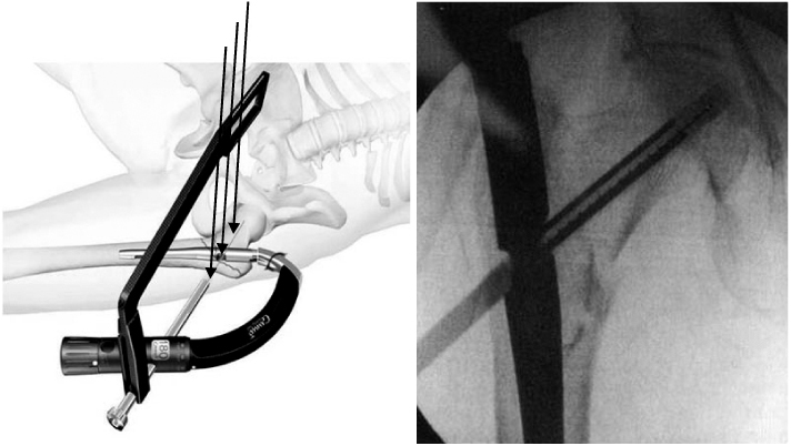
Fig. 2
(A) 80 year old male patient presented with intertrochanteric fracture as AO/ASIF classification A2.1.
(B) Postoperative radiographs show acceptable reduction and well positioned lag screw.
(C) Postoperative 6 months radiographs show minimal sliding of lag screw and well unite fracture site.
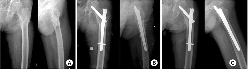
Fig. 3
(A) 75 year old female patient presented with intertrochanteric fracture as AO/ASIF classification A2.2.
(B) Postoperative radiographs show a minor split at the cortex of femur shaft and fixed with Gamma 3 long nail and roll wires.
(C) Postoperative 6 months radiographs show moderate sliding of lag screw and well united fracture site.
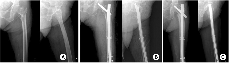
Fig. 4
(A) 86 year old female patient presented with intertrochanteric fracture as AO/ASIF classification A2.2.
(B) Postoperative radiographs show distal tip of nail contacts with anterior femur cortex.
(C) Postoperative 6 months radiographs show well united fracture site but the patient complains about thigh pain.

Fig. 5Gamma 3 nail, curved flexible target device allow small incisions, preventing soft tissue pressure and collision with iliac crest.
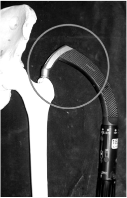
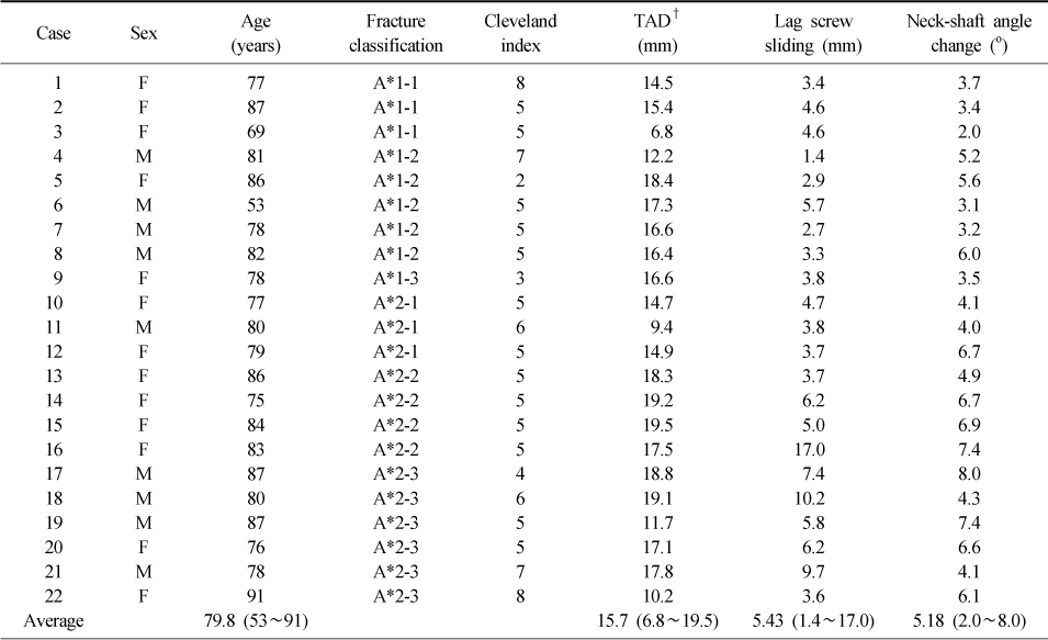

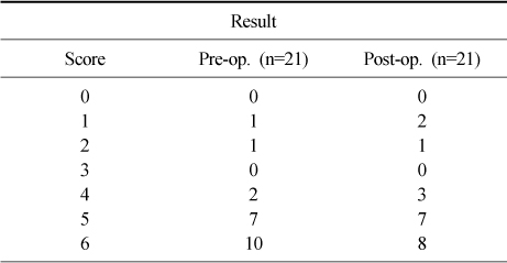
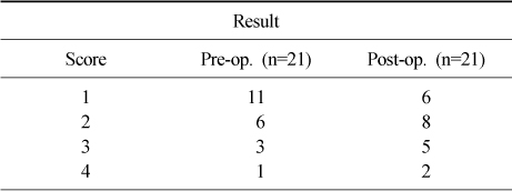
Figure & Data
REFERENCES
Citations
Citations to this article as recorded by 
- Changes in Tip-Apex Distance by Position and Film Distance Measured by Picture Archiving and Communication System (PACS)
Kyu Yeol Lee, Sung Soo Kim, Hyeon Jun Kim, Dong Ho Ha, Hyung Min Yoon, Hyun Su Do
Hip & Pelvis.2015; 27(1): 36. CrossRef - Results of Asian Type Gamma 3 Nail in Treatment of Trochanteric Fractures
Bing Zhe Huang, Yong Wook Park, Jin Su Park, Kyu Cheol Noh, Soung Yon Kim, Kook Jin Chung, Hong Kyun Kim, Hyong Nyun Kim, Yong Hyun Yoon, Ji Hyo Hwang
Journal of the Korean Fracture Society.2014; 27(3): 213. CrossRef - Treatment of Unstable Pertrochanteric Fractures with a Long Intramedullary Nail
Phil Hyun Chung, Suk Kang, Jong Pil Kim, Young Sung Kim, Ho Min Lee, Dae Jung Huh
Hip & Pelvis.2013; 25(1): 51. CrossRef - Comparative Study of Intertrochanteric Fracture Treated with the Proximal Femoral Nail Anti-Rotation and the Third Generation of Gamma Nail
Jae-Cheon Sim, Tae-Ho Kim, Ki-Do Hong, Sung-Sik Ha, Jong-Seong Lee
Journal of the Korean Fracture Society.2013; 26(1): 37. CrossRef
 Cite
CiteOperative Treatment with Gamma 3 Nail in Femur Intertrochanteric Fracture


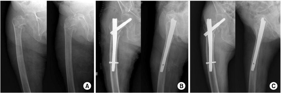

Fig. 1
Gamma 3 nail use sure shot guide to help determine correct nail positioning to allow proper placement of the lag screw.
Fig. 2
(A) 80 year old male patient presented with intertrochanteric fracture as AO/ASIF classification A2.1.
(B) Postoperative radiographs show acceptable reduction and well positioned lag screw.
(C) Postoperative 6 months radiographs show minimal sliding of lag screw and well unite fracture site.
Fig. 3
(A) 75 year old female patient presented with intertrochanteric fracture as AO/ASIF classification A2.2.
(B) Postoperative radiographs show a minor split at the cortex of femur shaft and fixed with Gamma 3 long nail and roll wires.
(C) Postoperative 6 months radiographs show moderate sliding of lag screw and well united fracture site.
Fig. 4
(A) 86 year old female patient presented with intertrochanteric fracture as AO/ASIF classification A2.2.
(B) Postoperative radiographs show distal tip of nail contacts with anterior femur cortex.
(C) Postoperative 6 months radiographs show well united fracture site but the patient complains about thigh pain.
Fig. 5
Gamma 3 nail, curved flexible target device allow small incisions, preventing soft tissue pressure and collision with iliac crest.
Fig. 1
Fig. 2
Fig. 3
Fig. 4
Fig. 5
Operative Treatment with Gamma 3 Nail in Femur Intertrochanteric Fracture
Patient data profile
*A: AO/ASIF classification, †TAD: Tip apex distance.
Ceder et al. mobility score & Jensen social function score
Clinical evaluation by Ceder et al. mobility score
Pre op. Mean score: 5.14, Post op. Mean score: 4.94 (-0.2).
Clinical evaluation by social function of Jensen
Pre op. Mean score: 1.57, Post op. Mean score: 2.17 (+0.6).
Table 1
Patient data profile
*A: AO/ASIF classification, †TAD: Tip apex distance.
Table 2
Ceder et al. mobility score & Jensen social function score
Table 3
Clinical evaluation by Ceder et al. mobility score
Pre op. Mean score: 5.14, Post op. Mean score: 4.94 (-0.2).
Table 4
Clinical evaluation by social function of Jensen
Pre op. Mean score: 1.57, Post op. Mean score: 2.17 (+0.6).

