 E-submission
 TOTA
TOTA TOTS
TOTS
Articles
- Page Path
- HOME > J Musculoskelet Trauma > Volume 28(1); 2015 > Article
-
Case Report
- Failure to Remove a Trochanteric Entry Femoral Nail and Its Cause in Adolescent Patients: Two Cases Report
- Ji-Hwan Kim, M.D., Seung-Oh Nam, M.D., Young-Soo Byun, M.D., Han-Sang Kim, M.D.
-
Journal of the Korean Fracture Society 2015;28(1):71-76.
DOI: https://doi.org/10.12671/jkfs.2015.28.1.71
Published online: January 20, 2015
Department of Orthopaedic Surgery, Daegu Fatima Hospital, Daegu, Korea.
- Address reprint requests to: Seung-Oh Nam, M.D. Department of Orthopaedic Surgery, Daegu Fatima Hospital, 99 Ayang-ro, Dong-gu, Daegu 701-724, Korea. Tel: 82-53-940-7324, Fax: 82-53-954-7417, nso1020@naver.com
• Received: November 4, 2014 • Revised: December 1, 2014 • Accepted: December 15, 2014
Copyright © 2015 The Korean Fracture Society. All rights reserved.
This is an Open Access article distributed under the terms of the Creative Commons Attribution Non-Commercial License (http://creativecommons.org/licenses/by-nc/3.0/) which permits unrestricted non-commercial use, distribution, and reproduction in any medium, provided the original work is properly cited.
- 1,015 Views
- 3 Download
Abstract
- Trochanteric entry femoral nails have been widely used for fixation of femoral shaft fractures because of easier identification of the entry point. Young patients usually request removal of the nail after healing of the fracture. We experienced a failure and difficulty in removal of the trochanteric entry nail in two adolescent patients. In the patient in which the nail could be removed with difficulty, dense compact bone was formed through the empty interlocking holes and the nail was held just like a latch. This finding was quite similar to the computed tomography findings of the patient in which the nail could not be removed. In order to remove the nail, the newly formed, dense compact bone in the interlocking holes must be broken and detached from the femur itself. We suggest that dense compact bone through the empty interlocking holes might be a clue for difficult removal of the trochanteric entry nail.
- 1. Ricci WM, Schwappach J, Tucker M, et al. Trochanteric versus piriformis entry portal for the treatment of femoral shaft fractures. J Orthop Trauma, 2006;20:663-667.ArticlePubMed
- 2. O'Malley DE, Mazur JM, Cummings RJ. Femoral head avascular necrosis associated with intramedullary nailing in an adolescent. J Pediatr Orthop, 1995;15:21-23.ArticlePubMed
- 3. Ansari Moein CM, Verhofstad MH, Bleys RL, van der Werken C. Soft tissue injury related to choice of entry point in antegrade femoral nailing: piriform fossa or greater trochanter tip. Injury, 2005;36:1337-1342.Article
- 4. Ha SH, Kim WH, Lee GC. Results of intramedullary nailing of femoral shaft fracture: trochanteric entry portal (Sirus Nail) versus Piriformis Entry Portal (M/DN Nail). J Korean Fract Soc, 2014;27:50-57.Article
- 5. Seligson D, Howard PA, Martin R. Difficulty in removal of certain intramedullary nails. Clin Orthop Relat Res, 1997;(340):202-206.Article
- 6. Stafford P, Norris BL, Nowotarski Im PJ. Hardware removal: tips & techniques in revision fracture surgery. Tech Orthop, 2002;17:522-530.Article
- 7. Lottes JO. Medullary nailing of the tibia with the triflange nail. Clin Orthop Relat Res, 1974;(105):53-66.Article
- 8. Takakuwa M, Funakoshi M, Ishizaki K, Aono T, Hamaguchi H. Fracture on removal of the ACE tibial nail. J Bone Joint Surg Br, 1997;79:444-445.ArticlePDF
- 9. Yoo CH, Byun YS, Kim HT, Kim HM, Park YM, Jeon SY. Intraoperative fracture of the tibia associated with removal of the interlocking intramedullary nail: report of 5 cases. J Korean Soc Fract, 1999;12:538-542.Article
REFERENCES
Fig. 1A 16-year-old female patient sustained a left femoral shaft fracture. (A) An initial anteroposterior radiograph shows a transverse fracture of the femoral shaft. (B) Radiographs five years after fixing the fracture using a Sirus femoral nail show fracture healing with remodeling of bridging callus. (C) An anteroposterior radiograph taken after failure to remove the nail shows the presence of the nail without interlocking screws.

Fig. 2Computed tomography scans show that dense compact bone had formed in the proximal and distal empty interlocking holes of the nail. Dense compact bone was more abundant in the dynamic interlocking hole.

Fig. 3A 17-year-old male patient sustained a right femoral shaft fracture. (A) An initial anteroposterior radiograph shows a transverse fracture of the femoral shaft. (B) Radiographs taken after exchange nailing using an A2FN show good fixation for hypertrophic nonunion. (C) An anteroposterior radiograph taken after removal of the nail shows fracture healing with bridging callus. (D) Photographs of the removed nail show that dense compact bone formed through the proximal and distal empty interlocking holes.

Figure & Data
REFERENCES
Citations
Citations to this article as recorded by 
 Cite
CiteFailure to Remove a Trochanteric Entry Femoral Nail and Its Cause in Adolescent Patients: Two Cases Report
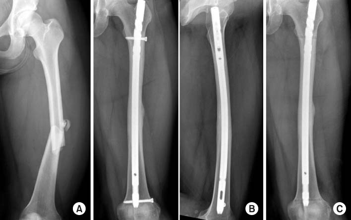
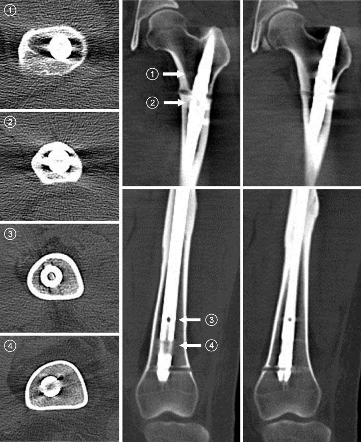
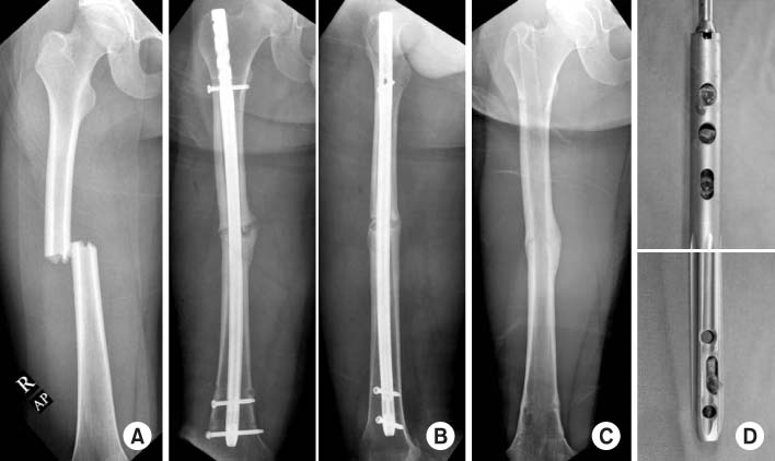
Fig. 1
A 16-year-old female patient sustained a left femoral shaft fracture. (A) An initial anteroposterior radiograph shows a transverse fracture of the femoral shaft. (B) Radiographs five years after fixing the fracture using a Sirus femoral nail show fracture healing with remodeling of bridging callus. (C) An anteroposterior radiograph taken after failure to remove the nail shows the presence of the nail without interlocking screws.
Fig. 2
Computed tomography scans show that dense compact bone had formed in the proximal and distal empty interlocking holes of the nail. Dense compact bone was more abundant in the dynamic interlocking hole.
Fig. 3
A 17-year-old male patient sustained a right femoral shaft fracture. (A) An initial anteroposterior radiograph shows a transverse fracture of the femoral shaft. (B) Radiographs taken after exchange nailing using an A2FN show good fixation for hypertrophic nonunion. (C) An anteroposterior radiograph taken after removal of the nail shows fracture healing with bridging callus. (D) Photographs of the removed nail show that dense compact bone formed through the proximal and distal empty interlocking holes.
Fig. 1
Fig. 2
Fig. 3
Failure to Remove a Trochanteric Entry Femoral Nail and Its Cause in Adolescent Patients: Two Cases Report

