 E-submission
 TOTA
TOTA TOTS
TOTS
Articles
- Page Path
- HOME > J Musculoskelet Trauma > Volume 24(3); 2011 > Article
-
Original Article
- Operative Treatment of Distal Humeral Comminuted Fractures with Orthogonal Plating
- Joong-Bae Seo, M.D., Jae-Sung Yoo, M.D.
-
Journal of the Korean Fracture Society 2011;24(3):243-248.
DOI: https://doi.org/10.12671/jkfs.2011.24.3.243
Published online: July 15, 2011
Department of Orthopedic Surgery, Dankook University College of Medicine, Cheonan, Korea.
- Address reprint requests to: Joong-Bae Seo, M.D. Department of Orthopaedic Surgery, Dankook University College of Medicine, 119, Dandae-ro, Dongnam-gu, Cheonan 330-715, Korea. Tel: 82-41-550-3060, Fax: 82-41-556-3238, ssjb1990@dku.edu
• Received: January 10, 2011 • Revised: March 1, 2011 • Accepted: June 20, 2011
Copyright © 2011 The Korean Fracture Society
- 1,010 Views
- 2 Download
Abstract
-
Purpose
- To analyze the results of operative treatment for Comminuted Fracture of Distal Humerus with Transolecranon approach and Orthogonal plating.
-
Materials and Methods
- The subjects were 22 patients with Comminuted fracture of humerus who were treated with Orthogonal plating. Patient's age, sex, type of fracture, surgical approach, method of fixation, time of operation, time of bony union, complication, range of motion were investigated, and Function of elbow was evaluated by functional evaluation of Riseborough and Radin, Mayo Elbow Performance Score (MEPS).
-
Results
- Age, sex, injuried arm, operation time were not related to postoperative result. Type C2 fractures showed better results in function and range of motion (ROM) than type C3 fractures. Also early rehabilitation was important to functional recovery and ROM. The postoperative ROM was average 110. Good were 16 cases, fair were 6 cases in functional evaluation of Riseborough and Radin. Excellent were 13 cases, good were 8 cases, fair was 1 case in MEPS.
-
Conclusion
- Operative treatment with Transolecranon approach and Orthogonal plating showed favorable result on its function. Intraarticular comminution and early rehabilitation were closely related to postoperative function of elbow.
- 1. Aitken GK, Rorabeck CH. Distal humeral fractures in the adult. Clin Orthop Relat Res, 1986;207:191-197.Article
- 2. Bryan RS, Morrey BF. Extensive posterior exposure of the elbow, A triceps sparing approach. Clin Orthop Relat Res, 1982;166:188-192.
- 3. Chen RC, Harris DJ, Leduc S, Borrelli JJ Jr, Tornetta P 3rd, Ricci WM. Is ulnar nerve transposition beneficial during open reduction internal fixation of distal humerus fractures? J Orthop Trauma, 2010;24:391-394.Article
- 4. Cho JH, Kim JY, Lee SY, Han KJ. Surgical treatment of intercondylar fractures of the humerus with posteior plates. J Korean Soc Surg Hand, 2008;13:212-216.
- 5. Eralp L, Kocaoglu M, Sar C, Atalar AC. Surgical treatment of distal intraarticular humeral fractures in adults. Int Orthop, 2001;25:46-50.ArticlePDF
- 6. Gupta R. Intercondylar fractures of the distal humerus in adults. Injury, 1996;27:569-572.Article
- 7. Gupta R, Khanchandani P. Intercondylar fractures of the distal humerus in adults: a critical analysis of 55 cases. Injury, 2002;33:511-515.Article
- 8. Helfet DL, Hotchkiss RN. Internal fixation of the distal humerus: a biomechanical comparison of methods. J Orthop Trauma, 1990;4:260-264.Article
- 9. Husband JB, Hastings H 2nd. The lateral approach for operative release of post-traumatic contracture of the elbow. J Bone Joint Surg Am, 1990;72:1353-1358.Article
- 10. Jupiter JB. Complex fractures of the distal part of the humerus and associated complications. J Bone Joint Surg Am, 1994;76:1252-1264.Article
- 11. Jupiter JB, David R. Orthopaedic knowledge update shoulder and elbow, 1997;1st ed. Philadelphia, AAOS. 397-404.
- 12. Jupiter JB, Neff U, Holzach P, Allgöwer M. Intercondylar fractures of the humerus. An operative approach. J Bone Joint Surg Am, 1985;67:226-239.Article
- 13. Kundel K, Braun W, Wieberneit J, Rüter A. Intraarticular distal humerus fractures. Factors affecting functional outcome. Clin Orthop Relat Res, 1996;332:200-208.
- 14. McCarty LP, Ring D, Jupiter JB. Management of distal humerus fractures. Am J Orthop (Belle Mead NJ), 2005;34:430-438.
- 15. Morrey BF, Sanchez J. Morrey BF. Functional evaluation of the elbow. In: The elbow and its disorders, 2009;4th ed. Philadelphia, WB Saunders. 87-88.Article
- 16. O'Driscoll SW. Optimizing stability in distal humeral fracture fixation. J Shoulder Elbow Surg, 2005;14:1 Suppl S. 186S-194S.
- 17. Pajarinen J, Björkenheim JM. Operative treatment of type C intercondylar fractures of the distal humerus: results after a mean follow-up of 2 years in a series of 18 patients. J Shoulder Elbow Surg, 2002;11:48-52.Article
- 18. Park JY, Seo JB, Chun JY, Kim MH, Min SH, Lee JH. Treatment of intercondylar fractures of humerus with Y-plate. J Korean Fract Soc, 2006;19:443-448.
- 19. Ring D, Jupiter JB. Complex fractures of the distal humerus and their complications. J Shoulder Elbow Surg, 1999;8:85-97.Article
- 20. Riseborough EJ, Radin EL. Intercondylar T fractures of the humerus in the adult. A comparison of operative and non-operative treatment in twenty-nine cases. J Bone Joint Surg Am, 1969;51:130-141.
- 21. Schemitsch EH, Tencer AF, Henley MB. Biomechanical evaluation of methods of internal fixation of the distal humerus. J Orthop Trauma, 1994;8:468-475.Article
- 22. Schuster I, Korner J, Arzdorf M, Schwieger K, Diederichs G, Linke B. Mechanical comparison in cadaver specimens of three different 90-degree double-plate osteosyntheses for simulated C2-type distal humerus fractures with varying bone densities. J Orthop Trauma, 2008;22:113-120.
- 23. Wang KC, Shih HN, Hsu KY, Shih CH. Intercondylar fractures of the distal humerus: routine anterior subcutaneous transposition of the ulnar nerve in a posterior operative approach. J Trauma, 1994;36:770-773.
REFERENCES
Fig. 1
(A) Anteroposterior and lateral radiographs of elbow in a 40-year-old man show intercondylar comminuted fracture of distal humerus (AO C3 type).
(B) Follow up anteroposterior and lateral radiographs 20weeks after operation show bony union without complication.


Figure & Data
REFERENCES
Citations
Citations to this article as recorded by 
 Cite
CiteOperative Treatment of Distal Humeral Comminuted Fractures with Orthogonal Plating
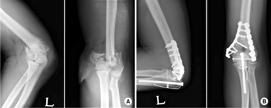
Fig. 1
(A) Anteroposterior and lateral radiographs of elbow in a 40-year-old man show intercondylar comminuted fracture of distal humerus (AO C3 type).
(B) Follow up anteroposterior and lateral radiographs 20weeks after operation show bony union without complication.
Fig. 1
Operative Treatment of Distal Humeral Comminuted Fractures with Orthogonal Plating
Summary of cases
*AO: AO classification, †OP day: period from injury to operation, ‡ROM: Range of motion, §MEPS: Mayo elbow performance score.
Comparison of clinical results between C2 fractures and C3 fractures
ROM: Range of motion, MEPS: Mayo elbow performance score.
Comparison of clinical results according to period of postoperativetime needed for spontaneous first elbow joint exercise
ROM: Range of motion, MEPS: Mayo elbow performance score.
Table 1
Summary of cases
*AO: AO classification, †OP day: period from injury to operation, ‡ROM: Range of motion, §MEPS: Mayo elbow performance score.
Table 2
Comparison of clinical results between C2 fractures and C3 fractures
ROM: Range of motion, MEPS: Mayo elbow performance score.
Table 3
Comparison of clinical results according to period of postoperativetime needed for spontaneous first elbow joint exercise
ROM: Range of motion, MEPS: Mayo elbow performance score.

