 E-submission
 TOTA
TOTA TOTS
TOTS
Articles
- Page Path
- HOME > J Musculoskelet Trauma > Volume 25(2); 2012 > Article
-
Review Article
- Lateral Condylar Fracture of the Humerus in Children: Is the Closed Pinning Saticfactory?
- Kwang-Soon Song, M.D., Ph.D.
-
Journal of the Korean Fracture Society 2012;25(2):163-168.
DOI: https://doi.org/10.12671/jkfs.2012.25.2.163
Published online: April 17, 2012
Department of Orthopedic Surgery, School of Medicine, Keimyung University, Daegu, Korea.
- Address reprint requests to: Kwang-Soon Song, M.D., Ph.D. Department of Orthopedic Surgery, School of Medicine, Keimyung University, 56, Dalseong-ro, Jung-gu, Daegu 700-712, Korea. Tel: 82-53-250-7250, Fax: 82-53-250-7205, skspos@dsmc.or.kr
Copyright © 2012 The Korean Fracture Society
- 2,020 Views
- 25 Download
- 1 Crossref
- 1. Badelon O, Bensahel H, Mazda K, Vie P. Lateral humeral condylar fractures in children: a report of 47 cases. J Pediatr Orthop, 1988;8:31-34.
- 2. Bast SC, Hoffer MM, Aval S. Nonoperative treatment for minimally and nondisplaced lateral humeral condyle fractures in children. J Pediatr Orthop, 1998;18:448-450.Article
- 3. Beaty JH, Kasser JR. Rockwood and Wilkins' fractures in children, 2001;6th ed. Philadelphia, Lippincott Williams & Wilkins. 592-610.
- 4. Canale ST. Campbell's operative orthopaedics, 2008;11th ed. St. Louis, Mosby. 1569-1575.
- 5. Conner AN, Smith MG. Displaced fractures of the lateral humeral condyle in children. J Bone Joint Surg Br, 1970;52:460-464.ArticlePDF
- 6. Crabbe WA. The treatment of fracture-separation of the capitular epiphysis. J Bone Joint Surg Br, 1963;45:722-726.ArticlePDF
- 7. Finnbogason T, Karlsson G, Lindberg L, Mortensson W. Nondisplaced and minimally displaced fractures of the lateral humeral condyle in children: a prospective radiographic investigation of fracture stability. J Pediatr Orthop, 1995;15:422-425.Article
- 8. Flynn JC. Nonunion of slightly displaced fractures of the lateral humeral condyle in children: an update. J Pediatr Orthop, 1989;9:691-696.
- 9. Flynn JC, Richards JF Jr. Non-union of minimally displaced fractures of the lateral condyle of the humerus in children. J Bone Joint Surg Am, 1971;53:1096-1101.Article
- 10. Flynn JC, Richards JF Jr, Saltzman RI. Prevention and treatment of non-union of slightly displaced fractures of the lateral humeral condyle in children. An end-result study. J Bone Joint Surg Am, 1975;57:1087-1092.Article
- 11. Foster DE, Sullivan JA, Gross RH. Lateral humeral condylar fractures in children. J Pediatr Orthop, 1985;5:16-22.Article
- 12. Hardacre JA, Nahigian SH, Froimson AI, Brown JE. Fractures of the lateral condyle of the humerus in children. J Bone Joint Surg Am, 1971;53:1083-1095.Article
- 13. Horn BD, Herman MJ, Crisci K, Pizzutillo PD, MacEwen GD. Fractures of the lateral humeral condyle: role of the cartilage hinge in fracture stability. J Pediatr Orthop, 2002;22:8-11.Article
- 14. Jakob R, Fowles JV, Rang M, Kassab MT. Observations concerning fractures of the lateral humeral condyle in children. J Bone Joint Surg Br, 1975;57:430-436.ArticlePDF
- 15. Kamegaya M, Shinohara Y, Kurokawa M, Ogata S. Assessment of stability in children's minimally displaced lateral humeral condyle fracture by magnetic resonance imaging. J Pediatr Orthop, 1999;19:570-572.Article
- 16. Launay F, Leet AI, Jacopin S, Jouve JL, Bollini G, Sponseller PD. Lateral humeral condyle fractures in children: a comparison of two approaches to treatment. J Pediatr Orthop, 2004;24:385-391.
- 17. Marzo JM, d'Amato C, Strong M, Gillespie R. Usefulness and accuracy of arthrography in management of lateral humeral condyle fractures in children. J Pediatr Orthop, 1990;10:317-321.Article
- 18. Milch H. Fractures and fracture-dislocations of humeral condyles. J Trauma, 1964;4:592-607.
- 19. Mirsky EC, Karas EH, Weiner LS. Lateral condyle fractures in children: evaluation of classification and treatment. J Orthop Trauma, 1997;11:117-120.Article
- 20. Noonan K. Resident review. Top five pediatric orthopedic conditions to diagnose in the emergency room. POSNA, 2011;02;5-6.
- 21. Rang M, Pring ME, Wenger DR. Rang's Children's Fractures, 2005;3rd ed. Philadelphia, Lippincott William, and Wilkins. 113-114.
- 22. Song KS, Kang CH, Min BW, Bae KC, Cho CH. Internal oblique radiographs for diagnosis of nondisplaced or minimally displaced lateral condylar fractures of the humerus in children. J Bone Joint Surg Am, 2007;89:58-63.Article
- 23. Song KS, Kang CH, Min BW, Bae KC, Cho CH, Lee JH. Closed reduction and internal fixation of displaced unstable lateral condylar fractures of the humerus in children. J Bone Joint Surg Am, 2008;90:2673-2681.Article
- 24. Water PM, Song KS. Latral condyle: Should We fix them all? Pediatric orthopaedic trauma: the cases for best treatment. Proceedings of the POSNA 2010 half daycourse, 2010;2010 May 4; Waikoloa (HI), Pediatric Orthopedic Society of North America. 43-46.
- 25. Song KS, Shin YW, Oh CW, Bae KC, Cho CH. Closed reduction and internal fixation of completely displaced and rotated lateral condyle fractures of the humerus in children. J Orthop Trauma, 2010;24:434-438.Article
- 26. Thönell S, Mortensson W, Thomasson B. Prediction of the stability of minimally displaced fractures of the lateral humeral condyle. Acta Radiol, 1988;29:367-370.Article
- 27. Vocke-Hell AK, Schmid A. Sonographic differentiation of stable and unstable lateral condyle fractures of the humerus in children. J Pediatr Orthop B, 2001;10:138-141.Article
- 28. Wadsworth TG. Injuries of the capitular (lateral humeral condylar) epiphysis. Clin Orthop Relat Res, 1972;85:127-142.Article
REFERENCES

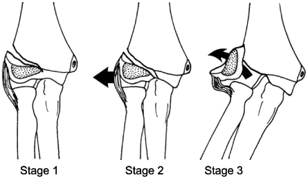

Fig. 4
(A) Anteroposterior radiograph of the elbow, showing a Finnbogason type A fracture (stable).
(B) Internal oblique radiograph of the same patient showing a Finnbogason type C (unstable) traversing the capitellum.

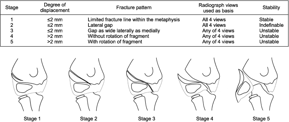
Fig. 6Algorithm of treatment. Closed reduction and internal fixation is suggested as first line treatment option for unstable fracture.
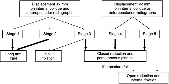
Fig. 7
(A) Anteroposterior radiograph showing a completely displaced fracture with rotation of the fracture fragment. Intra-operative (B) anteroposterior and (C) lateral radiographs from same patient showing reduction by pushing the fragment backward and medially with operator's thumb. Intra-operative (D) anteroposterior and lateral (E) radiographs showing final reduction of the fragment <2 mm of displacement with Kirschner wire. (F) Post-reduction aneteroposterior arthrogram showing fixation with three Kirschner wires, congruent reduction of the articular surface.

Figure & Data
REFERENCES
Citations
Citations to this article as recorded by 
- Diagnosis, management and complications of distal humerus lateral condyle fractures in children
Daniel A Shaerf, Ivor S Vanhegan, Rupen Dattani
Shoulder & Elbow.2018; 10(2): 114. CrossRef
 Cite
CiteLateral Condylar Fracture of the Humerus in Children: Is the Closed Pinning Saticfactory?
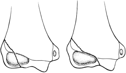

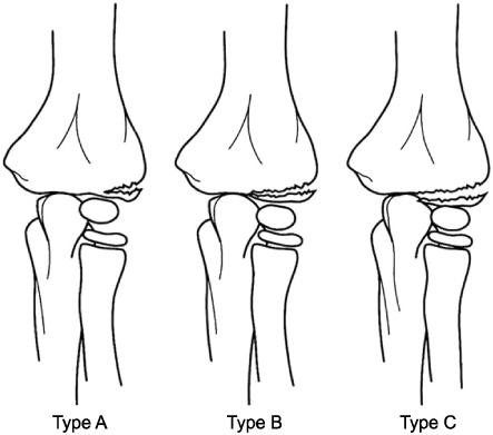
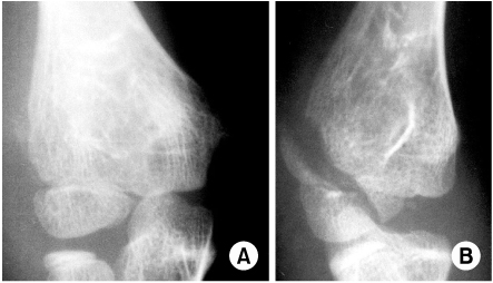


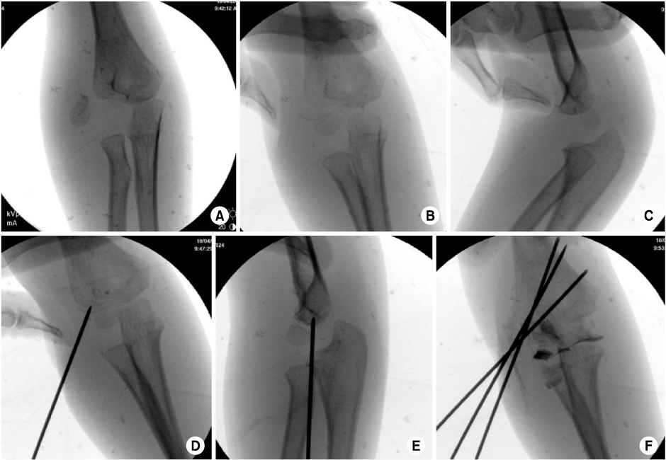
Fig. 1
Milch classification.
Fig. 2
Jakob classification.
Fig. 3
Finnbogason classification.
Fig. 4
(A) Anteroposterior radiograph of the elbow, showing a Finnbogason type A fracture (stable).
(B) Internal oblique radiograph of the same patient showing a Finnbogason type C (unstable) traversing the capitellum.
Fig. 5
Classification according to degree of displacement and fracture pattern.
Fig. 6
Algorithm of treatment. Closed reduction and internal fixation is suggested as first line treatment option for unstable fracture.
Fig. 7
(A) Anteroposterior radiograph showing a completely displaced fracture with rotation of the fracture fragment. Intra-operative (B) anteroposterior and (C) lateral radiographs from same patient showing reduction by pushing the fragment backward and medially with operator's thumb. Intra-operative (D) anteroposterior and lateral (E) radiographs showing final reduction of the fragment <2 mm of displacement with Kirschner wire. (F) Post-reduction aneteroposterior arthrogram showing fixation with three Kirschner wires, congruent reduction of the articular surface.
Fig. 1
Fig. 2
Fig. 3
Fig. 4
Fig. 5
Fig. 6
Fig. 7
Lateral Condylar Fracture of the Humerus in Children: Is the Closed Pinning Saticfactory?

