
 E-submission
 KOTA
KOTA TOTA
TOTA TOTS
TOTS
Search
- Page Path
- HOME > Search
Case Report
- Reconstruction of a 20-cm tibial bone defect after recurrent fracture-related infection using the induced membrane technique and rhBMP-2 augmentation with limited autogenous bone availability: a case report
- Whee Sung Son
- Received March 30, 2026 Accepted June 29, 2026 Published online August 21, 2026
- DOI: https://doi.org/10.12671/jmt.2026.00143 [Epub ahead of print]
-
 Abstract
 PDF
PDF - Fracture-related infection (FRI) poses the combined challenges of infection control and reconstruction of critical-sized bone defects. These challenges are amplified in recurrent cases with severely limited autogenous bone availability. We report a 61-year-old male patient with recurrent right tibial FRI after multiple operations and a 20-cm segmental defect. Suspected residual infectious foci were mapped by comparing preoperative fluorodeoxyglucose positron emission tomography/computed tomography (PET/CT) with post-debridement CT and were evaluated using site-specific pre- and post-debridement cultures. After a second targeted debridement, all post-debridement cultures were negative, and no clinical or laboratory evidence of active infection was observed during a 2-week antibiotic-free interval. The defect was subsequently reconstructed using the induced membrane technique with 30 cm³ of autogenous cancellous bone, 60 cm³ of demineralized bone matrix, and 9 mg of recombinant human bone morphogenetic protein-2 (rhBMP-2) delivered on a 20 cm³ hydroxyapatite carrier. Radiographic union was achieved, and no clinical evidence of infection recurrence was observed at 1 year. This case suggests the feasibility of combining PET/CT- and culture-guided anatomically targeted debridement with rhBMP-2-augmented induced membrane reconstruction when autogenous graft volume is severely limited. Level of evidence: V.
- 298 View
- 9 Download

Original Article
- Percutaneous anterior leverage technique for anteromedial cortical support in intertrochanteric femur fractures: a computed tomography-based validation study
- Whee Sung Son, Bum Jin Shim, Oog-jin Shon
- J Musculoskelet Trauma 2026;39(2):117-129. Published online March 27, 2026
- DOI: https://doi.org/10.12671/jmt.2025.00311
-
Abstract
PDF
- Background
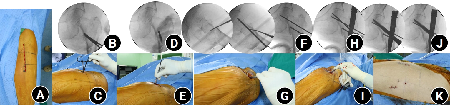
Anteromedial cortical support (AMCS) enhances stability in intertrochanteric femur fractures. However, reproducible, validated methods of achieving AMCS have not previously been reported. This study introduces a percutaneous anterior leverage technique and validates its AMCS effects using computed tomography (CT).
Methods
We retrospectively reviewed patients treated by a single surgeon between March 2022 and December 2024. The inclusion criteria were an AO/OTA classification of A1–A3, application of the percutaneous anterior leverage technique, available pre- and postoperative CT, and ≥6 months follow-up. Outcomes included CT-based AMCS (anterior on axial and medial on coronal images, classified as positive, neutral, or negative), time to union, union rate, changes in neck-shaft angle, and treatment failure (varus collapse, blade cut-through, or nonunion without the former two). The risk factors for failure were analyzed.
Results
Of 273 patients reviewed, 53 met the inclusion criteria. Follow-up was at least 6 months in all cases. Positive anterior support was achieved in 37 patients (69.8%) and positive medial support in 42 (79.25%). No patient demonstrated negative anterior support; one (1.9%) had negative medial support. Cortical support improved significantly after surgery. CT images demonstrated significant postoperative improvements (anterior P=0.026; medial P<0.001). Bone union was achieved in 50 patients (94.34%) at a mean of 3.93±1.48 months. The mean change in the neck-shaft angle at last follow-up was 1.75°±2.34° varus. Three patients (5.66%) experienced treatment failure. Anteromedial cortical breakage during follow-up differed between failure and nonfailure groups (P=0.002), but regression identified no independent predictors. No technique-related complications were observed.
Conclusions
Our percutaneous anterior leverage technique produced favorable CT-confirmed AMCS and high union with low failure, supporting its safety and effectiveness in intertrochanteric femur fractures. Level of evidence: IV. -
Citations
Citations to this article as recorded by
- Association of Long Versus Standard-Length Cephalomedullary Nail Constructs with Posterior Sagging in Intertrochanteric Fractures with Posteromedial Comminution: A Retrospective Comparative Study
Jun-Young Heu, Se-Won Lee, Ju-Yeong Kim
Journal of Clinical Medicine.2026; 15(18): 7208. CrossRef
- Association of Long Versus Standard-Length Cephalomedullary Nail Constructs with Posterior Sagging in Intertrochanteric Fractures with Posteromedial Comminution: A Retrospective Comparative Study
- 1,937 View
- 57 Download
- 1 Crossref
Technical Note
- Rim plate-assisted intramedullary nail and plate combination technique for complex tibial plateau-to-diaphysis fractures: a technical note and case series
- Whee Sung Son
- J Musculoskelet Trauma 2026;39(1):62-71. Published online December 4, 2025
- DOI: https://doi.org/10.12671/jmt.2025.00290

-
Abstract
PDF
- Complex tibial plateau-to-diaphysis fractures present a significant surgical challenge due to their intricate fracture patterns and frequent association with severe soft tissue damage and concomitant injuries. This technical note introduces a novel fixation strategy: the rim plate-assisted intramedullary nail-plate combination (NPC) technique. In this approach, a rim plate simplifies the conventional NPC procedure by unifying the tibial plateau fracture into a single structural segment. This modification eliminates the need to address the articular and diaphyseal components simultaneously while enhancing articular stability. Furthermore, the technique preserves soft tissue integrity and promotes early rehabilitation. Clinical case examples demonstrate its successful application in managing complex tibial plateau-to-diaphysis injuries. Level of evidence: V.
- 2,217 View
- 83 Download
 First
First Prev
Prev


