
 E-submission
 KOTA
KOTA TOTA
TOTA TOTS
TOTS
Search
- Page Path
- HOME > Search
Case Reports
- Progressive forearm instability after anatomical union of a radial head fracture: a case report of a challenging Essex-Lopresti injury
- SeongJu Choi
- Received April 6, 2026 Accepted May 26, 2026 Published online July 20, 2026
- DOI: https://doi.org/10.12671/jmt.2026.00157 [Epub ahead of print]
-
 Abstract
 PDF
PDF - Essex-Lopresti injuries remain challenging because anatomical restoration of bony structures does not always re-establish functional forearm stability. We report the case of a 51-year-old male patient who sustained an elbow dislocation, comminuted radial head and neck fracture, and distal radioulnar joint (DRUJ) dissociation after falling from a bicycle. He underwent open reduction and internal fixation of the radial head, interosseous membrane augmentation using a suture-button construct, and temporary DRUJ pinning. Immediate postoperative radiographs showed apparent restoration of radial length, and radiographic union of the radial head was achieved. However, ulnar-sided wrist pain initially improved but progressively recurred by 6 months, with increasing ulnar-positive variance and DRUJ widening. Elbow motion was preserved, but forearm rotation was limited, and computed tomography demonstrated heterotopic ossification around the radial head. Ulnar shortening osteotomy and radial head arthrolysis were performed as a second procedure, after which mechanical instability developed, with irreducible anterior dislocation of the radial head. A third operation, consisting of annular ligament reconstruction using a palmaris longus autograft, restored proximal radioulnar joint stability, although substantial limitation of forearm rotation persisted. This case highlights that even radiographic union and multistructural initial management may fail to restore balanced forearm mechanics in Essex-Lopresti injury. Level of evidence: V.
- 292 View
- 4 Download

- Radiocarpal subluxation after volar plating due to an insufficiently supported dorsal key fragment: a case report
- Yeongyoon Koh, Kanghun Yu, Jong Woong Park, In Cheul Choi
- J Musculoskelet Trauma 2026;39(3):269-274. Published online July 3, 2026
- DOI: https://doi.org/10.12671/jmt.2026.00185
-
Abstract
PDF
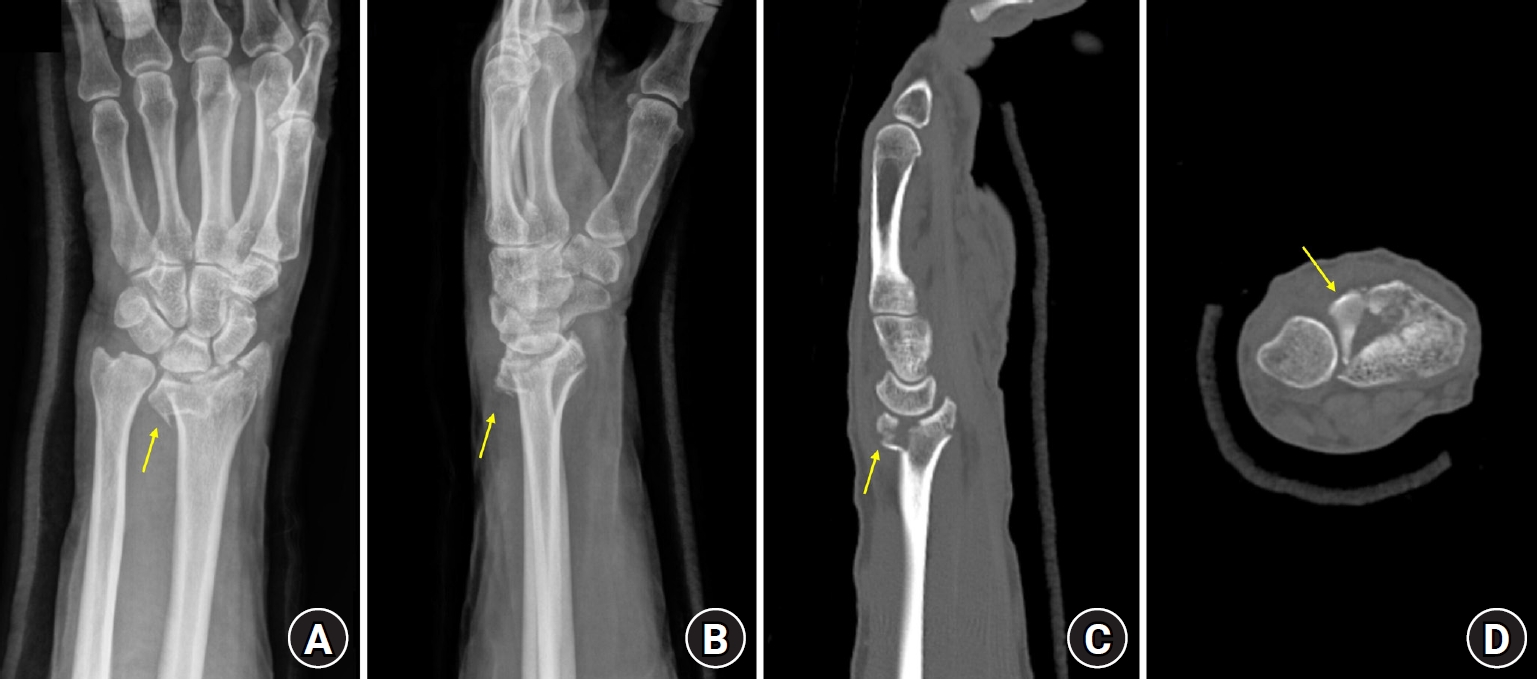
- Distal radius fractures are commonly managed using volar locking plate fixation, which provides stable fixation in most cases. However, certain fracture patterns involving dorsal key fragments may not be adequately stabilized with a volar approach alone, even when intraoperative reduction appears satisfactory. We report the case of a 58-year-old male patient with a complex intra-articular distal radius fracture involving a dorsoulnar corner (DUC) fragment. Preoperative computed tomography revealed a dorsal fragment associated with subtle dorsal radiocarpal subluxation. Despite this finding, the fragment was considered amenable to fixation through a volar approach because it was relatively large. Although satisfactory reduction was achieved intraoperatively, early postoperative imaging demonstrated progressive dorsal radiocarpal subluxation due to displacement of the DUC fragment, while overall alignment parameters remained preserved. Revision surgery with additional dorsal buttress fixation successfully restored stability. This case highlights the critical importance of recognizing dorsal key fragments and achieving adequate dorsal support during fixation, as failure to provide sufficient support may result in delayed instability despite acceptable initial reduction. Level of evidence: V.
- 345 View
- 12 Download
Review Article
- Definitive fixation for traumatic pelvic ring injuries: a dynamically informed, posterior-referenced framework
- Jeong-Hyun Koh, Seungyeob Sakong
- J Musculoskelet Trauma 2026;39(2):73-82. Published online March 24, 2026
- DOI: https://doi.org/10.12671/jmt.2026.00045
-
Abstract
PDF
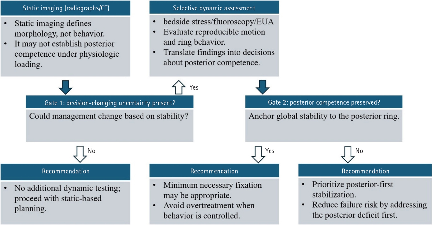
- Optimal definitive fixation for traumatic pelvic ring injuries remains challenging because static radiographs and computed tomography, although essential for defining morphology, do not consistently predict load-dependent behavior during early mobilization. This uncertainty contributes to substantial practice variation and continued reliance on simplified displacement thresholds, such as the 2.5 cm rule. Such rules can misclassify instability by underrepresenting posterior competence and by privileging static measurements over functional behavior. In this narrative review, we propose a dynamically informed, posterior- referenced framework composed of three linked elements: (1) decision-linked terminology that explicitly distinguishes dynamic instability, radiographic change, and clinical failure; (2) selective stress-based assessment when uncertainty is likely to alter management; and (3) escalation along a fixation continuum that weighs incremental stability against operative burden. When static imaging cannot establish posterior competence with confidence, we outline selective stress-based approaches to assess pelvic ring behavior and to translate demonstrated instability into fixation selection along a defined continuum. Across all steps, the framework emphasizes minimum necessary fixation and explicitly incorporates the cost of selection as a primary decision variable. The operative question, therefore, shifts from gap width alone to clinically relevant motion and preservation of posterior competence. In doing so, this approach aims to reduce both undertreatment and overtreatment and to improve the consistency and defensibility of definitive fixation strategies across diverse practice environments.
- 2,011 View
- 44 Download
 First
First Prev
Prev


