 E-submission
 TOTA
TOTA TOTS
TOTS
Search
- Page Path
- HOME > Search
Review Article
- Nonoperative management of distal radius fractures: when and how?
- Shin Woo Choi, Jae Kwang Kim
- J Musculoskelet Trauma 2026;39(2):93-102. Published online March 10, 2026
- DOI: https://doi.org/10.12671/jmt.2026.00024
-
 Abstract
 PDF
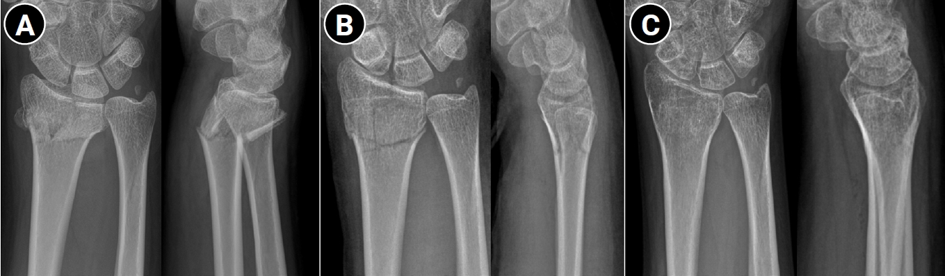
PDF - Distal radius fractures are among the most common injuries of the upper extremity, particularly in the elderly population. Although the use of volar locking plate fixation has increased in recent years, evidence from randomized and prospective studies demonstrates that, while operative treatment may achieve superior radiographic alignment and enable more rapid early recovery, these advantages tend to diminish over time and do not result in superior long-term patient-reported functional outcomes in elderly patients. In addition, radiographic parameters show only a limited correlation with functional recovery. Consequently, nonoperative treatment remains a valid and important treatment option for distal radius fractures. The decision to pursue nonoperative management should be based on a comprehensive assessment of radiographic parameters—including dorsal tilt, radial shortening, and intraarticular displacement—together with patient-specific factors such as age, activity level, comorbidities, and functional expectations. For stable or minimally displaced fractures, an immobilization period of 3‒4 weeks is generally recommended, whereas displaced fractures typically require immobilization for 5‒6 weeks. In cases requiring manual reduction, traditional treatment protocols recommend weekly radiographic follow-up during the first 2‒3 weeks to monitor for secondary displacement. Successful nonoperative management should also emphasize effective swelling control through limb elevation, as well as the initiation of early finger exercises to prevent hand stiffness. After removal of the cast or splint, active wrist mobilization is essential for restoring optimal range of motion and achieving functional recovery.
- 3,112 View
- 50 Download

Original Article
- Demographic and Radiographic Parameters as Predictors of Reduction Loss after Conservative Treatment of Distal Radius Fractures in Adults
- Kyu Jin Kim, Dae Won Shin, Seong Kee Shin
- J Korean Fract Soc 2023;36(2):45-51. Published online April 30, 2023
- DOI: https://doi.org/10.12671/jkfs.2023.36.2.45
-
Abstract
PDF
- Purpose
This study examined the demographic and radiological risk factors for later reduction loss of distal radius fractures treated conservatively. Materials and Methods This study enrolled patients treated for distal radius fractures between January 2017 and December 2019. Seventy-eight patients were included in the analysis and divided into two groups. The patients who showed minimal reduction loss within an acceptable radiologic angle after initial manual reduction were classified as Group A. The patients who showed reduction loss out of an acceptable radiologic angle and finally malunited or converted to surgical treatments were classified as Group B. The patient’s age and bone marrow density were used as demographic data. The initial X-ray images were evaluated to determine the fracture type. Various radiological parameters were measured. Results The 78-patient study cohort consisted of nine men and 69 women with a mean age of 67 years. Forty-eight cases were sorted into Group A, and 30 cases into Group B. On logistic regression analysis, the age of 80 or older was a risk factor for later fracture displacement among the demographic factors (p=0.037, odds ratio=4.937). Among the radiographic factors, the presence of distal ulnar fracture and dorsal cortical comminution were disclosed as risk factors of later displacement (p=0.049, 0.003, odds ratio=3.429, 7.196). Conclusion When conservative management for distal radius fracture is decided in patients more than 80 years of age or accompanied by a distal ulnar fracture or with dorsal cortical comminution, the possibility of later displacement of the distal radius should be considered.
- 829 View
- 4 Download
Review Articles
- Treatment Options of Osteoporotic Vertebral Compression Fractures
- Yu Mi Kim, Tae Kyun Kim, Dae Moo Shim, Kyeong Hoon Lim
- J Korean Fract Soc 2018;31(3):114-121. Published online July 31, 2018
- DOI: https://doi.org/10.12671/jkfs.2018.31.3.114
-
Abstract
PDF
- This paper reviews previous studies on the treatment of osteoporotic vertebral compression fractures in elderly patients to determine what factors should be considered for successful treatment. In osteoporotic vertebral compression fractures, the primary treatment is conservative treatments. Other treatments include osteoporosis treatment, pain control, orthosis, and physical therapy. Recently, percutaneous catheterization or balloon plasty is performed for rapid pain recovery and early ambulation. Percutaneous catheterization or balloon posterior plasty is effective in reducing pain and improving the activity ability. Surgical treatment should be considered in cases of nonunion or osteonecrosis, dent, deformation, and spinal cord compression after conservative treatment has failed. In surgical treatment, posterior spinal fixation and vertebroplasty are more advantageous in terms of the amount of bleeding, operation time compared to the anterior approach, but the most appropriate method should be selected through the patient's condition and understanding of each surgical method.
-
Citations
Citations to this article as recorded by
- Maigne Syndrome and Thoracolumbar Compression Fracture – An Overlooked Combination in Low Back Pain: A Case Report
Jae-Yong Shim, Myung-Hoon Shin
The Nerve.2025; 11(1): 21. CrossRef - Effects of Herbal Medicines on Bone Mineral Density Score in Osteoporosis or Osteopenia: Study Protocol for a Systematic Review and Meta-Analysis
Su Min Hong, Eun Jung Lee
Journal of Korean Medicine Rehabilitation.2021; 31(2): 49. CrossRef -
Spinal Stability Evaluation According to the Change in the Spinal Fixation Segment Based on Finite Element Analysis
Cheol-Jeong Kim, Seung Min Son, Jin-Young Heo, Chi-Seung Lee
Journal of the Computational Structural Engineering Institute of Korea.2020; 33(3): 145. CrossRef
- Maigne Syndrome and Thoracolumbar Compression Fracture – An Overlooked Combination in Low Back Pain: A Case Report
- 1,051 View
- 11 Download
- 3 Crossref
- Conservative Treatment of Proximal Humeral Fracture
- Hwansub Hyun, Jonghyun Ahn, Sang Jin Shin
- J Korean Fract Soc 2018;31(1):29-35. Published online January 31, 2018
- DOI: https://doi.org/10.12671/jkfs.2018.31.1.29
-
Abstract
PDF
- A proximal humeral fracture is an osteoporotic fracture that often occurs in elderly women. Approximately 80% of all proximal humeral fractures are non-displaced fractures, which can be treated with conservative treatment to achieve stable union. The treatment plan for fractures involving displaced and comminuted fractures is controversial. Malunion, avascular necrosis of the humeral head, and shoulder stiffness due to conservative treatment can occur but the functional deterioration is low and the patient satisfaction is high. The indications for the conservative management of proximal humeral fractures include a non-displaced fracture and a 2-part fracture, low-functional demanded 3-part fracture, and operative-limited 4-part fracture. Recently, the surgical indications have expanded as technological advances in surgical fixation methods and functional needs of elderly patients are increasing. Current treatment policy decisions tend to be determined by the personal preference and expert opinion rather than by evidence-based decision-making.
-
Citations
Citations to this article as recorded by- The Effect of Postoperative Korean Traditional Medicine for the of Proximal Humeral Fracture: A Case Report
Hyun Il Go, Hangyul Choi, Jieun Hong, Nam geun Cho
Journal of Acupuncture Research.2019; 36(1): 50. CrossRef
- The Effect of Postoperative Korean Traditional Medicine for the of Proximal Humeral Fracture: A Case Report
- 934 View
- 8 Download
- 1 Crossref
- Conservative Treatment of Mid-Clavicle Fractures
- Woong Kyo Jeong
- J Korean Fract Soc 2018;31(1):22-28. Published online January 31, 2018
- DOI: https://doi.org/10.12671/jkfs.2018.31.1.22
-
Abstract
PDF
- Clavicle fractures are very common injuries in adults and children and the majority of these fractures occur in the midshaft. Traditionally, mid-clavicle fractures have been treated with conservative methods and the clinical outcomes of this method are believed to be excellent. On the other hand, recent studies have shown that the clinical results of severe comminuted or markedly displaced fractures after conservative management were not as favorable as previously described. Despite these concerns, the conservative treatment of mid-clavicle fractures is still an efficient method, which can be applied to all patients as a primary care. This review focuses on the proper indication, technique, and limitations of conservative treatment of mid-clavicle fractures.
-
Citations
Citations to this article as recorded by- Two Patients Who Were Hospitalized for Clavicle Fracture Caused by a Traffic Accident and Improved with Korean Medicine Complex Treatment
Deok Kang, ByungSoo Kang, Hwe-Joon Jeong, Dong-Hoon Shin, Kyung-Moon Shin, Ji-Hoon O, Jae-Woo Yang
Journal of Korean Medicine Rehabilitation.2022; 32(3): 179. CrossRef
- Two Patients Who Were Hospitalized for Clavicle Fracture Caused by a Traffic Accident and Improved with Korean Medicine Complex Treatment
- 858 View
- 2 Download
- 1 Crossref
Case Report
- The Different Treatment Methods for Segmental Fractures of the Clavicle: Cases Report
- Sung Sik Ha, Ki Do Hong, Jae Cheon Sim, Yi Rak Seo, Tae Seok Nam
- J Korean Fract Soc 2017;30(3):151-155. Published online July 31, 2017
- DOI: https://doi.org/10.12671/jkfs.2017.30.3.151
-
Abstract
PDF
- Segmental fractures of the clavicle are very rare. Therefore, to date, there has not been a clear, standardized method of management of segmental clavicle fractures. Herein, two patients with a segmental fracture are described: One patient was treated conservatively, while another patient was treated operatively. Both patients showed excellent results. We discuss the various management options with a literature review.
-
Citations
Citations to this article as recorded by- Fratura segmentar da clavícula em paciente politraumatizado: Relato de caso
Carlos A. Sánchez, Pablo J. Coronel, Luisa F. García, Juan S. Afanador, Raúl Gonzalez
Revista Brasileira de Ortopedia.2024; 59(01): e139. CrossRef
- Fratura segmentar da clavícula em paciente politraumatizado: Relato de caso
- 1,662 View
- 5 Download
- 1 Crossref
Original Article
- The Result of Conservative Treatment of Proximal Humerus Fracture in Elderly Patients
- Seung Gil Baek, Chang Wug Oh, Young Soo Byun, Jong Keon Oh, Joon Woo Kim, Jong Pil Yoon, Hyun Joo Lee, Hyung Sub Kim
- J Korean Fract Soc 2013;26(4):292-298. Published online October 31, 2013
- DOI: https://doi.org/10.12671/jkfs.2013.26.4.292
-
Abstract
PDF
- PURPOSE
With the increase in the old age population, proximal humerus fractures have been increasing recently. However, complications after operative treatment, such as fixation failure, are common because of osteoporosis. We treated proximal humerus fractures in patients with osteoporosis conservatively, and evaluated the radiographic and functional results by analyzing the factors affecting the results.
MATERIALS AND METHODS
Nineteen out of 30 cases for whom the clinical follow-up was over 1 year were included in this retrospective study. There were 17 females and 2 males, and the mean age was 73.2 years. The causes were slip from a short height (18 cases) and a minor car accident (1 case). We evaluated the union period, nonunion, malunion and the Constant score and analyzed several factors affecting the functional result, such as age, fracture pattern, and malunion.
RESULTS
Seventeen cases (89.5%) obtained union within 12.8 weeks on average. Neck-shaft angle was 125.3degrees on average, with seven cases of malunion. The Constant score was 84.1 on average, and there were excellent scores in 11 cases, good scores in 4 cases, and fair scores in 2 cases. Fracture pattern, neck-shaft angle, or malunion did not affect the functional outcome, and elderly patients showed poorer shoulder function.
CONCLUSION
Proximal humeral fractures with osteoporosis may achieve a high rate of bony union when treated with conservative methods. Despite the common occurrence of malunion, a satisfactory functional outcome may be expected.
- 1,394 View
- 12 Download
Case Report
- Conservative Treatment of Valgus Impacted Four-Part Fracture of the Proximal Humerus: A Case Report
- Moon Chan Kim, Jae Lim Cho, Hung Tae Chung, Dong Jun Kim, In Bo Kim
- J Korean Fract Soc 2011;24(1):96-99. Published online January 31, 2011
- DOI: https://doi.org/10.12671/jkfs.2011.24.1.96
-
Abstract
PDF
- For valgus impacted four part fracture of the proximal humerus, surgical stabilization and early mobilization of the joint can produce the best clinical outcomes. But, we have experienced a case of conservative treatment and gained good clinical results. We have reported this case and included a review of the relevant literatures.
- 1,163 View
- 20 Download
Randomized Controlled Trial
- Comparison of the Result of Vertebroplasty and Conservative Treatment in Osteoporotic Vertebral Compression Fracture
- Ye Soo Park, Woo Jin Cho, Jae Lim Cho
- J Korean Fract Soc 2006;19(3):363-368. Published online July 31, 2006
- DOI: https://doi.org/10.12671/jkfs.2006.19.3.363
-
Abstract
- PURPOSE
To evaluate the results of vertebroplasty and conservative treatment in osteoporotic vertebral compression fractures.
MATERIALS AND METHODS
Patients were divided randomly into 2 groups; Group I (conservative treatment) and Group II (vertebroplasty). There are 14 cases in group I and 16 cases in group II. Radiologically, the progression of compression was observed. Clinical evaluation was done using Denis pain scale. In both groups, prolonged pain with nonunion or avascular necrosis that resulted in surgical intervention was evaluated as complication. In group II, the complication associated the procedures were evaluated.
RESULTS
Group II was superior to conservative treatment in terms of maintaining vertebral height radiologically. The characteristics of symptom improvement were the same in two groups. There were cement leakage among group II but they did not influence to the results. In group I, 2 subjects needed surgery due to prolonged pain. In group II, 1 subject needed surgery due to prolonged pain and there were 3 cement leakage cases which were insignificant.
CONCLUSION
In vertebroplasty group, complications associated the procedures were noted. In conservative treatment group, more patients needed operation. Therefore, we should be very prudent when we choose the treatment of the osteoporotic vertebral compression fracture. -
Citations
Citations to this article as recorded by- A Case Report of the Korean Medical Treatment of Dysphagia and Anorexia after Lumbar Compression Fracture
Hye-mi Jo, Eun-chang Lee, Hye-soo Youn, Choong-hyun Park, Da-young Han, Da-hae Jung, Jung-eun Lee
The Journal of Internal Korean Medicine.2022; 43(2): 219. CrossRef - A Retrospective Clinical Survey of Vertebral Compression Fractures
Ji Hye Oh, Yun Kyu Lee, Jae Soo Kim, Hyun Jong Lee, Sung Chul Lim
Journal of Acupuncture Research.2018; 35(4): 219. CrossRef - Lumbar Spine Fracture
Seung-Wook Back, Hyun-Joong Cho, Ye-Soo Park
Journal of the Korean Fracture Society.2011; 24(3): 277. CrossRef
- A Case Report of the Korean Medical Treatment of Dysphagia and Anorexia after Lumbar Compression Fracture
- 2,236 View
- 0 Download
- 3 Crossref
Original Articles
- Nonoperative Treatment of Isolated Lateral Malleolar Fracture
- Woo Chun Lee, Jong Ho Ahn
- J Korean Fract Soc 2005;18(3):291-293. Published online July 31, 2005
- DOI: https://doi.org/10.12671/jkfs.2005.18.3.291
-
Abstract
PDF
- PURPOSE
To evaluate the results of conservative treatment for isolated lateral malleolus fracture without medial ankle injury.
MATERIALS AND METHODS
From March 1999 to February 2003, 25 ankles in 25 patients were treated for isolated lateral malleolus fracture and followed for more than one year. Mean age was 46.9 years (range, 20~71 years). Cases without any swelling or tenderness on the deltoid area, or cases with minimal pain, swelling or tenderness on the deltoid area and medial clear space 1 mm or less on stress radiograph were included for the study. Immediate weight bearing was allowed with below-knee cast immobilization in all cases.
RESULTS
All were supinatin-external rotation stage II injury and mean duration of cast immobilization was 6.3+/-1.6 weeks after injury. There was no case which showed widening of medial clear space during routine radiographic follow-up. There was no change in the degree of displacement in spite of immediate weight bearing with short leg cast on.
CONCLUSION
Because the lateral malleolus fracture without medial injury can be managed nonoperatively, we need to differentiate this type of fracture to avoid unnecessary surgery, and for early return to normal daily activity. -
Citations
Citations to this article as recorded by- Posterior Plating in Distal Fibular Fracture
Choong-Hyeok Choi, Young-A Cho, Jae-Hoon Kim, Il-Hoon Sung
Journal of the Korean Fracture Society.2007; 20(2): 161. CrossRef
- Posterior Plating in Distal Fibular Fracture
- 1,393 View
- 5 Download
- 1 Crossref
- A Comparison of Vertebroplasty Versus Conservative Treatment in Osteoporotic Compression Fractures
- Sang Ho Moon, Dong Joon Kim, Chung Soo Hwang, Sang Eon Lee, Se Won Park
- J Korean Fract Soc 2004;17(4):374-379. Published online October 31, 2004
- DOI: https://doi.org/10.12671/jkfs.2004.17.4.374
-
Abstract
PDF
- PURPOSE
To compare clinical and radiological results between vertebroplasty and conservative treatment in osteoporotic compression fractures of thoracolumbar spine.
MATERIALS AND METHODS
34 patients were reviewed with at least 1 year follow up. Vertebroplasty was used in 14 and conservative treatment was done in 20 fractures. These groups were compared by clinical results which were evaluated by the scoring system according to pain, mobility and analgesic usage at preoperative, postoperative 1 month and postoperative 1 year. And also compared by the increment of kyphosis and loss of vertebral body height in lateral films at the same time. We compared duration of hospitalization between two groups.
RESULTS
Vertebroplasty group showed statistically significant less pain and mobility than conservative treatment (p<0.05), but there was no differences in analgesic usage at postoperative 1 year while significant difference at 1 month. In radiological comparison, vertebroplasty showed less increment of kyphosis and loss of body height significantly (p<0.05). Also vertebroplasty group had shorter hospitalization stay significantly (p<0.05).
CONCLUSION
Our retrospective analysis demonstrated that vertebroplasty provided significant pain relief, improvement of motion and reduction of analgesic usage and also provided considerable spinal stabilization that prevented further kyphosis and collapse. -
Citations
Citations to this article as recorded by- Outcome Comparison between Percutaneous Vertebroplasty and Conservative Treatment in Acute Painful Osteoporotic Vertebral Compression Fracture
Hwa-Yeop Na, Young-Sang Lee, Tae-Hoon Park, Tae-Hwan Kim, Kang-Won Seo
Journal of Korean Society of Spine Surgery.2014; 21(2): 70. CrossRef - Large Pulmonary Embolus after Percutaneous Vertebroplasty - A Case Report -
Sang Ho Moon, Soo Won Lee, Byoung Ho Suh, Sung Hwan Kim
Journal of Korean Society of Spine Surgery.2009; 16(1): 46. CrossRef
- Outcome Comparison between Percutaneous Vertebroplasty and Conservative Treatment in Acute Painful Osteoporotic Vertebral Compression Fracture
- 1,471 View
- 1 Download
- 2 Crossref
- Non-operative Treatment of Lateral Malleolar Fracture using Ankle Brace
- Nam Hong Choi, Ho Yoon Kwak, Baik Yong Song, Sang Wook Bae, In Mook Lee, Do Hyun Kim
- J Korean Soc Fract 2003;16(3):363-369. Published online July 31, 2003
- DOI: https://doi.org/10.12671/jksf.2003.16.3.363
-
Abstract
PDF
- PURPOSE
The purpose of this study was to evaluate the outcome of conservative treatment for minimal displaced lateral mallolar fracture using ankle brace.
MATERIALS AND METHODS
Eleven patients (eleven ankles) underwent conservative treatment with ankle brace for 8 weeks with full weight bearing ambulation. Inclusion criteria were minimal displacement (<3 mm) of fracture, no or mild tenderness or swelling on medial malleolar area and no lateral shift of talus. The patients were evaluated with AOFAS (the American Orthopedic Foot and Ankle society) Ankle-Hindfoot scale.
RESULTS
Average follow up was 103 weeks (36~192). All cases had normal range of motion of ankle. The average score of AOFAS Ankle-Hindfoot scale was 95 points.
CONCLUSION
The advantages of conservative treatment with ankle brace were early return to daily activity and work, comfort to the patients and a short period of rehabilitation. Conservative treatment with ankle brace for minimal displaced lateral malleolar fracture is recommended.
- 1,102 View
- 8 Download
- Treatment of femoral diaphyseal fractures in children: Comparison between conservative treatment and retrograde flexible intramedullary nailing
- Chang Wug Oh, Byung Chul Park, Joo Chul Ihn, Hyung Tae Soh, Seung Hoon Baek
- J Korean Soc Fract 2002;15(2):292-298. Published online April 30, 2002
- DOI: https://doi.org/10.12671/jksf.2002.15.2.292
-
Abstract
PDF
- PURPOSE
To compare clinical outcomes and complications between pediatric patients with femoral shaft fracture who had undergone conservative treatment and retrograde flexible intramedullary nailing.
MATERIALS AND METHODS
51 cases of 46 pediatric patients who had femoral shaft fracture were retrospectively studied. Hip spica cast was applied 3~6 weeks after traction in 24 cases of conservative treatment group and closed reduction and internal fixation with flexible nails were performed in 27 cases.
RESULT
Neither pain, limitation of joint motion, nor nonunion was reported in both groups. In radiologic evaluation, 4 cases of angulation more than 10 degrees were observed in conservative treatment group and none of surgical treatment group. In leg length discrepancy(LLD) over 10 mm, there was none in surgical treatment group, but 4 cases were seen in the conservative group. Two cases of limping were observed only in the conservative group. Mean time to weight bearing was earlier in surgical treatment group(7.5 weeks) than that in the conservative group(10.8 weeks).
CONCLUSION
As treatment of pediatric femoral shaft fracture, retrograde flexible intramedullary nailing had less complications such as LLD and angulation and enabled earlier rehabilitation than conservative treatment.
- 776 View
- 0 Download
- Ischial Weight Bearing Brace after Skin Traction for Treatment of Femoral Shaft Fractures in Children
- Dong Heon Kim, Jae Jin Oh, Joonho Yang
- J Korean Soc Fract 2000;13(4):1038-1043. Published online October 31, 2000
- DOI: https://doi.org/10.12671/jksf.2000.13.4.1038
-
Abstract
PDF
- PURPOSE
Most of the pediatric femoral shaft fractures are treated conservatively such as traction therapy followed by cast fixation. At Konkuk University Hospital, for those pediatric femoral shaft fractures that managed well with skin traction without having to perform bone traction, we utilized skin traction until callus appear on the radiologic studies. At this time, they wore ischial weight bearing braces were and forced on early ambulation with satisfactory result. MATERIAL AND METHODS: The pediatric patients between 2 to 10 years of age with femoral shaft fractures during January 1993 to January 1997 were selected for the study. They were treated with skin traction followed by wearing ischial weight-bearing braces. From the 39 selected cases, we selected 32 cases with 1-year follow-ups. For each case, results from before and after the treatment were studied RESULTS: The average post-therapy angular changes were that the varus angle change was 10.2, anterior 10.6. There were 7 cases of malunion, 6 cases of anterior angle change, 1 case of varus angle change. The average duration of skin traction was 4.3 weeks and initiation of weight-bearing was 5.8 weeks. We observed 6 cases of limblength discrepancy, but no signs of claudication in any cases.
CONCLUSION
In pediatric femoral shaft fractures, if the alignment is maintained well, then we can utilize skin traction followed by ischial weight-bearing braces, which enables earlier ambulation than the cast fixation. Also skin traction and weight-bearing braces has less complication than the cast fixation.
- 899 View
- 3 Download
- Comparison of the conservative and operative treatment of the intraarticular calcaneal fractures
- Chang Woo Kim, Min Young Chung, Ki Tae Jung, Eun Hwan Bae, Seong Ho Park, Ho Keun Park, Tae Hoon Jeong
- J Korean Soc Fract 1999;12(2):335-343. Published online April 30, 1999
- DOI: https://doi.org/10.12671/jksf.1999.12.2.335
-
Abstract
PDF
- The calcaneal fracture, which is considered to be the most common tarsal bone fracture, has rather difficulty in accurate diagnosis, classification and proper treatment. Furthermore, its prognosis is not good, either. The authors analysed 68 intraarticular calcaneal fractures (Sanders type II & III only) out of 147 cases, which were treated operatively or conservatively from June 1990 to May 1997, and found out that the results of conservative and operative treatment were approximately the same. The length of follow-up ranged from one year to four and half years (mean, 2.7years). The results were as follows: Of the 24 conservatively treated group, seven had excellent; eleven good; four fair; and two poor result. Of the 44 operatively treated group, eleven had excellent; twenty seven good; five fair; and one poor result. The sum of excellent and good results in conservative and operative treatment group were 75.0% and 86.4% each other, and these were not meaningful statistically (p=0.400). Therefore, the authors recommend a conservative treatment as an effective alternative method for the intraarticular calcaneal fracture.
-
Citations
Citations to this article as recorded by- Correlation Analysis of Reduction for Intra-Articular Calcaneal Fracture and Clinical Outcomes Using Postoperative Computed Tomography
Joon-Sang Eom, Young-Deuk Joo, Seong-Jun Kim, Min-Ho Shin, Dong-Oh Lee, Hong-Geun Jung
Journal of Korean Foot and Ankle Society.2014; 18(4): 165. CrossRef - Treatment of Intra-articular Calcaneal Fractures Using Minimally Invasive Sinus Tarsi Approach in Diabetic Patients
Hong-Moon Sohn, Sang-Ho Ha, Sang-Hong Lee, Jun-Young Lee, Jeong-Ho Kim, Sang-Jun Lee
Journal of the Korean Fracture Society.2008; 21(3): 195. CrossRef
- Correlation Analysis of Reduction for Intra-Articular Calcaneal Fracture and Clinical Outcomes Using Postoperative Computed Tomography
- 1,039 View
- 4 Download
- 2 Crossref
- Treatment of Acetabular Fractures -Comparison of Conservative Treatment and Surgical Treatment According to AO Comprehensive Classification-
- Chang Hyuk choi, Koing Woo Kwun, shin Kun Kim, Sang Wook Lee, seung Hee Kim
- J Korean Soc Fract 1998;11(2):288-295. Published online April 30, 1998
- DOI: https://doi.org/10.12671/jksf.1998.11.2.288
-
Abstract
PDF
- Acetabular fracture is a severe injury associated with other body injuries. they result in permanent disability due to management difficulty and its complications such as traumatic arthritis, avascular necrosis of femoral head, etc.. In order to restore excellent function of hip joint, anatomic reduction and secure internal fixation followed by early mobilization are neccessary. We analysed 21 patients who were diagnosed as type A1 acetabular fracture from Jan. of 1991 to Dec. of 1996, and compared the functional results of conservative treatment method with that of surgical treatment method. The results were as follows. 1. Conservative management was done at 8 cases, and surgical management was done at 13 cases with open reduction and internal fixation. 2. The functional result by Goodwin criteria was all satisfactory in conservative reatment method and 12 cases(92%) in surgical treatment method. 3. Associated injuries were found in 18 cases, among them pelvic bone fracture was the most common fractured site and knee ligament injury was the most common soft tissue injury. 4. In the cases of larger acetabular fragment or in the presence of associated injury and instability after closed reduction, faster rehabilitation was achieved by starting early range of motion exercise and weight-bearing after surgical treatment than classical conservative treatment.
- 595 View
- 1 Download
- Operative Treatment of the Displaced Clavicle Shaft Fracture in Adult
- Ig Gon Kim, Jae Hyek Kim, Chul Hyun Kim, Ryong Hwang
- J Korean Soc Fract 1998;11(2):273-280. Published online April 30, 1998
- DOI: https://doi.org/10.12671/jksf.1998.11.2.273
-
Abstract
PDF
- Clavicle fracture is one of the most common fracture, which had been managed via conservative methods with some exceptions such as nonunion. Operative treatment had been regarded as an important cause of nonunion and poor outcome. Nowadays, however, the goal of fracture treatment has become anatomical reduction, rigid fixation and early rehabilitation for better final results. We managed 43 clavicular shaft fracture which was displaced above 11mm, with conservative treatment (23 cases) and operative treatment (20 cases) since 1990 to 1995. All patients achieved good union in both group, except 5 nonunions of conservative treatment and no significant difference in union time. By functional evaluation of shoulder by Weitzman, final results were excellent in 17, good in 2 cases, fair in 1 case with operative treatment and excellent in 12, good in 4, fair in 3 and poor in 4 cases with conservative treatment. It was concluded that early operative treatment of clavicular shaft fracture showed better result than conservative treatment, especially in displaced and comminuted ones.
- 546 View
- 0 Download
Case Report
- Conservative Treatment of Moderately Displaced S-H type II Injury in Distal Radius : a Report of 5 Cases
- Tai Seung Kim, Ye Soo Park, Duck Keun Kim, Jae Lim Cho
- J Korean Soc Fract 1997;10(3):718-725. Published online July 31, 1997
- DOI: https://doi.org/10.12671/jksf.1997.10.3.718
-
Abstract
PDF
- Epiphyseal injury in children is most frequently developed in distal radius. Type II injury in Salter Harris classification is known to be most common. In most cases of Salter Harris type II injury, the conservative treatment such as closed reduction and cast immobilization is reported to be effective. However, in moderately displaced epiphyseal injury, repeated manipulation would give further damage to the epiphyseal plate and then results in premature closure of the epiphyseal plate and growth arrest. We experienced five cases of moderately displaced S-H type II injury of distal radius, which had been reduced immediately after injury by closed method at privatic clinic, but redisplaced. We did not try to get realignment because it had passed 6 to 14 days since fracture occurred. Rather, we thought maintaining of present position could be best. After the conservative treatment for five cases, we have evaluated follow up results over one year. The results were excellent. We hereby report it with literature review.
-
Citations
Citations to this article as recorded by- Epiphyseal Fractures of the Distal Radius in the Children
Hui Taek Kim, Myung Soo Youn, Jong Seo Lee, Young Jun Choi, Yoon Jae Seong
Journal of the Korean Fracture Society.2008; 21(3): 225. CrossRef
- Epiphyseal Fractures of the Distal Radius in the Children
- 915 View
- 0 Download
- 1 Crossref
Original Articles
- Conservative Treatment of Femoral Fractures in Children
- Min Young Chung, Chang Woo Kim, Won Chul Song, Sang Dong Suh, Soo Myoung Lee
- J Korean Soc Fract 1997;10(3):702-707. Published online July 31, 1997
- DOI: https://doi.org/10.12671/jksf.1997.10.3.702
-
Abstract
PDF
- Various forms of treatment for femoral fractures in children, which could give good results have been reported in the literatures. The purpose of this study is to evaluate the complication and related factors after conservative treatment of the femoral fracture in children and to suggest appropriate method of treatment. We reviewed 18 children between the ages of 1 and 11 years who had been treated by Russel or split Russel skin traction for femoral fracture at Dae-Han General Hospital from May, 1992 to December, 1995. All fractures united in average 10.2 weeks. The mean lengthening was 1.6mm. at the last follow-up. The angular deformities were up to 12 degrees in the sagittal plane and up to 10 degrees in the coronal plane at the time of bony union. The clinical results of treatment were compared with those from the literatures and other reports. Our conclusion from this study is that Russel or split Russel skin traction is a safe and effective method of treatment for femoral fractures in children.
- 720 View
- 6 Download
- Conservative Treatment for Distal Radius Fractures
- Sang Ho Ha, Sang Hong Lee, Dong Min Shin, Young Bae Pyo, Hyun Sik Mun
- J Korean Soc Fract 1996;9(2):319-325. Published online April 30, 1996
- DOI: https://doi.org/10.12671/jksf.1996.9.2.319
-
Abstract
PDF
- Fracture of the distal radius is one of the most common injuries Met in the orthopaedic field. Once it was thought that good function comes despite poor anatomic restoration in distal radius fractures. But now maximum recovery of wrist function is dependent on accurate and stable reduction of the radial articular surface. We reviewed thirty-nine cases of distal radius fracture that were treated with closed reduction under C-arm field and then the wrist was immobilized by a sugar tong cast splint and then a long arm cast, from Aug. 1992 to Aug. 1995 at the Department of Orthopaedic Surgery of Chosun University Hospital. The results of this study were as follows; 1. The main causes of these injuries were from slipping(51.3%) and falling down(28.2%). 2. Among the 39 cases of distal radius fracture, 12 cases(30.8% ) had unstable fractures. 3. In the 39 case, satisfactory results were shown in 25 cases but, in the unstable fracture group, 10 cases out of 12 cases showed unsatisfactory results. 4. When the articular surface of the radius was severely comminuted and the fracture site was severely displaced, the result of this study was poor. 5. Accurate and stable reduction of the radial articular surface & radial length was significantly correlated with the clinical results.
- 858 View
- 1 Download
- Treatment of the Clavicle Fracture
- Kwang Suk Lee, Jung Ho Park, Geol Choi
- J Korean Soc Fract 1995;8(3):461-466. Published online July 31, 1995
- DOI: https://doi.org/10.12671/jksf.1995.8.3.461
-
Abstract
PDF
- The clavicular fractures occur frequently and are treated conservatively, usually. But things are trending toward operative treatment in displaced cases due to nonunion. From June 1984 to November 1993, 153 patients(156 cases) among 297 patients with the clavicular fractures were analysed at Department of orthopedic surgery, Korea university hospital. The brief summary of the observations are as follows: 1. among 156 cases, the right side were 72 cases, the left side were 84 cases and both were 3 patients. The most common cause of injury was the the traffic accident and the most frequent site of the fracture was middle one-third. 2. According to the Allmans classification, the fractures were classified in three groups. Group Iwere 113 cases, Group II were 28 cases and Croup III were 15 cases and the average age of each groups were individually 28.7 yeara,35.4 years and 41.4 years respectively. 3. The average duration of the radiological union of the conservative treatment were 9.8 weeks in Group I ,9.7 weeks in Group II and 10.3 weeks in Group III. And of the operative treatment were 10.1 weeks in Group I , 10.1 weeks in Group II and 9.9 weeks in Group III Any difference between the conservative and the operative treatment was not observed. 4. The complications were nonunion in 2 cases, delayed union in 2 cases and refracture in 1 case after conservative treatment, and nonunion in 2 cases, delayed union in 1 case, refracture in 1 case and superficial wound infection in 1 case after operative treatment.
- 658 View
- 4 Download
- Conservative Treatment of Scapular Fractures
- Jae Won You, Hyun Jung Yoon
- J Korean Soc Fract 1994;7(2):364-370. Published online November 30, 1994
- DOI: https://doi.org/10.12671/jksf.1994.7.2.364
-
Abstract
PDF
- Fracture of the scapula is relatively uncommon injuries. It is often caused by violent direct trauma and associated injuries of the shoulder and thorax are very common. The purpose of this study is to evaluate the clinical results and the complications of the conservative treatment on the scapular fracture. We reviewed 42 cases of the scapular fracture treated conseuatively from 1987 to 1993. The follow up period ranged 14 to 30 months. The results were as follows. 1. The most common cause was traffic accident(30 out of 42 cases), especially pedestrian(15 cases). 2. Mostly associated injuries were ipsilateral clavicle fractures(25%), rib fractures(22.5%), humerus fractures(20%), hemopneumothorax(20%), brachial plexus injuries(20%), and head trauma(20%). 3. According to the classification by Ada and Miller, the neck fracture was most common(36.5%). 4. According to the criteria of functional result by Hardegger et al, excellent and good results are 80.9%(34 out of 42 cases). 5. The complications were the limited range of motion(3 cases), shoulder pain(2 cases), brachial plexus injury(2 cases), and malunion(2 cases). 6. In most cases of conservative treatment, we obtained satisfactory results but we think that the more active surgical treatment will be necessary in the cases of the intraarticular glenoid fracture, the combined fractures, the floating shoulder, and the double disruption of superior suspensory shoulder complex, especially in the active age.
-
Citations
Citations to this article as recorded by- Clinical Results of Lateral-Posterior Internal Fixation for the Treatment of Scapular Body Fractures
Yoon-Min Lee, Joo-Dong Yeo, Seok-Whan Song
Journal of the Korean Orthopaedic Association.2020; 55(1): 46. CrossRef
- Clinical Results of Lateral-Posterior Internal Fixation for the Treatment of Scapular Body Fractures
- 1,559 View
- 9 Download
- 1 Crossref
- The conservative treatment of acromioclaviclar dislocation
- Jae Do Kim, Jae Chang Lee, Kyu Yong Lee, Tae Jin Kim
- J Korean Soc Fract 1992;5(1):1-6. Published online May 31, 1992
- DOI: https://doi.org/10.12671/jksf.1992.5.1.1
- 628 View
- 1 Download
- Conservative Treatment of Ligamentous Injury of Knee in Head Trauma Patients
- Joon Young Kim, Young An Choi, Young Chul Choi, Bo Seok Kong
- J Korean Soc Fract 1989;2(1):42-48. Published online June 30, 1989
- DOI: https://doi.org/10.12671/jksf.1989.2.1.42
-
Abstract
PDF
- Operative treatment has been used in unstable ligamentous imjury of knee joint. We experienced three cases of ligamentous injury of knee that was accompained with head trauma and other organ injury. Despite of sugical candidate, we did only conservative treatment due to poor general condition and difficulty of anesthesia. The result was realtively better than we expected. We reported these cases.
- 611 View
- 0 Download
 First
First Prev
Prev

