Pseudoaneurysm of the Anterior Tibial Artery after Reduction with Pointed Bone Reduction Forceps on a Spiral Fracture of the Distal Tibia – A Case Report -
Article information
Abstract
Abstract
This paper reports a pseudoaneurysm of the anterior tibial artery after reduction with pointed bone reduction forceps on a spiral fracture of the distal tibia. Most reported injuries occurred at the proximal part of anterior tibial artery during drilling of the proximal tibia. To the best of the authors’ knowledge, injury of the distal part of anterior tibial artery has never been reported. This paper describes a 54-year-old woman with a pseudoaneurysm of the anterior tibial artery clinically detected 11 weeks after the index surgery. This report highlights the need for surgeons to be aware of and careful about this complication during and after surgical intervention.
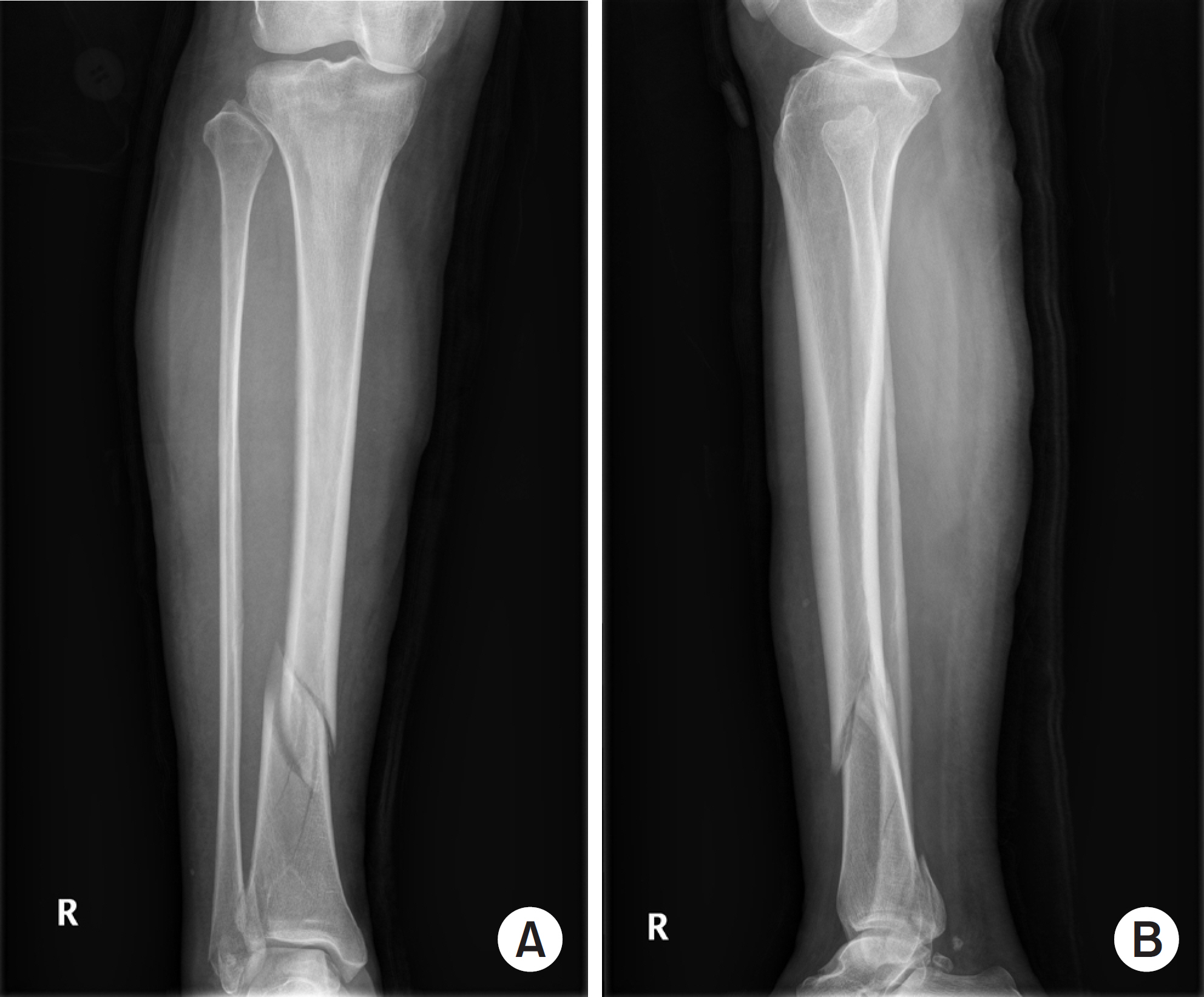
Anteroposterior (A) and lateral (B) plain radiographs of the right tibia and fibula show a spiral fracture with a wedge fragment.
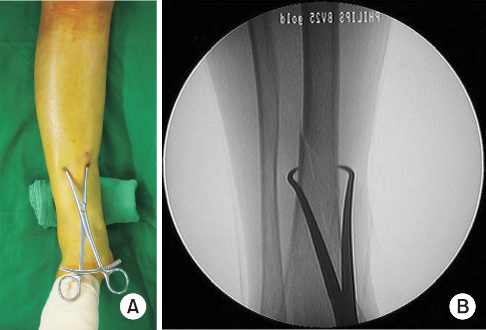
Intraoperative photograph (A) and fluoroscopic image (B) applied pointed bone reduction forceps.
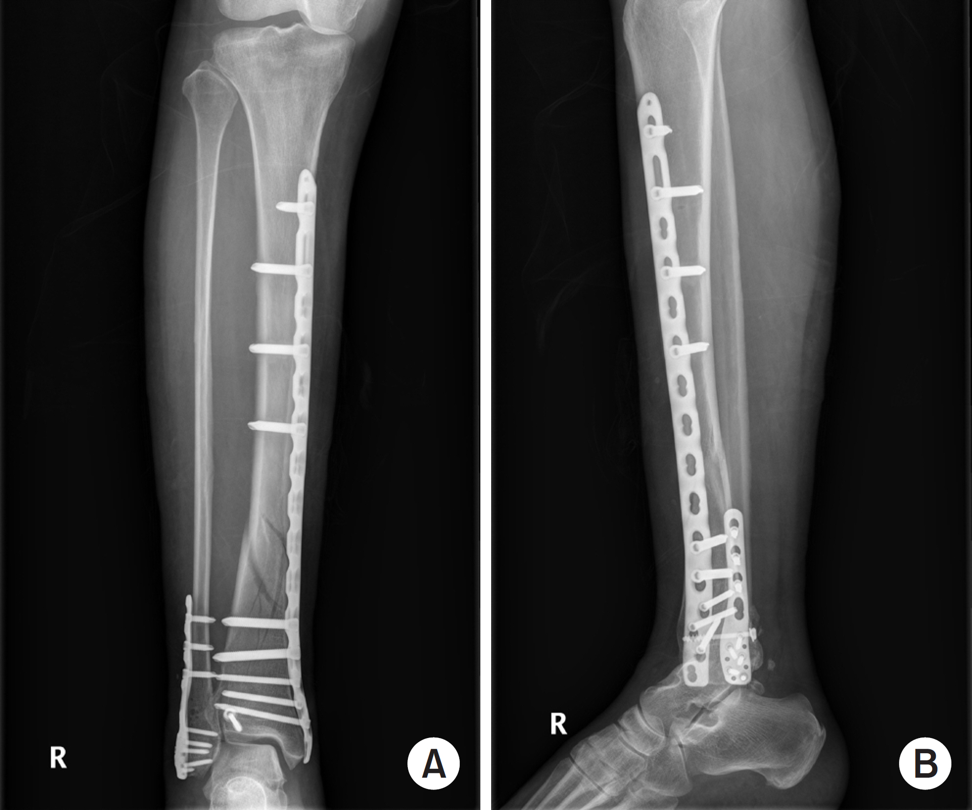
Immediate postoperative anteroposterior (A) and lateral (B) radiographs showed acceptable reduction of both the tibia and fibula.
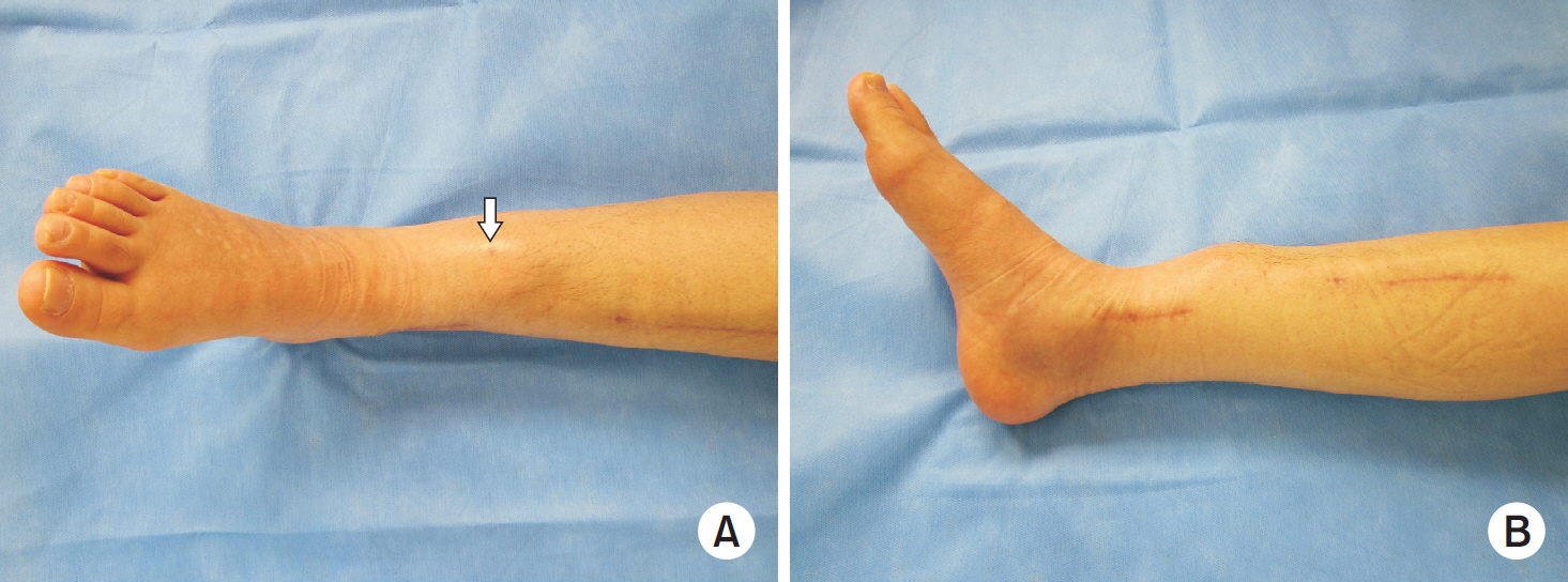
Frontal (A) and sagittal (B) photographs show a pulsating mass in the anterior aspect of the fracture site of the right lower leg. On top of the mass, there is a small scar indicating the insertion point of the pointed bone reduction forceps (arrow).
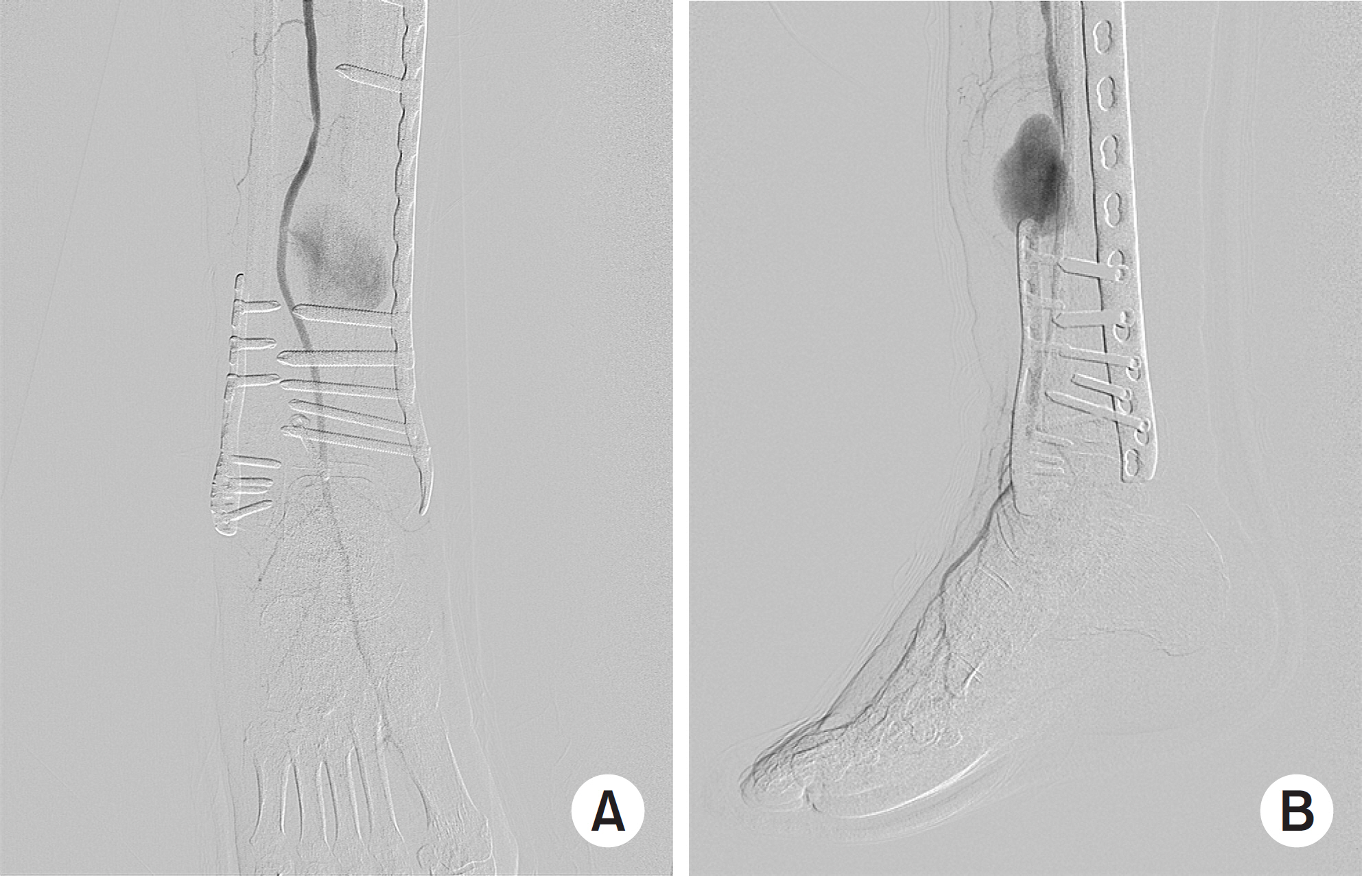
Anteroposterior (A) and lateral (B) arteriograms of the right leg show extravasation of the contrast media indicating pseudoaneurysm of the anterior tibial artery adjacent to the fracture site.